Original Article
Fasudil, an inhibitor of Rho-associated coiled-coil
kinase, attenuates hyperoxia-induced
pulmonary fibrosis in neonatal rats
Xiu-Jie Qi1,2, Wei Ning3, Feng Xu2, Hong-Xing Dang2, Fang Fang1, Jing Li2
1Ministry of Education Key Laboratory of Child Development and Disorders, Key Laboratory of Pediatrics in
Chongqing, Chongqing International Science and Technology Cooperation Center for Child Development and Disorders, Children’s Hospital of Chongqing Medical University, Chongqing 400014, China; 2Department of
Pediatric Intensive Care Unit, Children’s Hospital of Chongqing Medical University, Chongqing 400014, China;
3Daping Hospital and The Research Institute of Surgery of The Third Military Medical University, Chongqing
400042, China
Received August 19, 2015; Accepted September 24, 2015; Epub October 1, 2015; Published October 15, 2015
Abstract: Background: Oxygen therapy is important during the management of high-risk neonatal infants, such as those with preterm birth, low birth weight, and asphyxia. However, prolonged exposure to high oxygen concentra-tions can readily lead to diffuse nonspecific inflammation, which promotes airway remodeling and pulmonary fibro -sis. The Rho/Rho-associated coiled-coil kinase (Rho/ROCK) signaling pathway plays an important role in numerous developmental and proliferative diseases. This study was performed to determine the efficacy of ROCK inhibitor fa -sudil in blocking the development of hyperoxia-induced lung injury and fibrosis in neonatal rats. Methods: Neonatal rats were randomly divided into four groups: air + saline group, air + fasudil group, hyperoxia + saline group, and hyperoxia + fasudil group. The hyperoxia + saline and Hyp + fasudil groups were exposed to 95% oxygen for 21 days and administered intraperitoneal saline or fasudil once daily. The air + saline and air + fasudil group were exposed to 21% oxygen (room air) and administered the same volume of intraperitoneal saline or fasudil. Results: Fasudil-treated rats exhibited improved histopathological changes and decreased lung hydroxyproline content. Fasudil attenuated the protein level of alpha-smooth muscle actin, transforming growth factor-β1, and connective tissue growth factor. Additionally, fasudil reduced the activation of ROCK1 and myosin phosphatase targeting subunit 1 protein in the Rho/ROCK signaling pathway. Conclusions: Fasudil may be a potentially effective therapeutic drug for hyperoxia-induced pulmonary fibrosis.
Keywords: Hyperoxia, neonatal rats, pulmonary fibrosis, fasudil, Rho/ROCK signaling pathway
Introduction
Oxygen therapy is important during the man-agement of high-risk neonatal infants, such as those with preterm birth, low birth weight, and asphyxia. However, prolonged exposure to high oxygen concentrations can readily lead to dif-fuse nonspecific inflammation in the lung parenchyma and interstitium, which promotes airway remodeling and pulmonary fibrosis [1, 2]. Because of their pulmonary surfactant defi-ciency and premature antioxidant system, neo-natal infants (especially preterm infants) can also develop the additional complication of bronchopulmonary dysplasia (BPD), which is characterized by lung inflammation and fibrosis
[3]. Lung immaturity, acute and chronic lung injury, and pulmonary fibrosis are all known to contribute to the development of BPD [4]; how-ever, the pathogenesis of hyperoxia-induced BPD remains unclear.
mmatory cytokines induce the conversion of inactive GDP-bound Rho into active GTP-bound Rho. Active Rho then activates its downstream effector ROCK, which stimulates the phosphor-ylation of myosin phosphatase-targeting sub-unit 1 (MYPT1) at multiple amino acid loci. Phosphorylation of MYPT1 inactivates myosin light chain phosphatase (MLCP), thus affecting the level of cytoplasmic myosin light chain (MLC).
The Rho/ROCK signaling pathway has gained widespread attention in recent years. Specific ROCK inhibitors have become powerful tools in exploring the molecular role of Rho/ROCK sig-naling in various diseases. Fasudil, the only clinically approved ROCK inhibitor, was first used in Japan in 1995 as a novel and efficient vasodilator in patients with subarachnoid hem-orrhage-induced cerebral vasospasm. Since then, a large number of clinical trials have con-firmed its safety [9].
Various studies have suggested that fasudil is capable of inhibiting inflammation, blocking epithelial-mesenchymal transition (EMT), sup-pressing cell proliferation and migration, re- pressing organ reconstruction, and alleviating tissue fibrosis [10-12]. There are numerous reports on the efficacy of fasudil on pulmonary fibrosis in adult mice, nevertheless there are few in neonates. Activation of the Rho/ROCK signaling pathway in bleomycin-induced pulmo-nary fibrosis has suggested that this pathway is an important therapeutic target for fibrotic dis-eases [13]. We hypothesized that fasudil effec-tively blocks the development of hyperoxia-induced pulmonary fibrosis in neonatal rats. For this purpose, we exposed neonatal rats to 95% oxygen for 21 days to establish a hyperox-ia-induced lung fibrosis model [14]. Using this model, we observed the effect of fasudil on hyperoxia-induced lung fibrosis. We evaluated the histopathological changes and hydroxypro-line content of the lungs in hyperoxia-exposed rats. We also measured the protein level of alpha-smooth muscle actin (α-SMA), transform-ing growth factor-β1 (TGF-β1), and connective tissue growth factor (CTGF) in lung tissue to elu-cidate the molecular mechanisms involved in the blocking of hyperoxia-induced lung fibrosis by fasudil in neonatal rats. Finally, we mea-sured the ROCK1 and MYPT1 phosphorylation levels in the Rho/ROCK signaling pathway.
Materials and methods
Animals and treatments
This animal study was approved by our insti- tutional animal research ethics committee. Twenty-four full-term neonatal Sprague-Dawley rats (male and female; body weight, 5.0 ± 0.5 g) were purchased from the experimental animal center of Daping Hospital of the Third Military Medical University, Chongqing, China. All rats were randomly divided into four groups of six rats each: those administered air + saline (Air + NS group), air + fasudil (Air + FAS group), hyper-oxia + saline (Hyp + NS group), and hyperhyper-oxia + fasudil (Hyp + FAS group). Rats in the Hyp + NS and Hyp + FAS groups were placed in animal oxygen cage, into which 100% oxygen was con-stantly flowed. The oxygen concentration was monitored by a digital oxygen concentration meter (CY12C; Xin’an Jiang Analytical In- strument Factory, Jiande, Zhejiang, Hangzhou, China) to ensure that it remained at > 95%; the concentration of carbon dioxide remained at < 5%. Rats in the Air + NS and Air + FS groups were exposed to room air (21% oxygen). Fasudil (Tianjin Chase Sun Pharmaceutical Co., Ltd., Tianjin, China) was intraperitoneally inject-ed at 20 mg/kg once daily for 21 days; the same volume of saline was injected daily as a vehicle. The mother rats were exchanged between the air and hyperoxia groups at 10:00 each morning to prevent the oxygen exposure from affecting their lactation ability. Breeding pads, food, and water were replaced daily. Animal survival rates and body weights were recorded every other day. The oxygen cage was open for less than 1 hour each day. The ambi-ent temperature was 22°C to 26°C, and the humidity was 60% to 70%.
All rats were sacrificed under anesthesia by intraperitoneal injection of 3% chloral hydrate on day 21 following the treatments. The lungs were harvested from each rat. The left lungs were soaked in 4% paraformaldehyde overnight and embedded in paraffin, and the right lungs were frozen in liquid nitrogen for western blot analysis.
Lung histology
development [15], was measured using a verti-cal line drawn from the center of a respiratory bronchiole to the nearest pleura. Ten different fields were randomly selected from each slide under light microscopy (100× magnification).
Hydroxyproline assay by alkali hydrolysis method
The total collagen content of the lung tissue was determined by analysis of hydroxyproline
body to α-SMA (1:200 dilution; Abcam, Cam- bridge, MA, USA). Phosphate-buffered saline was used as a negative control. The next day, the slides were incubated with horseradish per-oxidase-labeled goat anti-rabbit secondary antibody for 1 hour at room temperature. The slides were then stained with Dolichos biflorus
agglutinin (DAB) (Zhongshan Jinqiao Biotechno- logy Co.) and hematoxylin-eosin, dehydrated, clarified, and mounted. Cells with brown
gran-Figure 1. Body weights and mortality rates at 21 days. Rats in the hyperoxia groups were exposed to 95% oxygen for 21 days. Rats in the air groups were exposed to room air (21% oxygen). Fasudil was intraperitoneally injected at 20 mg/kg once daily for 21 days. All rats were sacrificed under anesthesia on day 21. A. Body weights on day 21. B. Survival rates. NS, saline; FAS, fasudil; Hyp, hyperoxia. #P < 0.05 compared with Air + NS group.
on day 21 after hyperoxia exposure as previously described [13]. The hy- droxyproline assay was conducted using a hydroxy-proline test kit (A030-2; Jiancheng Biological Engi- neering Institute, Nanjing, China) according to the manufacturer’s instructi- ons. Briefly, 1 ml of hydroly-sate was added to 30 to 100 mg of lung tissue. The tissue was boiled for 20 minutes and then cooled using flowing tap water. Reaction reagents were added to the lung tissue fol-lowing adjustment of the pH value, and the absor-bance at 550 nm was mea-sured. The content of hydroxyproline in the lung tissue of rats from each group was then calculated according to the manufac-turer’s instructions.
Immunohistochemistry
[image:3.612.89.384.69.519.2]ules in the cell membrane and cytoplasm were considered to be α-SMA-positive cells.
Western blot analysis
The frozen lung tissue was homogenized with radioimmunoprecipitation assay lysis buffer containing phenylmethylsulfonyl fluoride. The total protein was extracted, and the protein concentration was determined using a bicin-choninic acid assay. Fifty micrograms of protein from each sample was loaded into 10% sodium dodecyl sulfate polyacrylamide gel electropho-resis and transferred to a polyvinylidene difluo-ride membrane. After blocking in 5% bovine serum albumin at room temperature for 1 hour,
the membrane was then incubated with specif-ic primary antibodies overnight at 4°C. The next day, the membrane was incubated with a sec-ondary antibody followed by development using an enhanced chemiluminescence kit (Catalog No. KGP1123; Key Gen Biotech, Nanjing, China). The primary antibodies included anti-α-SMA antibody (1:200), anti-TGF-β1 antibody (1:1000) (GeneTex, Inc., Irvine, CA, USA), anti-CTGF antibody (1:1000) (GeneTex), anti-ROCK1 antibody (1:500), anti-p-MYPT1 antibody (1: 1000), anti-MYPT1 antibody (1:1000) (Cell Sig- naling Technology, Inc., Beverly, MA, USA), and rabbit anti-β-actin polyclonal antibody (1:2000) (Si Zhen Bai Biological Technology, Beijing, China).
Statistical analysis
Statistical analyses were performed using SPSS Statistics for Windows, Version 19.0 (IBM Corp., Armonk, NY, USA). Data are expressed as mean ± standard deviation. Comparison of dif-ferences among groups was performed using one-way analysis of variance followed by LSD test. The cumulative survival rate was calculat-ed using Kaplan-Meier survival analysis. Sta- tistically significant differences were indicated by P values of < 0.05.
Results
Effects of fasudil on body weight and mortality
Compared to air groups, the body weights of hyperoxia groups were significantly reduced fol-lowing 21 days of hyperoxia treatments (P < 0.05). The body weight of Hyp + FAS group was even lower than that Hyp + NS group; however, there was no significant difference between the two groups (P > 0.05) (Figure 1A).
The animal survival rates were 100.0% in the Air + NS and Air + FAS groups, 66.7% in the Hyp + NS group, and 58.3% in the Hyp + FAS group. The survival rates were significantly lower in the hyperoxia than air groups (P < 0.05). The sur-vival rate in the Hyp + FAS group was lower than that in the Hyp + NS group; however, there was no significant difference between the two groups (P > 0.05) (Figure 1B).
Effects of fasudil on histopathological changes
The effect of fasudil on the development of hyperoxia-induced inflammation and fibrosis was examined on day 21 (Figure 2A-H). A
[image:5.612.89.288.73.205.2]nor-Figure 3. Radial alveolar count. Rats were exposed to hyperoxia and treated with fasudil for 21 days. All rats were sacrificed on day 21, and their lungs were harvested. The radial alveolar count was calculated as described in the Materials and methods. NS, sa -line; FAS, fasudil; Hyp, hyperoxia. #P < 0.05 com-pared with Air + NS group.
[image:5.612.90.285.307.433.2]mal, well-alveolarized lung structure was ob- served in the Air + NS and Air + FAS groups. In contrast, hyperoxia treatment induced the development of a disordered lung tissue struc-ture, alveolar collapse, obvious alveolar wall thickening, and numerous blue-stained stripes
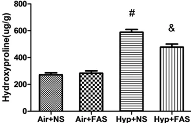
[image:6.612.90.387.69.549.2]trast, hyperoxia treatment resulted in signifi-cantly elevated lung tissue hydroxyproline lev-els (P < 0.05). The content of hydroxyproline was significantly lower in rats treated with fasudil than in those exposed to hyperoxia alone (P < 0.05).
Figure 5. α-SMA protein levels in lung tissue. Rats were exposed to hyperoxia and treated with fasudil for 21 days. All rats were sacrificed on day 21, and their lungs were harvested. A-D. Immunohistochemical staining for α-SMA in lung tis -sue (200× magnification). Vascular smooth muscle cell. Bronchial smooth muscle cell. Myofibroblast. E. Western blot for expression of α-SMA protein in lung tissue. a. Air + NS group; b, Air + FAS group; c, Hyp + NS group; d, Hyp + FAS group. #P < 0.05 compared with Air + NS group; &P < 0.05 compared with Hyp + NS group.
and flakes indicating colla-gen deposition. Although fibrotic lesions were obser- ved in the Hyp + FAS group, both the extent and inten-sity of the lesions were less severe than those of the Hyp + NS group.
Effects of fasudil on RAC
We measured the RAC va- lue on day 21 to observe the effect of fasudil on the development of neonatal rat lungs (Figure 3). RAC values were similar be- tween the Air + NS and Air + FAS groups. However, hyperoxia treatment signifi-cantly lowered the RAC val-ues in the Hyp + NS group (P < 0.05). The RAC value in the Hyp + FAS group was slightly higher than that in the Hyp + NS group, but there was no statistically significant difference.
Effects of fasudil on hy-droxyproline level
con-Effects of fasudil on α-SMA
Myofibroblasts play important roles in promot-ing inflammation, tissue repair, and pulmonary fibrosis. Therefore, we examined the expres-sion and location of the myofibroblast-specific marker α-SMA in lung tissue by immunohisto-chemistry and measured the protein content by western blot analysis (Figure 5A-E). Immu- nohistochemical staining of lung sections showed that α-SMA-positive cells were mainly expressed in the bronchial smooth muscle cells and vascular smooth muscle cells of the rats in the air groups. Only small numbers of α-SMA-positive cells were observed in the alveolar septa, alveolar surfaces, and bronchiolar epi-thelium. Conversely, after exposure to hyperox-ia, large numbers of α-SMA-positive cells were noted in the alveolar septa and alveolar surfac-es. Substantially fewer α-SMA-positive cells were present in the hyperoxia-exposed lung tis-sue after fasudil treatment.
Semiquantitative western blotting revealed that the α-SMA protein levels were similar between the Air + NS and Air + FAS groups. In contrast, the α-SMA protein levels were signifi-cantly higher in the hyperoxia groups than in the air groups (P < 0.05). However, the levels were significantly lower after fasudil treatment (P < 0.05) (Figure 5E).
Effects of fasudil on TGF-β1 and CTGF
The profibrotic cytokines TGF-β1 and CTGF play key roles in the occurrence and development of pulmonary fibrosis. We examined the protein levels of TGF-β1 and CTGF in lung tissue on day 21 by western blotting (Figure 6A and 6B). The protein levels of both TGF-β1 and CTGF were similar between the Air + NS and Air + FAS groups. In contrast, hyperoxia treatment was associated with significantly higher TGF-β1 and CTGF protein levels in both the Hyp + NS and Hyp + FAS groups (P < 0.05). However, the lev-els were significantly lower in the Hyp + FAS group than in the Hyp + NS group (P < 0.05).
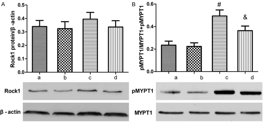
Effects of fasudil on p-MYPT1 and ROCK1 pro-tein levels
[image:7.612.93.519.72.282.2]The expression of ROCK1 and p-MYPT1 in lung tissue after hyperoxia exposure was measured by western blotting (Figure 7A and 7B). The lev-els of ROCK1 and phosphorylated MYPT1 exhibited no differences between the two air groups. Hyperoxia exposure stimulated a rising trend in the ROCK1 protein level (P > 0.05), but a significantly increased MYPT1 phosphoryla-tion level. Fasudil treatment reduced the expression of ROCK1 protein (P > 0.05) and sig-nificantly decreased the MYPT1 phosphoryla-tion level (P < 0.05).
Discussion
To the best of our knowledge, this is the first report of the attenuation of hyperoxia-induced pulmonary fibrosis by fasudil, a ROCK inhibitor, in neonatal rats. In this study, fasudil signifi-cantly (1) improved hyperoxia-induced patho-logical changes and reduced the content of hydroxyproline in the lungs of neonatal rats, (2) inhibited both expression of the myofibroblast-specific marker α-SMA and production of the profibrotic cytokines TGF-β1 and CTGF, and (3) inhibited the activation of ROCK1 and MYPT1 in the Rho/ROCK signaling pathway.
Transformation of fibroblasts into myofibro-blasts plays a pivotal role in the pathogenesis of fibrosis [16]. Myofibroblasts are a special type of fibroblasts that possess the functions of both fibroblasts and smooth muscle cells. α-SMA is considered to be a characteristic marker of myofibroblasts [17]. Known sources of myofibroblasts include local fibroblasts, bone marrow-derived fibrocytes, and resident epithelia cells (through epithelial mesenchymal transition) [18, 19].
[image:8.612.91.522.71.271.2]Myofibroblasts secrete cytokines, chemokines, growth factors, extracellular matrix (ECM), and protease and play important roles in inflamma-tion, remodeling, and fibrosis in various tissues [20, 21]. The Rho/ROCK signaling pathway is
capable of inducing EMT, regulating the produc-tion of myofibroblasts, and participating in the development of fibrosis [22]. Studies have shown that fasudil blocks bleomycin-induced pulmonary fibrosis by inhibition of EMT and downregulation of α-SMA mRNA and protein levels [13]. Y-27632, another ROCK inhibitor, inhibits renal fibrosis through similar mecha-nisms [23]. Our findings are consistent with these reports [16], in which hyperoxia signifi-cantly upregulated and fasudil signifisignifi-cantly reduced the expression of α-SMA on the alveo-lar surface. These results imply that hyperoxia may promote the transition of alveolar epitheli-al cells to myofibroblasts. We speculate that the antifibrotic effect of fasudil in neonatal rats may be mediated by inhibition of EMT.
TGF-β1 plays important regulatory roles in vari-ous pathological processes involving inflamma-tory responses, tissue repair, and organ fibrosis [24-26]. As a downstream target of TGF-β1, CTGF collaboratively promotes fibroblast prolif-eration and accumulation, stimulates the syn-thesis of collagen and fibronectin, and acceler-ates deposition of the ECM [27]. Yin et al. [28] reported that both TGF-β1 and CTGF were sig-nificantly upregulated in rats with bleomycin-induced pulmonary fibrosis. Application of anti-TGF-β1 antibody and anti-CTGF antibody signifi-cantly inhibits secretion and synthesis of colla-gen and other ECM proteins. In the present
study, hyperoxia upregulated the protein levels of both TGF-β1 and CTGF in lung tissue, and fasudil treatment significantly decreased these levels. This finding suggests that both TGF-β1 and CTGF play important roles in the pathogen-esis of hyperoxia-induced pulmonary fibrosis. Inhibition of the profibrotic cytokines TGF-β1 and CTGF is likely to be an important molecular mechanism that contributed to the antifibrotic effect of fasudil in this study.
Rho/ROCK signaling is a ubiquitous pathway in various tissues of the body and mediates the process of tissue fibrogenesis through its func-tional contributions to the inflammatory res- ponse, its ability to block EMT, and its regula-tion of the cytoskeleton and cell migraregula-tion, as well as other activities [23, 29, 30]. Nagatoya et al. [31] reported that the RhoB mRNA and protein levels were significantly upregulated in unilateral ureteral obstruction-induced fibrotic kidneys. Shimizu et al. [8] showed that the ROCK2 mRNA and protein levels were increased in bleomycin-induced fibrotic lungs. These stud-ies suggest that the Rho/ROCK signaling path-way plays important roles in the pathogenesis of organ fibrosis. In the present study, the expression of ROCK1 protein in lung tissue slightly increased and the expression of its downstream active target p-MYPT1 protein was significantly upregulated following 21 days of hyperoxia exposure. Fasudil treatment slightly reduced the ROCK1 protein levels and signifi-cantly reduced the p-MYPT1 expression. These results suggest that the Rho/ROCK pathway was activated at the protein level in this hyper-oxia-induced pulmonary fibrosis model. The Rho/ROCK signaling pathway may be involved in hyperoxia-induced pulmonary fibrosis.
In addition to its profibrotic role, ROCK also plays an important role in lung development in neonates; e.g., it is involved in the formation of pulmonary gas exchange units and expansion of the lungs [32, 33]. We examined the RAC val-ues following fasudil treatment to study the role of fasudil in lung development in neonatal rats. Our data suggest that fasudil has no apparent effects on RAC values and that inhibition of the ROCK pathway by fasudil may have little effect on normal lung development in neonatal rats. This finding is in line with that reported by Ziino et al. [34]. One explanation is that fasudil can-not completely inhibit ROCK activity as
previ-ously reported [32]. Therefore, the remaining ROCK activity is able to maintain the normal lung development. We also noted that fasudil had no obvious effect on the body weight or mortality of the normoxic neonatal rats in the present study. Under hyperoxic conditions, however, fasudil appeared to have an amplified effect on hyperoxia-induced body weight loss and increased mortality in rats. The underlying mechanism of this phenomenon is unclear. One possible mechanism involves the low tis-sue perfusion caused by inhibition of the ROCK pathway.
In summary, fasudil blocks the development of hyperoxia-induced pulmonary fibrosis in neona-tal rats. The antifibrotic effect is possibly medi-ated by decreasing the levels of the profibrotic cytokines TGF-β1 and CTGF and inhibiting the development of myofibroblasts in lung tissue, thereby inhibiting ECM synthesis. The regulato-ry mechanism of fasudil may be related to its inhibition of activation of the Rho/ROCK signal-ing pathway. Targetsignal-ing the Rho/ROCK signalsignal-ing may represent a novel therapy for hyperoxia-induced pulmonary fibrosis. The effect of fasudil on lung development and other organs remains to be further studied. Clinically, we may be able to use local therapy specifically tar-geting lung, such as inhalation of fasudil to reduce pulmonary fibrosis so as to avoid the systemic effects of fasudil on human body.
Acknowledgements
This study was supported by funding from National Natural Science Foundation of China (NO. 81101442). We thank Medjaden Biosci- ence Limited for assisting in the preparation of this manuscript.
Disclosure of conflict of interest
None.
Authors’ contributions
JL, FX and HXD conceived and designed the experiments. XJQ and WN performed the exper-iments and analyzed the data. XJQ wrote the paper. FF contributed reagents, materials and laboratory environment.
of Chongqing Medical University, Chongqing 400-014, China. Tel: +8613883077978; E-mail: lijing-wangyi@126.com; lijing12555@163.com
References
[1] Kallet RH and Matthay MA. Hyperoxic acute lung injury. Respir Care 2013; 58: 123-141. [2] Lv R, Zheng J, Ye Z, Sun X, Tao H, Liu K, Li R, Xu
W, Liu W and Zhang R. Advances in the therapy of hyperoxia-induced lung injury: findings from animal models. Undersea Hyperb Med 2014; 41: 183-202.
[3] Dieperink HI, Blackwell TS and Prince LS. Hy-peroxia and apoptosis in developing mouse lung mesenchyme. Pediatr Res 2006; 59: 185-190.
[4] Ali Z, Schmidt P, Dodd J and Jeppesen DL. Bronchopulmonary dysplasia: A review. Arch Gynecol Obstet 2013; 288: 325-333.
[5] Amin E, Dubey BN, Zhang SC, Gremer L, Dvor -sky R, Moll JM, Taha MS, Nagel-Steger L, Piekorz RP, Somlyo AV and Ahmadian MR. Rho-kinase: regulation, (dys)function, and inhibi-tion. Biol Chem 2013; 394: 1399-1410. [6] Amano M, Nakayama M and Kaibuchi K.
Rho-kinase/ROCK: A key regulator of the cytoskel-eton and cell polarity. Cytoskelcytoskel-eton (Hoboken) 2010; 67: 545-554.
[7] de Godoy MA and Rattan S. Role of rho kinase in the functional and dysfunctional tonic smooth muscles. Trends Pharmacol Sci 2011; 32: 384-393.
[8] Shimizu Y, Dobashi K, Iizuka K, Horie T, Suzuki K, Tukagoshi H, Nakazawa T, Nakazato Y and Mori M. Contribution of small GTPase Rho and its target protein rock in a murine model of lung fibrosis. Am J Respir Crit Care Med 2001; 163: 210-217.
[9] Mohri M, Shimokawa H, Hirakawa Y, Masumo -to A and Takeshita A. Rho-kinase inhibition with intracoronary fasudil prevents myocardial ischemia in patients with coronary microvascu-lar spasm. J Am Coll Cardiol 2003; 41: 15-19. [10] Righetti RF, Pigati PA, Possa SS, Habrum FC,
Xisto DG, Antunes MA, Leick EA, Prado CM, Martins Mde A, Rocco PR and Tiberio Ide F. Ef -fects of Rho-kinase inhibition in lung tissue with chronic inflammation. Respir Physiol Neu -robiol 2014; 192: 134-146.
[11] van Beuge MM, Prakash J, Lacombe M, Gos -ens R, Post E, Reker-Smit C, Beljaars L and Poelstra K. Reduction of fibrogenesis by selec -tive delivery of a Rho kinase inhibitor to hepat-ic stellate cells in mhepat-ice. J Pharmacol Exp Ther 2011; 337: 628-635.
[12] Zhang H, Liu X, Liu Y, Yi B and Yu X. Epithelial-mesenchymal transition of rat peritoneal me-sothelial cells via Rhoa/Rock pathway. In Vitro Cell Dev Biol Anim 2011; 47: 165-172.
[13] Jiang C, Huang H, Liu J, Wang Y, Lu Z and Xu Z. Fasudil, a rho-kinase inhibitor, attenuates bleomycin-induced pulmonary fibrosis in mice. Int J Mol Sci 2012; 13: 8293-8307.
[14] Dang H, Wang S, Yang L, Fang F and Xu F. Up -regulation of Shh and Ptc1 in hyperoxiain-duced acute lung injury in neonatal rats. Mol Med Rep 2012; 6: 297-302.
[15] Lavezzi AM, Corna MF, Alfonsi G and Matturri L. Possible role of the alpha7 nicotinic receptors in mediating nicotine’s effect on developing lung - implications in unexplained human peri-natal death. BMC Pulm Med 2014; 14: 11. [16] Ni J, Dong Z, Han W, Kondrikov D and Su Y. The
role of RhoA and cytoskeleton in myofibroblast transformation in hyperoxic lung fibrosis. Free Radic Biol Med 2013; 61: 26-39.
[17] Phan SH. Genesis of the myofibroblast in lung injury and fibrosis. Proc Am Thorac Soc 2012; 9: 148-152.
[18] Lee R, Perry B, Heywood J, Reese C, Bonner M, Hatfield CM, Silver RM, Visconti RP, Hoffman S and Tourkina E. Caveolin-1 regulates chemo-kine receptor 5-mediated contribution of bone marrow-derived cells to dermal fibrosis. Front Pharmacol 2014; 5: 140.
[19] Lee R, Reese C, Bonner M, Tourkina E, Hajdu Z, Riemer EC, Silver RM, Visconti RP and Hoff -man S. Bleomycin delivery by osmotic mini-pump: similarity to human scleroderma inter-stitial lung disease. Am J Physiol Lung Cell Mol Physiol 2014; 306: L736-748.
[20] Kendall RT and Feghali-Bostwick CA. Fibro-blasts in fibrosis: novel roles and mediators. Front Pharmacol 2014; 5: 123.
[21] Reese C, Lee R, Bonner M, Perry B, Heywood J, Silver RM, Tourkina E, Visconti RP and Hoff -man S. Fibrocytes in the fibrotic lung: altered phenotype detected by flow cytometry. Front Pharmacol 2014; 5: 141.
[22] He J, Xu Y, Koya D and Kanasaki K. Role of the endothelial-to-mesenchymal transition in renal fibrosis of chronic kidney disease. Clin Exp Nephrol 2013; 17: 488-497.
[23] Liu M, Gu M, Wu Y, Zhu P, Zhang W, Yin C and Zhang WJ. Therapeutic effect of Y-27632 on chronic allograft nephropathy in rats. J Surg Res 2009; 157: e117-127.
[24] Gagliardo R, Chanez P, Gjomarkaj M, La Grutta S, Bonanno A, Montalbano AM, Di Sano C, Al -bano GD, Gras D, Anzalone G, Riccobono L and Profita M. The role of transforming growth fac -tor-beta1 in airway inflammation of childhood asthma. Int J Immunopathol Pharmacol 2013; 26: 725-738.
[26] Boothe DL, Coplowitz S, Greenwood E, Barney CL, Christos PJ, Parashar B, Nori D, Chao KS and Wernicke AG. Transforming growth factor beta-1 (TGF-beta1) is a serum biomarker of ra -diation induced fibrosis in patients treated with intracavitary accelerated partial breast ir-radiation: preliminary results of a prospective study. Int J Radiat Oncol Biol Phys 2013; 87: 1030-1036.
[27] Qi W, Chen X, Poronnik P and Pollock CA. Transforming growth factor-beta/connective tissue growth factor axis in the kidney. Int J Bio-chem Cell Biol 2008; 40: 9-13.
[28] Yin Q, Nan HY, Zhang WH, Yan LF, Cui GB, Huang XF and Wei JG. Pulmonary microvascu -lar endothelial cells from bleomycin-induced rats promote the transformation and collagen synthesis of fibroblasts. J Cell Physiol 2011; 226: 2091-2102.
[29] Mong PY and Wang Q. Activation of Rho kinase isoforms in lung endothelial cells during in-flammation. J Immunol 2009; 182: 2385-2394.
[30] Komers R. Rho kinase inhibition in diabetic ne-phropathy. Curr Opin Nephrol Hypertens 2011; 20: 77-83.
[31] Nagatoya K, Moriyama T, Kawada N, Takeji M, Oseto S, Murozono T, Ando A, Imai E and Hori M. Y-27632 prevents tubulointerstitial fibrosis in mouse kidneys with unilateral ureteral ob-struction. Kidney Int 2002; 61: 1684-1695. [32] Cloutier M, Tremblay M and Piedboeuf B.
ROCK2 is involved in accelerated fetal lung de-velopment induced by in vivo lung distension. Pediatr Pulmonol 2010; 45: 966-976.
[33] Wan H, Liu C, Wert SE, Xu W, Liao Y, Zheng Y and Whitsett JA. CDC42 is required for struc -tural patterning of the lung during develop-ment. Dev Biol 2013; 374: 46-57.