Introduction
Macrophages are mature myeloid cells, mostly derived from the differentiation of circulating monocytes after tissue infiltration. Far to be a homogeneous population, macrophages display a wide range of phenotypes and physiological properties depending on the cytokines inducing their maturation [1]. Moreover, macrophages are very plastic cells, able to switch from one functional sub-population to another depending on the stimulus received [2]. Another aspect of macrophage heterogeneity is the tissue speciali-sation of resident macrophages, as microglial cells in the brain, Kupffer cells in the liver or alveolar macrophages in the lung. However, it is still not clear if these resident macrophages are derived in steady state condition from circulat-ing monocytes and if they are terminally differ-entiated cells [1]. After birth, macrophages are known to affect different stages of skin wound healing, modulating the function of the different cell types involved in this process. The benefit of inflammation and inflammatory cells in the wound healing is a matter of debate as in nor-mal conditions it promotes wound closure but
also the fibrosis associated with scar formation. Wound Associated Macrophages (WAM) have a central role in the control of wound inflamma-tion. Here, we will present an overview of the main functions of WAM and their influence on the other major cell types present in the wound during the healing process and how modulating these processes might result in better wound healing.
Wound healing
In humans, and more widely in all mammalian species, the wound healing process can be sub-divided in three consecutive and overlapping stages: inflammation, new tissue formation and remodelling [3]. The transition from one stage to another depends on the maturation and dif-ferentiation of the main cell populations in-volved, among which the keratinocytes, the fi-broblasts and the macrophages.
Inflammation
The first event occurring after injury is the for-mation of a blood clot by activated platelets.
Review Article
Skin wound healing modulation by macrophages
Mathieu P. Rodero, Kiarash Khosrotehrani
University of Queensland Centre for Clinical Research, Experimental Dermatology Group, Brisbane, Australia
Received July 5, 2010; accepted July 23, 2010; available online July 25, 2010
Abstract: Skin wound healing is a multi stage phenomenon that requires the activation, recruitment or activity of nu-merous cell types as keratinocytes, endothelial cells, fibroblast and inflammatory cells. Among the latter, macro-phages appear to be central to this process. They colonize the wound at its very early stage and in addition to their protective immune role seem to organize the activity of other cell types at the following stages of the healing. Their benefit to this process is however controversial, as macrophages are described to promote the speed of healing but may also favour the fibrosis resulting from it in scars. Moreover wound healing defects are associated with abnormali-ties in the inflammatory phase. In this review, we summarise our knowledge on what are the Wound Associated Macrophages, and how they interact with the other cell types to control the reepithelialisation, angiogenesis and the extracellular matrix remodelling. We believe this knowledge may open new avenues for therapeutic intervention on skin wounds.
The blood plug will be composed of various cell types including platelet, red and white blood cells. The initial plug is stabilised by fibrin fibres and will be a scaffold for the various infiltrating cells. The first inflammatory cells recruited are the neutrophils [4]. They infiltrate massively the wound during the first 24h post injury [5] at-tracted by the numerous inflammatory cytokines produced by the activated platelets, endothelial cells, as well as by the degradation products from pathogens. Neutrophils enter apoptosis soon after infiltrating the wound and the release of cytokines during this apoptotic process is an important component in macrophage recruit-ment. Macrophages infiltrate the wound mas-sively 2 days post injury and exacerbate at this stage an intense phagocytic activity [6].
New tissue formation
The reepithelialisation process begins few hours after the wound formation. Keratinocytes from the wound edges migrate over the wound bed at the interface between the wound dermis and the fibrin clot. This migration is facilitated by the production of specific proteases such as the collagenase by the epidermal cells to degrade the extracellular matrix [7]. Activated fibroblasts also migrate to the wound bed and form, with the macrophages, the granulation tissue. A massive angiogenesis allowing the supply of oxygen and nutrients necessary for the healing process also occurs within this tissue [8]. Later, some of the fibroblasts differentiate into myofi-broblasts. These contractile cells will help bridge the gap between the wound edges [9]. During the same time, growth factors produced by the granulation tissue will favour proliferation and differentiation of epithelial cells restoring the epithelial barrier integrity.
Remodelling
The last stage of the wound healing process consists in a gradual involution of the granula-tion tissue and dermal regeneragranula-tion. This step is associated with the apoptosis of myofibroblasts, endothelial cells and macrophages. The remain-ing tissue is therefore composed mostly of ex-tracellular matrix proteins, essentially collagen type III that will be remodelled by the metallo-proteinase produced by the epidermal cells, endothelial cells, fibroblasts and the macro-phages remaining in the scar and be replaced by collagen type I [8].
Macrophages and wound healing
Wound macrophage phenotypes
Macrophages used to be divided in several sub-populations depending of the way they had been activated, their cell surface markers or their functionality. [1, 10,11]. Schematically, macrophages activated by microbial agents and cytokines like Interferon gamma (IFNγ) are clas-sified as M1 macrophages. These macrophages produce an important level of Nitric Oxide (NO) and pro inflammatory cytokines such as Tumor Necrosis Factor alfa (TNFα), IL-1β, IL-6, or IL-12, and overexpress MHC class II molecules. They bear microbicidal and antitumoral properties. M2 macrophages are a much more heterogene-ous population composed of all macrophages that do not correspond to M1 characteristics [12]. M2 macrophages could be divided in 3 sub-populations. The alternatively activated macrophages or M2a, that promote a Th2 type of inflammation resulting in increased IgE as observed in allergy and parasite immunity, the M2b macrophages, that promote Th2 inflamma-tion and bear some immunoregulainflamma-tion proper-ties, and the deactivated macrophages or M2c, able to control the inflammation and implicated in tissue remodelling [12]. In another classifica-tion, M1 macrophages are considered as the classically activated macrophages as compared to the alternatively activated macrophages. The best described alternative activation consists in the stimulation of the IL-4R by IL-4 and IL-13 that induces pro Th2 macrophages [13]. Of course, all these sub populations should not be considered as distinct populations in vivo but more as different stages of a continuum of acti-vation and differentiation of macrophage popu-lations [14]. The phenotype of skin wound infil-trating macrophages is not yet fully character-ised, but it already appears that it changes dur-ing the healdur-ing process suggestdur-ing that macro-phages have different roles in the diverse phases of skin repair [15, 16].
macrophages as cells bearing both classical activation markers, like TNFα expression, and alternatively activated markers like the man-nose receptor [15]. The authors also described the evolution of the macrophage phenotype. Day 1 macrophages produced more TNFα and IL6 and less Tumor Growth Factor beta (TGFβ) compared to day 7 macrophages suggesting a transition from an inflammatory to an immu-noregulatory or tissue remodelling state. How-ever, it is important to consider that neither IL4 nor IL13 were detectable in this model, perhaps because of the aseptic experimental condition. However, it is not clear to what extent the bacte-rial colonization of the wound would modify Wound Associated Macrophage phenotypes and healing functions, as it is clear that parasitic and bacterial component are major macro-phage activators [13].
Macrophage importance in wound healing
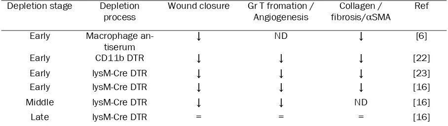
In order to show the implication of macrophages in the control of wound healing, several studies have analysed skin wound healing upon macro-phage depletion (Table 1). The first attempt con-sisted in corticosteroids associated with anti macrophage sera to deplete macrophages in wounded guinea pigs. This treatment resulted in a delayed infiltration of the wound by fibroblasts and decreased fibrosis [6]. These results are in accordance with observations that fetal wounds heal without fibrosis and are not infiltrated by macrophages. In a model of PU.1 null mice where animals lacked macrophages, mast cells and functional neutrophils due to defective mye-lopoiesis, wounds performed on newborns addi-tionally treated with antibiotic healed at the same speed as wild types, but without scar for-mation, suggesting that inflammatory cells are
not needed for wound closure [21]. However, several recent models of specific inducible macrophage depletion, based on genetically modified mice resulted in detrimental effect of pre-injury depletion of macrophages [16, 22, 23]. Mice depleted before injury typically show a defect in re-epithelisation, granulation tissue formation, angiogenesis, wound cytokine pro-duction and myofibroblast associated wound contraction. Recently, the effect of a post-injury depletion of the WAM has also been investi-gated. Macrophage depletion during the granu-lation tissue formation, about 3 days after in-jury, is associated with vascularisation defect, delay in wound closure as well as in granulation tissue maturation [16]. Finally, in their study, Lucas et al did not find any morphological or biological differences between mice that were or were not depleted 9 days after injury suggest-ing no further implication of macrophages at later stages.
During the healing process numerous cytokines and growth factors are produced by the various cell types present in the wound or at the wound edge [24]. The level of production of the differ-ent cytokines depends on the regulation of the cross talk between the major cell populations in the wound: epithelial cells, endothelial cells, fibroblasts and inflammatory cells. However, the literature clearly lacks in vivo characterisation of the cytokine production kinetics by the various cell types during skin wound healing. Most of our current knowledge concerning the ability of a cell population to produce or to respond to a specific cytokine is based on in vitro studies.
Cross talk with keratinocyte
[image:3.612.79.533.100.228.2]Injured and inflamed keratinocytes produce Table 1. Impact of macrophages depletion strategy on wound healing
Depletion stage Depletion
process Wound closure Gr T fromation / Angiogenesis fibrosis/Collagen / αSMA Ref Early Macrophage
an-tiserum ↓ ND ↓ [6]
Early CD11b DTR ↓ ↓ ↓ [22]
Early lysM-Cre DTR ↓ ↓ ↓ [23]
Early lysM-Cre DTR ↓ ↓ ↓ [16]
Middle lysM-Cre DTR ↓ ↓ ND [16]
Late lysM-Cre DTR = = = [16]
several cytokines allowing the recruitment and activation of the WAM, including chemokines, interleukins and growth factors [25, 26]. On the other hand, the ability of immune cells to pro-duce factors able to regulate keratinocyte growth in vitro has been described since 1988 [27]. Interestingly, conditioned medium from co-culture of macrophages and allogenic T cells were much more efficient in inducing keratino-cyte proliferation compared to conditioned me-dium from macrophages alone. However, the direct implication of WAM in producing cyto-kines or growth factors to influence directly or indirectly keratinocyte migration and prolifera-tion is not clear in vivo. Several cytokines and growth factors, that can be produced by macro-phages, have been associated with reepitheliali-sation, mostly in vitro. Among the Epithelial growth Factor family (EGF), the main members involved in wound healing are EGF, TGFα and Heparin Bound EGF (EGF-HB) [28-32]. The acti-vation of the EGF Receptor (EGFR) on keratino-cytes promotes cell migration and proliferation. Although, only TGFα and EGF-HB have been described to be produced by macrophages in vitro, their production by WAM in vivo is more enigmatic [33, 34]. TGFα has been detected in macrophages collected from sub epidermal wound cylinders 6 days after implantation in mice [35]. However, no significant wound heal-ing abnormalities were observed in TGFα-/- mice [36]. In addition, all EGFR ligands are syn-thetised as membrane-anchored forms, which can be released as a soluble form after prote-olysis by MMP [37]. Therefore, another possible role for WAM might be the regulation of EGF family release by the production of MMPs. Other cytokines as IL-6, IL1 and TNF-α produced by macrophages are also associated with reepi-thelisation. Even if the secretion of these cyto-kines by WAM has not been clearly demon-strated yet, the ability of macrophages activated by the inflammatory cytokine IFNγ and/or bacte-rial products via Toll Like Receptor 4 (TLR4) to produce IL-6, IL1β and TNFα makes this hy-pothesis probable [13]. The implication of IL-1 and IL-6 in the reepithelialisation is indirect and is mediated by other cell types present in the wound, at least the fibroblast [38, 39]. On the contrary, TNFα directly stimulates transcription of genes associated with numerous cell func-tions including inflammation, mobility, cell divi-sion and survival in keratinocytes [25]. Of note, in a model of sponge implanted under aseptic conditions in the dermis, macrophages
ex-tracted on day 1 produced more TNFα com-pared to day 7 [15]. Finally, the other cytokine known to be massively produced by macro-phages, and highly implicated in reepithelialisa-tion, is TGFβ, although its effect on keratinocyte proliferation and migration remains controver-sial [36, 40-43]. Cultured WAM extracted from implanted sponges start producing TGFβ from the very first day of the wound [15].
Influence on fibroblasts and myofibroblast
fibro-blasts [54, 55]. It induces the production of sev-eral growths factors by fibroblasts, including Connective Tissue Growth Factor (CTGF) [56], that results in their proliferation by an autocrine loop [57]. But the main action of TGFβ on fibro-blast is to promote their differentiation into myo-fibroblast and to favour collagen production [58]. The secretion of TGFβ and MMP by the macrophages is critical in the control of the ECM composition. Accordingly, macrophage depletion is associated with a defect of Alpha Smouth Muscle Actin (αSMA) positive cell in the granulation tissue [23]. However, it is estimated that 30 to 50% of the granulation tissue αSMA+ cells do not derive from fibroblasts, but more likely from bone marrow derived fibrocytes or mesenchymal stem cells [59-61]. The implica-tion of macrophage produced cytokines in the recruitment, activation and differentiation of these marrow derived fibrocytes in the wound bed is unknown and has to be further investi-gated.
Cross talk with endothelial cell
Neo-angiogenesis in the granulation tissue is an important process allowing the supply of nutri-ents necessary for the healing process. The main angiogenic factor in the wound is the Vas-cular Endothelial Growth Factor (VEGF) and its reduced expression results in a wound healing defect [62]. VEGF promotes wound vascularisa-tion by multiple mechanisms targeting directly or indirectly endothelial cells as reviewed by
Eming et al [63]. VEGF promotes endothelial cell and precursor recruitment [64] but it also acts as a mitogen and survival factor on the endothelial cells [65-67]. The increase of VEGF production in the wound is described in migra-tory keratinocytes and in macrophages infiltrat-ing the granulation tissue [62]. The keratinocyte production of VEGF is indirectly promoted by macrophages as well through the secretion of cytokines as TNFα or TGFβ [68]. CTGF produced by fibroblasts [56], also favours endothelial cell proliferation and new vessel formation [69, 70]. VEGF family members, mostly VEGF-C and VEGF -D, have also been recognized for their ability to modulate lymphangiogenesis [71]. Decreased macrophage numbers and activation has been associated with reduced lymphatic vessel for-mation in diabetic mouse wounds [72]. It is im-portant to note that double positive F4/80/Lyve -1 cells have been described to be integrated in lymphatic vessels in several models of neo-lymphangiogenesis suggesting that macro-phages could be directly implicated in their for-mation [72, 73].
The other main pro-antigenic molecule during the wound healing is the Placental Growth Fac-tor (PlGF), a member of the VEGF family [67] [74]. PlGF expression is induced in vitro by TGFα
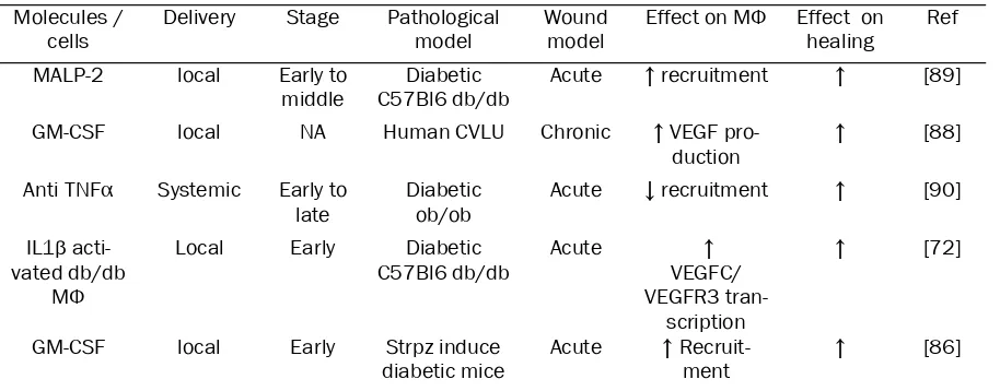
[image:5.612.81.538.96.275.2]and TGFβ, and is produced during the angio-genic stage of the healing process [75]. The effects of PlGF are similar to those of VEGF, promoting monocytes and endothelial precursor cell migration [76] and favouring endothelial Table 2. Targeting of macrophages to control healing of pathological wound
Molecules /
cells Delivery Stage Pathological model Wound model Effect on MΦ Effect on healing Ref
MALP-2 local Early to
middle C57Bl6 db/dbDiabetic Acute ↑ recruitment ↑ [89]
GM-CSF local NA Human CVLU Chronic ↑ VEGF
pro-duction ↑ [88]
Anti TNFα Systemic Early to
late Diabeticob/ob Acute ↓ recruitment ↑ [90] IL1β
acti-vated db/db MΦ
Local Early Diabetic
C57Bl6 db/db Acute VEGFC/↑ VEGFR3
tran-scription
↑ [72]
GM-CSF local Early Strpz induce
diabetic mice Acute ↑ Recruit-ment ↑ [86] CVLU: Chronic Veinous Leg Ulcer. Strpz : streptozotocin
cell survival [77]. PlGF stimulates also the se-cretion of VEGF by the monocyte/macrophages [78]. Interestingly, even if PlGF is described to have chemotactic properties by his own on VEGFR1+ expressing cells, the synergy between PlGF and VEGF is important in angiogenesis [79]: i) VEGFR1 homodimers may be a decoy receptor. Binding of PlGF homodimer to VEGFR1 homodimer make more VEGF available to bind to activated VEGFR2. ii) VEGF/PlGF erodimers bind to VEGFR1/VEGFR2 het-erodimer and induce more potent angiogenic signals iii) binding of PlGF homodimers to VEGFR1 homodimers favours VEGFR2 homodimer phosphorylation [80]. Overall, PlGF seems to potentiate the effect of VEGF in wound angiogenesis. These results altogether point to the important role of macrophages in coordinat-ing the angiogenic signal durcoordinat-ing wound healcoordinat-ing.
Macrophages and defective wound healing
[image:6.612.102.506.335.694.2]Despite their heterogeneous aetiology, most chronic wounds have in common a defect in the progression from the inflammatory to the tissue formation stage. Loots et al reported increased infiltration of chronic and diabetic wounds by WAM compared to control acute wounds. In addition they observed higher amounts of ECM in the wound edge [81]. Similarly, in diabetic db/db mice, wounds are characterising by a prolonged expression of inflammatory cytokines and larger infiltration and persistence of the WAM [82]. However, these cells seem to have altered sensitivity towards exogenous signals such as VEGF or IGF1 [83, 84] as well as al-tered ability to release cytokine [85]. Several studies have targeted macrophages, by differ-ent strategies in order to improve defective wound healing (Table 2). In the obese diabetic
db/db mouse model, the injection of peritoneal macrophages activated by IL-1β at the wound site was associated with an increased produc-tion of prolymphangiogenic molecules such as VEGF-C, resulting in improved lymphangiogene-sis and wound healing [72]. Local application of GM-CSF is associated with better healing both in human and mouse pathological wounds in a clinical setting. In Streptozotocin induced dia-betic mice, GM-CSF is associated with a stronger infiltration of the wound by macro-phages, increased angiogenesis and a better healing, while no effect was observed in normal wound healing [86] [87]. Similarly, in a pilot study in humans, GM-CSF treatment improves chronic vascular ulcers, probably by promoting the secretion of VEGF by macrophages [88]. With a similar strategy, Deiters et al injected Macrophage-Activating Lipopeptide-2 (MALP-2) in wounds on obese diabetic db/db mice. MALP-2 induced the transcription of several genes associated with wound healing, including GM-CSF and IL-1 [89] and resulted in increased infiltration by WAM and more rapid wound clo-sure. In opposition to these activating ap-proaches, Goren et al used a depleting strategy based on the systemic injection of a neutralizing anti-TNFα in ob/ob mice [90]. The treatment resulted in a systemic and local depletion of macrophages that was associated with a faster healing. Taken together, these results sustain the hypothesis that in chronic or diabetic wounds different strategies that might affect macrophage phenotype might modify the heal-ing.
Conclusions
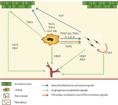
Our current knowledge points to macrophages as key players in skin wound healing after birth. Healing is a complex and evolving process, and because of their plasticity, WAM progress with the wound, adapting their cytokine expression profile [15]. Because of this plasticity and of their central position, WAM seem to be the ideal target for clinical interventions in chronic wounds, to accelerate closure or to attenuate fibrosis (Figure 1). For this purpose, there is still a need to further characterise this population. It seems obvious that, similar to other inflamma-tion models, WAM are essentially composed of infiltrating blood monocytes that change from an inflammatory phenotype to a repair one [15, 91, 92]. It is not clear if the multiple functions of WAM are exclusive to a unique macrophage
population or fulfilled by specialised sub popula-tions at different time points? Should the future therapeutic strategies consist in the control of the WAM polarisation or in the specific recruit-ment or depletion of sub populations? These strategies will also have to define the appropri-ate timing for their effectiveness as WAM bear different functions during the healing time course [16].
Acknowledgement
This work was supported by the Ramaciotti foundation.
Please address correspondence to: Kiarash Khosro-tehrani, MD, PhD, University of Queensland Centre for Clinical Research, Building 71/918, Royal Brisbane & Women's Hospital Campus, Herston, QLD, 4029, Tel: +6173346 6077, Fax : +61733465598, E-mail: k.khosrotehrani@uq.edu.au
References
[1] Gordon S and Taylor PR. Monocyte and macro-phage heterogeneity. Nat Rev Immunol 2005; 5: 953-964.
[2] Stout RD and Suttles J. Functional plasticity of macrophages: reversible adaptation to changing microenvironments. J Leukoc Biol 2004; 76: 509-513.
[3] Gurtner GC, Werner S, Barrandon Y and Lon-gaker MT. Wound repair and regeneration. Na-ture 2008; 453: 314-321.
[4] Ross R and Odland G. Human wound repair. II. Inflammatory cells, epithelial-mesenchymal in-terrelations, and fibrogenesis. J Cell Biol 1968; 39: 152-168.
[5] Kim MH, Liu W, Borjesson DL, Curry FR, Miller LS, Cheung AL, Liu FT, Isseroff RR and Simon SI. Dynamics of neutrophil infiltration during cutane-ous wound healing and infection using fluores-cence imaging. J Invest Dermatol 2008; 128: 1812-1820.
[6] Leibovich SJ and Ross R. The role of the macro-phage in wound repair. A study with hydrocorti-sone and antimacrophage serum. Am J Pathol 1975; 78: 71-100.
[7] Pilcher BK, Dumin JA, Sudbeck BD, Krane SM, Welgus HG and Parks WC. The activity of colla-genase-1 is required for keratinocyte migration on a type I collagen matrix. J Cell Biol 1997; 137: 1445-1457.
[8] Singer AJ and Clark RA. Cutaneous wound heal-ing. N Engl J Med 1999; 341: 738-746.
[9] Gabbiani G. The myofibroblast in wound healing and fibrocontractive diseases. J Pathol 2003; 200: 500-503.
[11] Mantovani A, Sozzani S, Locati M, Allavena P and Sica A. Macrophage polarization: tumor-associated macrophages as a paradigm for po-larized M2 mononuclear phagocytes. Trends Immunol 2002; 23: 549-555.
[12] Mantovani A, Sica A, Sozzani S, Allavena P, Vec-chi A and Locati M. The chemokine system in diverse forms of macrophage activation and polarization. Trends Immunol 2004; 25: 677-686.
[13] Martinez FO, Helming L and Gordon S. Alterna-tive activation of macrophages: an immunologic functional perspective. Annu Rev Immunol 2009; 27: 451-483.
[14] Stout RD, Jiang C, Matta B, Tietzel I, Watkins SK and Suttles J. Macrophages sequentially change their functional phenotype in response to changes in microenvironmental influences. J Immunol 2005; 175: 342-349.
[15] Daley JM, Brancato SK, Thomay AA, Reichner JS and Albina JE. The phenotype of murine wound macrophages. J Leukoc Biol 2010; 87: 59-67. [16] Lucas T, Waisman A, Ranjan R, Roes J, Krieg T,
Muller W, Roers A and Eming SA. Differential Roles of Macrophages in Diverse Phases of Skin Repair. J Immunol 2009;
[17] Mosser DM and Edwards JP. Exploring the full spectrum of macrophage activation. Nat Rev Immunol 2008; 8: 958-969.
[18] Raes G, Noel W, Beschin A, Brys L, de Baetselier P and Hassanzadeh GH. FIZZ1 and Ym as tools to discriminate between differentially activated macrophages. Dev Immunol 2002; 9: 151-159. [19] Wynes MW and Riches DW. Induction of
macro-phage insulin-like growth factor-I expression by the Th2 cytokines IL-4 and IL-13. J Immunol 2003; 171: 3550-3559.
[20] Torocsik D, Bardos H, Nagy L and Adany R. Iden-tification of factor XIII-A as a marker of alterna-tive macrophage activation. Cell Mol Life Sci 2005; 62: 2132-2139.
[21] Martin P, D'Souza D, Martin J, Grose R, Cooper L, Maki R and McKercher SR. Wound healing in the PU.1 null mouse--tissue repair is not dependent on inflammatory cells. Curr Biol 2003; 13: 1122-1128.
[22] Mirza R, DiPietro LA and Koh TJ. Selective and specific macrophage ablation is detrimental to wound healing in mice. Am J Pathol 2009; 175: 2454-2462.
[23] Goren I, Allmann N, Yogev N, Schurmann C, Linke A, Holdener M, Waisman A, Pfeilschifter J and Frank S. A transgenic mouse model of in-ducible macrophage depletion: effects of diph-theria toxin-driven lysozyme M-specific cell line-age ablation on wound inflammatory, angio-genic, and contractive processes. Am J Pathol 2009; 175: 132-147.
[24] Chen L, Tredget EE, Wu PY and Wu Y. Paracrine factors of mesenchymal stem cells recruit macrophages and endothelial lineage cells and enhance wound healing. PLoS One 2008; 3:
e1886.
[25] Banno T, Gazel A and Blumenberg M. Effects of tumor necrosis factor-alpha (TNF alpha) in epi-dermal keratinocytes revealed using global tran-scriptional profiling. J Biol Chem 2004; 279: 32633-32642.
[26] McKay IA and Leigh IM. Epidermal cytokines and their roles in cutaneous wound healing. Br J Dermatol 1991; 124: 513-518.
[27] Hancock GE, Kaplan G and Cohn ZA. Keratino-cyte growth regulation by the products of im-mune cells. J Exp Med 1988; 168: 1395-1402. [28] Rheinwald JG and Green H. Epidermal growth
factor and the multiplication of cultured human epidermal keratinocytes. Nature 1977; 265: 421-424.
[29] Li Y, Fan J, Chen M, Li W and Woodley DT. Trans-forming growth factor-alpha: a major human serum factor that promotes human keratinocyte migration. J Invest Dermatol 2006; 126: 2096-2105.
[30] Schultz G, Rotatori DS and Clark W. EGF and TGF -alpha in wound healing and repair. J Cell Bio-chem 1991; 45: 346-352.
[31] Marikovsky M, Vogt P, Eriksson E, Rubin JS, Tay-lor WG, Joachim S and Klagsbrun M. Wound fluid -derived heparin-binding EGF-like growth factor (HB-EGF) is synergistic with insulin-like growth factor-I for Balb/MK keratinocyte proliferation. J Invest Dermatol 1996; 106: 616-621.
[32] Shirakata Y, Kimura R, Nanba D, Iwamoto R, Tokumaru S, Morimoto C, Yokota K, Nakamura M, Sayama K, Mekada E, Higashiyama S and Hashimoto K. Heparin-binding EGF-like growth factor accelerates keratinocyte migration and skin wound healing. J Cell Sci 2005; 118: 2363-2370.
[33] Edwards JP, Zhang X and Mosser DM. The ex-pression of heparin-binding epidermal growth factor-like growth factor by regulatory macro-phages. J Immunol 2009; 182: 1929-1939. [34] Hallbeck AL, Walz TM and Wasteson A.
Inter-leukin-6 enhances transforming growth factor-alpha mRNA expression in macrophage-like hu-man monocytoid (U-937-1) cells. Biosci Rep 2001; 21: 325-339.
[35] Rappolee DA, Mark D, Banda MJ and Werb Z. Wound macrophages express TGF-alpha and other growth factors in vivo: analysis by mRNA phenotyping. Science 1988; 241: 708-712. [36] Grose R and Werner S. Wound-healing studies in
transgenic and knockout mice. Mol Biotechnol 2004; 28: 147-166.
[37] Massague J and Pandiella A. Membrane-anchored growth factors. Annu Rev Biochem 1993; 62: 515-541.
[38] Gallucci RM, Sloan DK, Heck JM, Murray AR and O'Dell SJ. Interleukin 6 indirectly induces kerati-nocyte migration. J Invest Dermatol 2004; 122: 764-772.
expression by interleukin 1. J Biol Chem 1994; 269: 10753-10757.
[40] Ashcroft GS, Yang X, Glick AB, Weinstein M, Let-terio JL, Mizel DE, Anzano M, Greenwell-Wild T, Wahl SM, Deng C and Roberts AB. Mice lacking Smad3 show accelerated wound healing and an impaired local inflammatory response. Nat Cell Biol 1999; 1: 260-266.
[41] Li AG, Wang D, Feng XH and Wang XJ. Latent TGFbeta1 overexpression in keratinocytes re-sults in a severe psoriasis-like skin disorder. EMBO J 2004; 23: 1770-1781.
[42] Sellheyer K, Bickenbach JR, Rothnagel JA, Bund-man D, Longley MA, Krieg T, Roche NS, Roberts AB and Roop DR. Inhibition of skin development by overexpression of transforming growth factor beta 1 in the epidermis of transgenic mice. Proc Natl Acad Sci U S A 1993; 90: 5237-5241. [43] Werner S and Grose R. Regulation of wound
healing by growth factors and cytokines. Physiol Rev 2003; 83: 835-870.
[44] Werner S, Krieg T and Smola H. Keratinocyte-fibroblast interactions in wound healing. J Invest Dermatol 2007; 127: 998-1008.
[45] Hubner G, Brauchle M, Smola H, Madlener M, Fassler R and Werner S. Differential regulation of pro-inflammatory cytokines during wound healing in normal and glucocorticoid-treated mice. Cytokine 1996; 8: 548-556.
[46] Brauchle M, Angermeyer K, Hubner G and Werner S. Large induction of keratinocyte growth factor expression by serum growth factors and pro-inflammatory cytokines in cultured fibro-blasts. Oncogene 1994; 9: 3199-3204.
[47] Takehara K. Growth regulation of skin fibro-blasts. J Dermatol Sci 2000; 24 Suppl 1: S70-77.
[48] Beer HD, Florence C, Dammeier J, McGuire L, Werner S and Duan DR. Mouse fibroblast growth factor 10: cDNA cloning, protein characteriza-tion, and regulation of mRNA expression. Onco-gene 1997; 15: 2211-2218.
[49] Wang X, Waldeck H and Kao WJ. The effects of TGF-alpha, IL-1beta and PDGF on fibroblast ad-hesion to ECM-derived matrix and KGF gene expression. Biomaterials 31: 2542-2548. [50] Mori R, Shaw TJ and Martin P. Molecular
mecha-nisms linking wound inflammation and fibrosis: knockdown of osteopontin leads to rapid repair and reduced scarring. J Exp Med 2008; 205: 43-51.
[51] Stramer BM, Mori R and Martin P. The inflamma-tion-fibrosis link? A Jekyll and Hyde role for blood cells during wound repair. J Invest Dermatol 2007; 127: 1009-1017.
[52] Annes JP, Munger JS and Rifkin DB. Making sense of latent TGFbeta activation. J Cell Sci 2003; 116: 217-224.
[53] Occleston NL, Fairlamb D, Hutchison J, O'Kane S and Ferguson MW. Avotermin for the improve-ment of scar appearance: a new pharmaceutical in a new therapeutic area. Expert Opin Investig
Drugs 2009; 18: 1231-1239.
[54] Postlethwaite AE, Keski-Oja J, Moses HL and Kang AH. Stimulation of the chemotactic migra-tion of human fibroblasts by transforming growth factor beta. J Exp Med 1987; 165: 251-256. [55] Border WA and Noble NA. Transforming growth
factor beta in tissue fibrosis. N Engl J Med 1994; 331: 1286-1292.
[56] Grotendorst GR, Okochi H and Hayashi N. A novel transforming growth factor beta response element controls the expression of the connec-tive tissue growth factor gene. Cell Growth Differ 1996; 7: 469-480.
[57] Igarashi A, Okochi H, Bradham DM and Groten-dorst GR. Regulation of connective tissue growth factor gene expression in human skin fibroblasts and during wound repair. Mol Biol Cell 1993; 4: 637-645.
[58] Desmouliere A, Geinoz A, Gabbiani F and Gabbi-ani G. Transforming growth factor-beta 1 induces alpha-smooth muscle actin expression in granu-lation tissue myofibroblasts and in quiescent and growing cultured fibroblasts. J Cell Biol 1993; 122: 103-111.
[59] Direkze NC, Forbes SJ, Brittan M, Hunt T, Jeffery R, Preston SL, Poulsom R, Hodivala-Dilke K, Al-ison MR and Wright NA. Multiple organ engraft-ment by bone-marrow-derived myofibroblasts and fibroblasts in bone-marrow-transplanted mice. Stem Cells 2003; 21: 514-520.
[60] Ishii G, Sangai T, Sugiyama K, Ito T, Hasebe T, Endoh Y, Magae J and Ochiai A. In vivo charac-terization of bone marrow-derived fibroblasts recruited into fibrotic lesions. Stem Cells 2005; 23: 699-706.
[61] Herdrich BJ, Lind RC and Liechty KW. Multipo-tent adult progenitor cells: their role in wound healing and the treatment of dermal wounds. Cytotherapy 2008; 10: 543-550.
[62] Brown LF, Yeo KT, Berse B, Yeo TK, Senger DR, Dvorak HF and van de Water L. Expression of vascular permeability factor (vascular endothe-lial growth factor) by epidermal keratinocytes during wound healing. J Exp Med 1992; 176: 1375-1379.
[63] Eming SA, Brachvogel B, Odorisio T and Koch M. Regulation of angiogenesis: wound healing as a model. Prog Histochem Cytochem 2007; 42: 115-170.
[64] Li B, Sharpe EE, Maupin AB, Teleron AA, Pyle AL, Carmeliet P and Young PP. VEGF and PlGF pro-mote adult vasculogenesis by enhancing EPC recruitment and vessel formation at the site of tumor neovascularization. FASEB J 2006; 20: 1495-1497.
[65] Dvorak HF, Brown LF, Detmar M and Dvorak AM. Vascular permeability factor/vascular endothe-lial growth factor, microvascular hyperpermeabil-ity, and angiogenesis. Am J Pathol 1995; 146: 1029-1039.
antiapoptotic proteins Bcl-2 and A1 in vascular endothelial cells. J Biol Chem 1998; 273: 13313 -13316.
[67] Odorisio T, Cianfarani F, Failla CM and Zambruno G. The placenta growth factor in skin angiogene-sis. J Dermatol Sci 2006; 41: 11-19.
[68] Frank S, Hubner G, Breier G, Longaker MT, Greenhalgh DG and Werner S. Regulation of vascular endothelial growth factor expression in cultured keratinocytes. Implications for normal and impaired wound healing. J Biol Chem 1995; 270: 12607-12613.
[69] Babic AM, Chen CC and Lau LF. Fisp12/mouse connective tissue growth factor mediates endo-thelial cell adhesion and migration through in-tegrin alphavbeta3, promotes endothelial cell survival, and induces angiogenesis in vivo. Mol Cell Biol 1999; 19: 2958-2966.
[70] Shimo T, Nakanishi T, Nishida T, Asano M, Kan-yama M, Kuboki T, Tamatani T, Tezuka K, Take-mura M, MatsuTake-mura T and Takigawa M. Connec-tive tissue growth factor induces the prolifera-tion, migraprolifera-tion, and tube formation of vascular endothelial cells in vitro, and angiogenesis in vivo. J Biochem 1999; 126: 137-145.
[71] Nagy JA, Vasile E, Feng D, Sundberg C, Brown LF, Detmar MJ, Lawitts JA, Benjamin L, Tan X, Man-seau EJ, Dvorak AM and Dvorak HF. Vascular permeability factor/vascular endothelial growth factor induces lymphangiogenesis as well as angiogenesis. J Exp Med 2002; 196: 1497-1506.
[72] Maruyama K, Asai J, Ii M, Thorne T, Losordo DW and D'Amore PA. Decreased macrophage num-ber and activation lead to reduced lymphatic vessel formation and contribute to impaired diabetic wound healing. Am J Pathol 2007; 170: 1178-1191.
[73] Schledzewski K, Falkowski M, Moldenhauer G, Metharom P, Kzhyshkowska J, Ganss R, Demory A, Falkowska-Hansen B, Kurzen H, Ugurel S, Geginat G, Arnold B and Goerdt S. Lymphatic endothelium-specific hyaluronan receptor LYVE-1 is expressed by stabilin-LYVE-1+, F4/80+, CDLYVE-1LYVE-1b+ macrophages in malignant tumours and wound healing tissue in vivo and in bone marrow cul-tures in vitro: implications for the assessment of lymphangiogenesis. J Pathol 2006; 209: 67-77. [74] Ribatti D. The discovery of the placental growth
factor and its role in angiogenesis: a historical review. Angiogenesis 2008; 11: 215-221. [75] Failla CM, Odorisio T, Cianfarani F, Schietroma C,
Puddu P and Zambruno G. Placenta growth fac-tor is induced in human keratinocytes during wound healing. J Invest Dermatol 2000; 115: 388-395.
[76] Hattori K, Heissig B, Wu Y, Dias S, Tejada R, Ferris B, Hicklin DJ, Zhu Z, Bohlen P, Witte L, Hendrikx J, Hackett NR, Crystal RG, Moore MA, Werb Z, Lyden D and Rafii S. Placental growth factor reconstitutes hematopoiesis by recruiting VEGFR1(+) stem cells from bone-marrow
micro-environment. Nat Med 2002; 8: 841-849. [77] Adini A, Kornaga T, Firoozbakht F and Benjamin
LE. Placental growth factor is a survival factor for tumor endothelial cells and macrophages. Can-cer Res 2002; 62: 2749-2752.
[78] Bottomley MJ, Webb NJ, Watson CJ, Holt L, Buk-hari M, Denton J, Freemont AJ and Brenchley PE. Placenta growth factor (PlGF) induces vascular endothelial growth factor (VEGF) secretion from mononuclear cells and is co-expressed with VEGF in synovial fluid. Clin Exp Immunol 2000; 119: 182-188.
[79] Carmeliet P, Moons L, Luttun A, Vincenti V, Compernolle V, De Mol M, Wu Y, Bono F, Devy L, Beck H, Scholz D, Acker T, DiPalma T, Dewerchin M, Noel A, Stalmans I, Barra A, Blacher S, Van-dendriessche T, Ponten A, Eriksson U, Plate KH, Foidart JM, Schaper W, Charnock-Jones DS, Hicklin DJ, Herbert JM, Collen D and Persico MG. Synergism between vascular endothelial growth factor and placental growth factor contributes to angiogenesis and plasma extravasation in pathological conditions. Nat Med 2001; 7: 575-583.
[80] Autiero M, Waltenberger J, Communi D, Kranz A, Moons L, Lambrechts D, Kroll J, Plaisance S, De Mol M, Bono F, Kliche S, Fellbrich G, Ballmer-Hofer K, Maglione D, Mayr-Beyrle U, Dewerchin M, Dombrowski S, Stanimirovic D, Van Hum-melen P, Dehio C, Hicklin DJ, Persico G, Herbert JM, Shibuya M, Collen D, Conway EM and Carme-liet P. Role of PlGF in the intra- and intermolecu-lar cross talk between the VEGF receptors Flt1 and Flk1. Nat Med 2003; 9: 936-943.
[81] Loots MA, Lamme EN, Zeegelaar J, Mekkes JR, Bos JD and Middelkoop E. Differences in cellular infiltrate and extracellular matrix of chronic dia-betic and venous ulcers versus acute wounds. J Invest Dermatol 1998; 111: 850-857.
[82] Wetzler C, Kampfer H, Stallmeyer B, Pfeilschifter J and Frank S. Large and sustained induction of chemokines during impaired wound healing in the genetically diabetic mouse: prolonged persis-tence of neutrophils and macrophages during the late phase of repair. J Invest Dermatol 2000; 115: 245-253.
[83] Tchaikovski V, Olieslagers S, Bohmer FD and Waltenberger J. Diabetes mellitus activates sig-nal transduction pathways resulting in vascular endothelial growth factor resistance of human monocytes. Circulation 2009; 120: 150-159. [84] Goren I, Muller E, Pfeilschifter J and Frank S.
Severely impaired insulin signaling in chronic wounds of diabetic ob/ob mice: a potential role of tumor necrosis factor-alpha. Am J Pathol 2006; 168: 765-777.
[85] Zykova SN, Jenssen TG, Berdal M, Olsen R, Myk-lebust R and Seljelid R. Altered cytokine and nitric oxide secretion in vitro by macrophages from diabetic type II-like db/db mice. Diabetes 2000; 49: 1451-1458.
and Bao S. Granulocyte-macrophage colony-stimulating factor enhances wound healing in diabetes via upregulation of proinflammatory cytokines. Br J Dermatol 2009;
[87] Ure I, Partsch B, Wolff K and Petzelbauer P. Granulocyte/macrophage colony-stimulating factor increases wound-fluid interleukin 8 in normal subjects but does not accelerate wound healing. Br J Dermatol 1998; 138: 277-282. [88] Cianfarani F, Tommasi R, Failla CM, Viviano MT,
Annessi G, Papi M, Zambruno G and Odorisio T. Granulocyte/macrophage colony-stimulating factor treatment of human chronic ulcers pro-motes angiogenesis associated with de novo vascular endothelial growth factor transcription in the ulcer bed. Br J Dermatol 2006; 154: 34-41.
[89] Deiters U, Barsig J, Tawil B and Muhlradt PF. The macrophage-activating lipopeptide-2 accelerates wound healing in diabetic mice. Exp Dermatol 2004; 13: 731-739.
[90] Goren I, Muller E, Schiefelbein D, Christen U, Pfeilschifter J, Muhl H and Frank S. Systemic anti -TNFalpha treatment restores diabetes-impaired skin repair in ob/ob mice by inactivation of macrophages. J Invest Dermatol 2007; 127:
2259-2267.
[91] Nahrendorf M, Swirski FK, Aikawa E, Stangen-berg L, Wurdinger T, Figueiredo JL, Libby P, Weissleder R and Pittet MJ. The healing myocar-dium sequentially mobilizes two monocyte sub-sets with divergent and complementary func-tions. J Exp Med 2007; 204: 3037-3047. [92] Cochain C, Rodero MP, Vilar J, Recalde A, Richart