REVIEW
The sarcomeric cytoskeleton: from molecules to motion
Mathias Gautel1,* and Kristina Djinović-Carugo2,3ABSTRACT
Highly ordered organisation of striated muscle is the prerequisite for the fast and unidirectional development of force and motion during heart and skeletal muscle contraction. A group of proteins, summarised as the sarcomeric cytoskeleton, is essential for the ordered assembly of actin and myosin filaments into sarcomeres, by combining architectural, mechanical and signalling functions. This review discusses recent cell biological, biophysical and structural insight into the regulated assembly of sarcomeric cytoskeleton proteins and their roles in dissipating mechanical forces in order to maintain sarcomere integrity during passive extension and active contraction.α-Actinin crosslinks in the Z-disk show a pivot-and-rod structure that anchors both titin and actin filaments. In contrast, the myosin crosslinks formed by myomesin in the M-band are of a ball-and-spring type and may be crucial in providing stable yet elastic connections during active contractions, especially eccentric exercise.
KEY WORDS: Striated muscle, Sarcomere, Cytoskeleton, Titin,
Obscurin,α-Actinin, Myomesin, Z-disk, M-band
Introduction
Muscles convert chemical energy into mechanical work, thus enabling the generation of force and movement. A subset of muscles, the striated skeletal and heart muscles, act as linear motors where force and movement are generated in a highly directional way. Striated muscle owes its striped appearance under the microscope to the remarkably regular, repetitive assembly of contractile structures, the sarcomeres, into myofibrils. In sarcomeres, the contractile actin and myosin filaments are integrated in paracrystalline order by the action of accessory cytoskeletal proteins, forming what is often termed the sarcomeric cytoskeleton (Agarkova and Perriard, 2005; Fürst and Gautel, 1995; Gautel, 2011). The completely regular, parallel and repetitive array of actin and myosin allows force and motion to be generated only in the direction of the filament axis, which is amplified along the length of a myofibril, and ultimately a muscle fibre, by millions of sarcomeres acting in unison over distances of millimetres to centimetres. This, together with the sophisticated coupling to the cellular excitation and calcium release machinery via T-tubules and the sarcoplasmic reticulum (SR: see Fig. 1), enables the rapid and vectorial generation and relaxation of force that is absent in other contractile actomyosin systems like smooth muscle or non-muscle cells. Indeed, the loss of regular myofibrillar order or efficient coupling to the excitation–contraction
machinery is associated with many severe human muscle diseases, for example hypertrophic cardiomyopathy, where myofibril disarray is one of the defining hallmarks of the pathology.
Most proteins of the sarcomeric cytoskeleton are organised by specific interactions with the giant blueprint protein of the sarcomere, the ∼3 MDa titin (reviewed in Tskhovrebova and Trinick, 2003), encoded by theTTNgene. Titin, the biggest protein of the human body (over 1.5 µm) (Kontrogianni-Konstantopoulos et al., 2009), combines elastic, architectural and signalling functions. Titin consists of hundreds of specific ca. 100 amino acid immunoglobulin or fibronectin domains (see below), which interact with similar domains in the sarcomeric cytoskeletal proteins myomesin, myosin-binding protein, obscurin and obsl1 (Gautel, 2011; Lange et al., 2006; Linke and Kruger, 2010) as well as with myosin. Additional unique motifs or specialised repeats form binding sites for other proteins, notablyα-actinin. By virtue of their tight interactions in the Z-disk and M-band, where overlapping titin molecules are crosslinked, titin filaments form an uninterrupted connective, elastic link along the myofibres, hence the alternative name connectin (Maruyama et al., 1977).
Recently, mechanistic and structural insight has revealed a wealth of detailed information on how titin assembles the sarcomeric cytoskeleton. In addition to integrating the contractile filaments in ordered sarcomeres, these many interactions also coordinate the position and possibly mechanical activation of signalling domains and metabolic enzymes, thus providing a direct link between mechanical activity and cellular signalling in muscle (Gautel, 2011).
The term cytoskeleton is normally used to describe the cellular network of protein fibres or filaments within the cytoplasm or nucleoplasm that fulfils such diverse functions as the maintenance and dynamics of cell shape, organelle positioning, cellular transport, cell motility and locomotion including cell division, to name but a few major functions. The major and best-characterised generic cytoskeletal filaments are microfilaments ( polymeric filaments composed of actin), intermediate filaments ( polymeric filaments composed of intermediate filament proteins from a family of over 60 members) and microtubules ( polymers of tubulin dimers). The term sarcomeric cytoskeleton therefore requires some explanation of its historical background, as it is generally used to describe only a subset of structural proteins within the contractile sarcomeres of striated muscle that are directly involved in sarcomere assembly. Importantly, two of these proteins–nebulin and titin– form long filaments that are composed of single polypeptides, and as such, the sarcomeric cytoskeleton is a unique specialisation of the actomyosin system found only in higher animals.
What is a sarcomeric cytoskeleton, therefore?
In the striated muscles of animals, actin filaments of nearly identical lengths are arranged in interdigitating arrays with myosin filaments of identical length in the largest highly ordered multi-protein assemblies known to biology: the sarcomeres (Fig. 1). They can be regarded as a highly specialised form of the actin cytoskeleton. 1
King’s College London BHF Centre of Research Excellence, Randall Division for Cell and Molecular Biophysics, and Cardiovascular Division, New Hunt’s House, London SE1 1UL, UK.2Department of Structural and Computational Biology, Max F. Perutz Laboratories, University of Vienna, Campus Vienna Biocenter 5, Vienna A-1030, Austria.3Department of Biochemistry, Faculty of Chemistry and Chemical Technology, University of Ljubljana, Aškerčeva 5, Ljubljana 1000, Slovenia.
*Author for correspondence ([email protected])
This is an Open Access article distributed under the terms of the Creative Commons Attribution License (http://creativecommons.org/licenses/by/3.0), which permits unrestricted use,
distribution and reproduction in any medium provided that the original work is properly attributed.
Journal
of
Experimental
However, for the purposes of clarity, this review will not focus on the commonalities of sarcomeres with less specialised and organised forms of the actin–myosin cytoskeleton such as stress fibres or the actomyosin filaments in smooth muscle (which lack regular lateral or axial assembly and hence the ability of rapid, unidirectional contraction). Rather, we focus on the particular set of structural proteins that are specific to sarcomeres and unique in many of their functional and architectural features. These proteins, which provide sarcomeres with mechanical stability, elasticity, spatial organisation and long-range communication capacity, are summarised as the sarcomeric cytoskeleton in the following. This term may be as imprecise and misleading as that of cytoskeleton itself, as the sarcomeric cytoskeleton, too, is not a dead and stiff skeleton but a dynamic protein network. However, it may suffice as a summary term for a group of structurally, functionally and evolutionarily related proteins in the sarcomere.
The main characteristic of the sarcomeric cytoskeleton is the remarkable axial and transverse order of actin and myosin filaments forming the paracrystalline lattice of the sarcomeres (Figs 1, 2). For this, the axial distance of actin and myosin filaments, their lengths and the interfilament distances need be kept constant by links that are both mechanically stable and able to yield appropriately upon the stresses and strains of contraction and relaxation (Figs 1, 2). Myofibrils are exposed to, and have to withstand, both axial and lateral forces during active contraction. The activation of myosin motor domains on the bipolar myosin filament leads to the development of stochastic force imbalances and hence shear forces between neighbouring myosin filaments (reviewed in Agarkova et al., 2003; Gautel, 2011). These shear forces lead to the rapid but normally reversible deformation of M-bands, with
myosin filament displacements at least of the order of 14 nm, while Z-disks show significantly higher mechanical stability, rapidly deforming the M-band at the onset of isometric contractions (Huxley et al., 1982) and ultimately leading to their rupture (Hirose and Wakabayashi, 1993; Horowits and Podolsky, 1987). The mechanical properties of the Z-disk have also been tested using atomic force microscopy (AFM) of myofibrils, which confirmed the Z-disk to be the stiffest of the anchoring planes in both activated and relaxed myofibrils (Akiyama et al., 2006; Wakayama et al., 2000; Yamada et al., 2003). Intriguingly, eccentric exercise, one of the strongest stimulants of hypertrophic muscle growth and performance endurance (Vogt and Hoppeler, 2014), not only induces marked deformation of sarcomere structure (Friden and Lieber, 1992) and is the primary cause of injury (Lieber and Fridén, 1999) but also induces the translocation of Z-disk components (myotilin) to M-bands (Carlsson et al., 2007). This latter observation, together with others on similar ‘multicompartment proteins’(Lange et al., 2006), supports an as yet poorly understood interplay between the Z-disk and M-band in exercise-induced muscle remodelling, either as mechanosensors or as passively regulated signalling scaffolds.
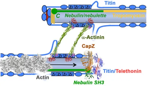
[image:2.612.55.294.52.277.2]Most proteins of the sarcomeric cytoskeleton share common architectural principles, being composed predominantly of 100 amino acid domains of two types, the intracellular immunoglobulin (Ig) and the fibronectin type-III (Fn3) superfamilies. These domains confer specificity to ligand interactions and the stability required for
Fig. 1. A schematic view of the sarcomeric cytoskeleton showing some of
the links between membranes and sarcomeres.T-tubules (Tt) link the
sarcolemma to the sarcoplasmic reticulum (SR) at the triads via
dihydropyridine receptors (small orange cylinder) and ryanodine receptors (small green cylinder). The figure shows myosin (turquoise), actin (light blue),
α-actinin Z-links (brown), M-band crosslinks (green), titin (red) and nebulin (dark blue, shown only for one thin filament). Sarcomeres are linked by other extra-sarcomeric cytoskeletal structures at the Z-disk and M-band, comprising desmin, synemin, plectin and nesprin (summarised as green wavy lines). Links from the sarcomere to T-tubules or the SR are formed by obscurin (light blue wavy lines). Principal sarcomere regions are marked by Z, I, A and M.
A
B
Fig. 2. Schematic representation of the transverse Z-disk and M-band
lattice.(A) Schematic representation of the transverse Z-disk structure as it
appears under the electron microscope, with the tetragonal lattice of antiparallel actin filaments (opposite polarity of actin filaments is represented by dark blue and purple circles) linked predominantly byα-actinin struts (Z-links, light blue). This structure undergoes reversible conformational changes to the basket-weave form (right) upon myosin binding and a shift of tropomyosin during thin-filament activation as interfilament distance increases. (B) Schematic representation of the M-band lattice as it appears in transverse sections under the electron microscope. The links reflect the projections of M-bridges and M-links (see Agarkova et al., 2003), whose molecular identity is still unclear; a model for their possible molecular interpretation was proposed by Lange et al. (2005a). Large circles, myosin filaments; small circles, components of the linking filaments. The M-band can undergo both axial and transverse conformational changes in the contracting sarcomere.
Journal
of
Experimental
[image:2.612.320.555.60.285.2]mechanical integration. The elucidation of the molecular structure of the sarcomeric cytoskeleton began with the identification of repetitive patterns of Ig and Fn3 domains in the nematode protein twitchin, followed by the identification of titin and unc-89 (Benian et al., 1989, 1996). Twitchin and unc-89 can be considered as analogues of titin and obscurin, respectively, although important differences exist regarding the interactions and activity of their signalling domains. It is likely that ancestral molecules of similar composition evolved with the first striated muscles and thus predate the Cambrian radiation, as the striated muscles of all extant animals contain sarcomeric cytoskeleton proteins composed of Ig and Fn3 superfamily domains (Benian et al., 1989, 1996; Burkart et al., 2007; Fürst and Gautel, 1995; Higgins et al., 1994; Tskhovrebova and Trinick, 2003). This is despite considerable differences in the composition of actin and myosin filaments ( paramyosin only in invertebrates), the differences in lattice structures of sarcomeric sub-regions like the Z-disk (hexagonal in insect flight muscle, tetragonal in all vertebrate striated muscles) and, lastly, the presence of additional signalling domains and the domain patterns in the sarcomeric cytoskeleton proteins. This review will focus on striated muscles in these animals, but will refer to the situation in models organisms likeCaenorhabditis elegans
andDrosophila melanogasterfor relevant parallels.
The sarcomeric cytoskeleton is central in coordinating myofibril assembly, although many details still remain to be unravelled. A scaffold of actin filaments, integratingα-actinin and titin, seems to be the core component for the subsequent integration of M-band modules, sarcomeric myosin and accessory proteins (Ehler et al., 2004, 1999; Sanger et al., 2010; Van der Ven et al., 1999; White et al., 2014).In vivo, primary sarcomere assembly occurs close to the cell membrane and seems to involve as yet poorly understood links to the membrane cytoskeletal system. Clathrin-coated vesicles have recently been found to play an unexpected role in the assembly and maintenance of sarcomeres (Vassilopoulos et al., 2014), and as phospholipid regulation seems essential for the activation of several Z-disk proteins, such membrane links might be functionally important. Sarcomere-like regular arrangements of actin and myosin filaments under the cell membrane have also recently been observed in striking images of the contractile structures in epithelial cells, where non-muscle myosin-II is arranged underneath the plasmalemma in regular, repetitive patterns (Ebrahim et al., 2013). The organising principles of these sarcomere-like structures are currently elusive, although non-muscle isoforms of sarcomeric proteins might be involved.
Below, we discuss recent developments in the cytoskeletal biology of the sarcomere from a historical perspective, but focusing on the Z-disk and M-band as the main mechanical integrators of actin and myosin filaments.
Z-disk cytoskeleton: form follows function?
Myofibril assembly begins at the Z-disk (reviewed in Ehler and Gautel, 2008; Sanger et al., 2010). Here, antiparallel actin filaments are crosslinked and capped at their barbed ends, leading to tetragonal arrays of precisely tailored thickness (Figs 1–3). Crosslinking requires predominantly the actin-crosslinking protein
α-actinin, which forms the mechanical links between the antiparallel actin filaments from two sarcomere halves. The N-termini of titin also enter the Z-disk in an antiparallel arrangement from two half-sarcomeres (Fig. 3), with titin filaments from opposite sarcomere halves crosslinked in an antiparallel way by
α-actinin, and titin filaments crosslinked by telethonin, both filaments probably from the same half-sarcomere. This tetragonal
array is stiff (as measured in AFM, see above) but not inflexible: a transition occurs from a small-square lattice pattern seen in electron microscopy (EM) cross-sections to a basket-weave pattern (Goldstein et al., 1990; Perz-Edwards and Reedy, 2011). This transition was initially believed to be directly induced by mechanical force (Goldstein et al., 1987), but has recently been shown to result from conformational changes induced by tropomyosin movement on the actin filament (Perz-Edwards and Reedy, 2011) (Fig. 2), with the small-square lattice occurring when tropomyosin is in the closed position (Fig. 2A). However, tropomyosin does not appear to be a component of the Z-band, terminating just outside it (Trombitás et al., 1990). How these changes are therefore transmitted from the I- and A-band regions of the thin filament to the Z-disk remains a mystery. Possibly, other thin-filament-associated proteins play a role, and the giant nebulin and its small cardiac homologue, nebulette, would be plausible candidates, as they span from the Z-disk into the I-band (see review by Chu et al., 2016, in this issue; see also below). A structural model of the core Z-disk that reconciles known protein-binding sites and mapped protein and epitope positions, but not the axial repeat observed in EM, is shown in Fig. 3. However, it should be noted that Z-disks contain large numbers of additional proteins as well as bound phospholipids (Fukami et al., 1992; Li and Russell, 2013). Some of these components may only be transiently associated (Lange et al., 2006), and because of this complexity and the optical density of Z-disks, their high-resolution analysis is highly challenging.
[image:3.612.317.562.59.203.2]Z-disks are of highly variable thickness, with those in the fast glycolytic fibres of fish white muscles or avian breast muscle showing only two layers of transverse Z-disk crosslinks (or Z-links), while slow postural and cardiac muscles have up to seven
Fig. 3. Schematic representation of the Z-disk.Top: from the model; bottom:
actual structure.α-Actinin crosslinks actin filaments via its N-terminal actin-binding domain (red), while its C-terminal calmodulin-like domain ( purple/blue) crosslinks titin Z-repeats (Zr, blue squares). The mechanical crosslink is formed by the antiparallel, stiff dimer of spectrin repeats in the rod domain (green). Titin filaments are crosslinked (‘capped’) in an antiparallel array by single telethonin molecules (red), while the barbed ends (arrow) of actin filaments are capped by the capZ complex (beige trapezium). The actin filament regulator nebulin (and its small cardiac homologue nebulette; green) cooperates in actin filament regulation and capZ capping, with its C-terminal SH3 domain (green circle) near the capping complex. This model agrees with the ultrastructural position of titin and nebulin domains (Young et al., 1998), telethonin andα-actinin as well as the proposed interactions with capZ (Witt et al., 2006); the conformation of titin interdomain linkers is unknown. Titin domains are shown in blue. The exact position of accessory proteins like myotilin, LDB3 (ZASP/cypher) and MYOZ (myozenin/FATZ/calsarcin) is not yet known. PDB structure accession numbers for solved Z-disk proteins in this scheme are: actin, 3J8A;α-actinin, 4D1E; capZ, 1IZN; nebulin SH3 domain, 1NEB; and titin/telethonin, 1YA5.
Journal
of
Experimental
Z-link layers (Luther, 2009). In mammals, sarcomericα-actinin is transcribed from two genes,ACTN2andACTN3, withα-actinin-3 being specifically expressed in fast-twitch fibres. Intriguingly, while there seems to be a correlation between Z-disk thickness and
α-actinin isoforms, the contribution ofα-actinin isoforms to Z-disk architecture is not strictly correlated to contractile properties (Schachat et al., 1985). In contrast, titin may control the thickness of the Z-disk by determining the number ofα-actinin crosslinks via differentially spliced binding motifs, the titin Z-repeats (Gautel et al., 1996; Sorimachi et al., 1997). Z-repeats bind to EF-hand motifs 3–4 of the C-terminal calmodulin-like domain (CaM) of
α-actinin (Atkinson et al., 2001). The interaction of Z-repeats with
α-actinin has been studied in detail at the cellular, biochemical, biophysical and structural level, confirming and characterising the interaction of the helical Z-repeats with theα-actinin CaM domain (Atkinson et al., 2000, 2001; Ribeiro et al., 2014; Sorimachi et al., 1997; Young et al., 1998). There is a strict correlation of the number of titin Z-repeats with the number of Z-links: chicken pectoralis major muscle, a fast glycolytic muscle with thin Z-disks, expresses titin with two Z-repeats (Peckham et al., 1997), while cardiac and slow skeletal muscles in mammals with thick Z-disks express up to seven Z-repeats (Gautel et al., 1996; Sorimachi et al., 1997). However, it is currently unclear how their α-actinin-binding properties on the molecular level are translated into the assembled structure, as there appears to be a mismatch in the length of the repeat and the periodicity of the Z-disk as measured by EM (Luther and Squire, 2002). It seems unlikely, however, that Z-disk thickness is determined byα-actinin isoforms alone. In humans, a common truncating mutation (R577X) in the ACTN3 gene leads to homozygous loss of α-actinin-3 in about 18% of healthy white individuals and is detrimental to sprint performance (replicated in 14 other cohorts), while the presence of the intact gene correlates highly with athletic sprint performance (Garton and North, 2015; Kim et al., 2014; Yang et al., 2003). There appears to be no change in Z-disk thickness, however. Whether the sprint performance advantage of wild-type ACTN3 carriers is due to subtle differences in molecular mechanical properties or in the interaction with any of the many α-actinin interacting proteins that link to cellular signalling and metabolic pathways (Djinovic-Carugo et al., 2002; Lange et al., 2006; Linke and Kruger, 2010; Sanger et al., 2010) is currently unclear. However, these observations underscore the role of the sarcomeric cytoskeleton as not only a passive scaffold but also a determinant of muscle performance and metabolism (Berman and North, 2010), which has recently been linked to differential transcriptional regulation by calcineurin via calsarcin-2/myozenin-1/FATZ-1 at theα-actinin scaffold (Seto et al., 2013). Form and function seem to be truly interdependent.
When reflecting on the function of Z-disks as stable and quite stiff mechanical integrators of titin and actin filaments, it is amazing to realise how dynamic the exchange of protein subunits of this apparently stable structure is. All major Z-disk proteins except telethonin show rapid dynamic exchange in cardiac and skeletal myocytes, with half-lives of about 25 s for α-actinin-2 (Ribeiro et al., 2014; Stout et al., 2008; Wang et al., 2005). This suggests that the fast-exchanging components may be in a dynamically regulated equilibrium between a high-affinity Z-disk-bound conformation and a low-affinity cytoplasmic conformation. Theα-actinin–titin interaction is regulated by an intramolecular pseudoligand mechanism, in which a titin Z-repeat-like sequence between the actin-binding domain and the central rod domain interacts with EF-hand motifs 3–4 of the juxtaposed CaM domain. This interaction can be released by the binding of acidic phospholipids, especially
phosphatidylinositol 4,5-bisphosphate (PIP2), to the actin-binding domain, thus activating titin binding (Ribeiro et al., 2014; Young and Gautel, 2000).
As outlined above,α-actinin as a component of Z-links is likely to undergo conformational changes during the transition of small-square Z-disks to the basket-weave form, as this conversion clearly requires plasticity on the side of the Z-links. Until recently, the exact localisation of the required flexible links was a matter of speculation, with bending of theα-actinin rod domain a plausible possibility (Luther, 2009; Perz-Edwards and Reedy, 2011). The recent complete structure ofα-actinin and separate rod structures (Ribeiro et al., 2014; Ylänne et al., 2001) suggests that this is unlikely: the central dimerisation domain, formed from eight spectrin-like repeats in an antiparallel dimer, is a rigid and inflexible rod. However, flexible segments link the actin- and titin-binding domains at the N- and C-termini. In particular, the pseudoligand neck region is likely to undergo structural rearrangement in the open, titin-binding form, leading to substantial rotational freedom of the rod versus the actin-binding domain (Ribeiro et al., 2014). This pivot will allow the stiff rod to rotate against the actin-binding domains, thus ensuring not only their appropriate relative position but also substantial angular movement between the actin-attached actin-binding domains (Ribeiro et al., 2014). α-Actinin can therefore be considered to have a pivot-and-rod structure, with its mechanical plasticity residing in the pivot regions (Fig. 4A). Future
A
[image:4.612.316.562.349.599.2]B
Fig. 4. The molecular architecture and mechanics of the main sarcomeric
crosslinkersα-actinin and myomesin.α-Actinin is an antiparallel dimer with
one actin- and one titin-binding domain at either end of a long, stiff rod formed from eight interacting spectrin-like repeats. Movement between the domains is likely to occur around the neck domain in a pivot-like manner, allowing rotation of the actin-binding domains around the rod (red arrows). The links between C-terminal myomesin domains My9 to My13 resemble a ball-and-spring arrangement, with highly extensible, rapidly refolding helices providing significant extensibility and the ability to buffer considerable forces (red arrows) while linking the tightly folded Ig domains. Shown here is the structure of C-terminal domains My12–My13, which also illustrates the antiparallel dimerisation of myomesin via the My13 domain. PDB structure accession numbers: My12-13, 2R15 (Pinotsis et al., 2008);α-actinin complete structure,
4D1E (Ribeiro et al., 2014).
Journal
of
Experimental
reconstructions of basket-weave and small-square lattice forms of the Z-disk now have a reliable starting template.
Apart from the requirement for actin filaments to be crosslinked at the Z-disk, they need to be capped to prevent their fast-growing barbed ends from expanding into the next half-sarcomere. This involves the action of the major muscle actin-capping protein complex, CapZ orβ-actinin (Cooper and Schafer, 2000; Maruyama, 2002). Actin filament capping and crosslinking need to be spatiotemporally coordinated during myofibril turnover and dynamic remodelling to achieve and maintain Z-disks of precisely tailored width. The involvement of giant scaffold proteins, especially titin and nebulin, is an attractive solution to this problem. Nebulin, the giant actin-associated protein whose C-terminus crosses the Z-disk, was implicated in binding and targeting CapZ to the Z-disk (Pappas et al., 2008; Witt et al., 2006), although conflicting data exist regarding the exact binding site. While the C-terminal SH3 domain of nebulin was suggested to interact with both CapZ and titin and thereby to attach actin filament barbed ends to the Z-disk periphery (Witt et al., 2006), genetic deletion of the nebulin SH3 domain has no impact on Z-disk width (Yamamoto et al., 2013). This could be explained if the major binding site of nebulin for CapZ were to reside in nebulin modules 160–164 and not the SH3 domain, as proposed (Pappas et al., 2008). CapZ was also identified as a ligand of theα -actinin rod (Papa et al., 1999). This is even more mysterious as an explanation for spatially defined capping activity in the Z-disk, as
α-actinin crosslinks actin across the entire Z-disk. Directly defining the distance between pointed- and barbed-end capping proteins would be an appealing mechanism for nebulin to act as a regulator of thin filament length regulation. This concept is currently under intense scrutiny because of the mismatch of the pointed-end capping protein tropomodulin and the N-terminus of nebulin, which do not coincide. Accordingly, alternative explanations via a ‘two-segment’ model that challenge the paradigm of the nebulin controller have been proposed by the team of V. Fowler (Castillo et al., 2009; Fowler et al., 2006; Gokhin and Fowler, 2013; Littlefield and Fowler, 2008) and are further discussed in the review by Chu et al. (2016) in this issue. Capping and uncapping of actin filaments in the Z-disk is a dynamic process that seems to rapidly respond to external stimuli, presumably to remodel and renew sarcomeres in response to physiological requirements. CapZ was also found to be regulated by PIP2, where PIP2 decreases its affinity to F-actin (Heiss and Cooper, 1991). Exercise-induced changes in PIP2 levels dynamically regulate the exchange of CapZ at the Z-disk (Hartman et al., 2009; Lin et al., 2013), not dissimilar to the regulation ofα-actinin (Ribeiro et al., 2014). But are these PIP2-regulated proteins alone in regulating actin dynamics at the nascent or remodelling Z-disk? Analysis of the dynamic regulation of the Arp2/3 complex (a major regulator of actin nucleation of branching) suggested that insulin-like growth factor-1 (IGF-1) induces phosphatidylinositol 3-kinase–Akt signalling, which in turn promotes the formation of a complex of nebulin and N-WASP via the nebulin SH3 domain (Takano et al., 2011). As mentioned, the nebulin SH3 domain is unexpectedly not required for normal skeletal muscle structure, although it might be necessary to support active contraction in mouse (Yamamoto et al., 2013).
Have we, therefore, overlooked major players in actin dynamics in muscle? Recently, the formin-homology family of proteins (FHOD) has been implicated in regulating actin dynamics (Iskratsch and Ehler, 2011; Iskratsch et al., 2010, 2013), suggesting that we are still remote from understanding the details of Z-disk dynamics.
Titin filaments from presumably the same sarcomere are crosslinked at the Z-disk by the small telethonin/tcap protein encoded by theTCAPgene. This unique 167-residue protein (Valle et al., 1997) forms an antiparallel complex with the N-terminal domains of titin at the Z-disk edge (Fig. 3) that is of extraordinary mechanical stability (Bertz et al., 2009; Zou et al., 2006). It was proposed that telethonin could act as a ‘cap’ for titin based on knockdown data in primary skeletal and cardiac myocytes. These data suggested that telethonin was required for the structural integrity of sarcomeres and titin-regulated sarcomere assembly (Gregorio et al., 1998; Mues et al., 1998). However, recent studies in myocytes (Wang et al., 2005) and animal models revealed that telethonin enters the Z-disk late and is therefore not involved in primary myofibril assembly (Zhang et al., 2009) or required to maintain sarcomere integrity under baseline conditions (Knoll et al., 2011). Rather, it appears that, at least in cardiomyocytes, the titin– telethonin complex is somehow implicated in the organisation or maintenance of T-tubules near the Z-disk (Fig. 1) (Ibrahim et al., 2013; Zhang et al., 2009), involving phosphorylation of its C-terminus (Candasamy et al., 2014). Deletion of theTCAPgene in fish and mouse leads to disruption of the sarcomere–T-tubule interaction, but not of sarcomere assembly as such. These intriguing observations need to be reconciled with the fact that in skeletal muscle, T-tubules end at the A-band–I-band boundary and not at the Z-disk, as in cardiac muscle. The functions of telethonin at the Z-disk in skeletal muscle are therefore likely to be independent of T-tubule assembly. That such functions are essential for normal skeletal muscle function in humans is highlighted by recessive truncating mutations in the human TCAP gene that lead to limb-girdle muscular dystrophy (Moreira et al., 2000). As truncated telethonin can be incorporated into Z-disks and binds titin (Barresi et al., 2015; Zou et al., 2006), the deleted C-terminus must fulfil crucial, as yet unidentified functions.
The integration of sarcomeres into larger assemblies of myofibrils and their lateral alignment involves additional cytoskeletal links, especially to the intermediate filament (IF) proteins at the Z-disk periphery. Connections to the network formed by the IF protein desmin that surrounds the myofibrils (Granger and Lazarides, 1979) can be made by nebulin (Tonino et al., 2010), which, together with another IF, synemin, contributes to the linkage between costameres and the contractile apparatus (García-Pelagio et al., 2015; Li et al., 2014). Further cytoskeletal protein links at the Z-disk periphery exist via the actin crosslinker filamin-C (van der Ven et al., 2000), the actin–IF–tubulin crosslinker plectin (Castañóon et al., 2013), spectrin and the spectrin-like protein nesprin (Banerjee et al., 2014; Chapman et al., 2014). These cytoskeletal connections outside the sarcomere cannot be comprehensively discussed within this article.
M-band cytoskeleton: all’s well that ends well
The C-terminal end of titin is integrated into the M-band, and is essential for sarcomere integrity, as will be discussed. The M-band crosslinks adjacent myosin filaments in their central bare zone in a hexagonal lattice (Fig. 2B), ensuring that the antiparallel arrays of myosin motor domains are precisely aligned. Ultrastructurally, the M-band shows up to five strong M-bridge lines, and its structure correlates roughly with heart frequency in vertebrates (Pask et al., 1994). The arrangement of myosin filaments can follow two different lattice types, a‘simple lattice’and a‘super lattice’that is found in most tetrapods (Luther and Squire, 2014). Myosin filaments are crosslinked at the M-band in a ternary complex involving the myosin crosslinking protein myomesin, obscurin (an ∼800 kDa analogue of the nematode UNC89; Benian and Mayans,
Journal
of
Experimental
2015) or its small homologue obscurin-like-1 (obsl1), and the C-terminal domain of titin (Fukuzawa et al., 2008).
The known integral sarcomeric cytoskeleton proteins of the M-band comprise three closely related M-M-band proteins: myomesin-1, M-protein (myomesin-2) and myomesin-3 that are encoded by three mammalianMYOMgenes (Schoenauer et al., 2008). In addition, titin, the giant protein obscurin and obsl1 are involved in organising the transverse crosslinks between myosin filaments that are essential to maintain the axial alignment of adjacent myosin filaments. A number of further constituents of the M-band are known, which are, however, involved in non-structural functions by scaffolding of metabolic enzyme complexes, signalling complexes or components of the protein turnover machinery.
Myomesin isoforms correlate with the contractile properties in different fibre types: while myomesin-1 is constitutively expressed in all striated muscles, myomesin-2 is expressed only in the adult heart and fast fibres, whereas myomesin-3 is expressed in skeletal muscle intermediate fibre types (Agarkova et al., 2004; Schoenauer et al., 2011, 2008). Of the threeMYOMisogenes,MYOM1appears to be the one that is essential in all muscle types: not only is it constitutively co-expressed with other myomesins but also the cellular knockdown of myomesin-1 completely disrupts myofibril integrity (Fukuzawa et al., 2008).
Myomesin-1 is identical to skelemin (Price, 1987) and is encoded by the same gene,MYOM1, with skelemin being a splice variant of the MYOM1 gene resulting from differential splicing of a single exon (Steiner et al., 1999), leading to insertion of a serine– proline-rich domain between Ig domains My6 and My7 (Price and Gomer, 1993). This splice variant is identical to EH-myomesin (Agarkova et al., 2000). Whereas skelemin was initially reported to be a peri-myofibrillar protein that might be involved in the linkage between M-bands and the desmin intermediate filament network, recent analysis suggests myomesin-1 and EH-myomesin-1 completely overlap in the M-band. The use of a different protein name for a splice variant of the same gene should be avoided, but a rational nomenclature of the twoMYOM1splice variants (myomesin, and skelemin7/EH-myomesin) awaits agreement. By principle of first description (Eppenberger et al., 1981) and agreement with the HUGO-approved gene nomenclature, myomesin should, however, be the accepted name. The reported localisation of skelemin epitopes at the sarcolemma and the sub-membranous cytoskeleton in non-muscle cells is currently irreconcilable with the strictly sarcomeric localisation that is observed with the well-characterised monoclonal antibodies that recognise constitutive epitopes in the
MYOM1protein: B4 (Grove et al., 1984) and BB78 (Vinkemeier et al., 1993) and the polyclonal antibody My190Nrt, specific for the N-terminal domain (Obermann et al., 1996), as well as with antibodies recognising the EH-myomesin insertion (serine– proline-rich insertion) (Schoenauer et al., 2011). It is also surprising given the highly specific interactions with muscle-specific proteins (see below) and the strictly myogenic regulation of the MYOM1
promoter, which is inactive even in proliferating myoblasts (Steiner et al., 1999). Clearly, further analysis of gene expression patterns and standardised reagents will be required to resolve these questions.
In muscle sarcomeres, the unequivocal myosin-crosslinking function of myomesin requires its assembly into antiparallel dimers. This is achieved via homotypic Ig–Ig interaction of the C-terminal domain (My13), which self-assembles in an antiparallel orientation (Lange et al., 2005a; Pinotsis et al., 2012, 2008). This ability of My13 to mediate antiparallel dimerisation is shared between all MYOM isoforms. However, despite the high homology
of the My13 domains in myomesin, M-protein and myomesin-3, there is no evidence for heterodimer formation (Schoenauer et al., 2008), suggesting that each myomesin form fulfils separate and distinct functions. The interaction of the N-terminal myosin-binding domain is not yet fully understood: this region is highly divergent between the three myomesin isoforms, possibly reflecting their different positions in the bare zone of the myosin filament, and contains multiple phosphorylation sites indicative of dynamic regulation.
Myomesin is also an elastic protein that displays a unique architecture in its C-terminal region from My9 to My13 that could be called a ‘ball-and-spring’ pattern. Here, the stably folded Ig domains are interspersed by short helices, of which the N-terminal two-thirds form a hydrophobic interface with the preceding domain; the last third of the helix is a free-folding helix with a hydrophilic surface. As AFM and molecular dynamics simulations have revealed, these helices can undergo rapidly reversible unfolding– refolding transitions at forces between 15 and 40 pN, much below the unfolding forces of individual myomesin Ig domains (over 80 pN; Schoenauer et al., 2005) or the dissociation force of the myomesin dimer (over 130 pN; Berkemeier et al., 2011; Pinotsis et al., 2012; Xiao and Gräter, 2014). As a consequence, myomesin-1 can extend by about 50 nm by reversible unfolding of the C-terminal ball-and-spring segment alone (Berkemeier et al., 2011; Pinotsis et al., 2012) (Fig. 4B).
Furthermore, the serine–proline-rich insertion in EH-myomesin (skelemin) was shown by biophysical analyses to be an intrinsically disordered protein sequence that can behave as an entropic spring, with similar rubber-like elastic properties to the PEVK region in titin (Schoenauer et al., 2005). Why embryonic hearts and skeletal slow fibres would require this additional capacity to elongate by approximately 36 nm is currently unclear, but it has been proposed that the re-expression of the EH-myomesin isoform in some forms of cardiomyopathy might aid in dissipating the effects of unfavourable mechanical stress on the integrity of the M-band (Schoenauer et al., 2011). It was also suggested that the softer and‘fuzzier’EH-myomesin-containing M-bands provide a design that is more stable in eccentric contractions, as EH-myomesin is expressed not only in embryonic hearts but also significantly in slow, postural muscles (Agarkova et al., 2003). This passive extensibility of the EH-insertion seems to disagree with its proposed function as a binding site for desmin filaments for skelemin.
Apart from its central myosin-crosslinking role, myomesin-1 (but not myomesin-2 and -3) also interacts with the Ig domain Ob3 of the related proteins obscurin and its small homologue obscurin-like-1 (obsl1) (Fukuzawa et al., 2008). Obscurin was initially identified as a ligand of the Z9–Z10 domains of the Z-disk portion of titin, showing an intriguing, developmentally regulated change in preferential sarcomeric localisation from Z-disk to M-band (Young et al., 2001). Its M-band localisation could be satisfactorily resolved by identifying its integration in a ternary complex with M-band titin and myomesin (Fig. 5). The C-terminal M10 Ig domain of titin interacts with the N-terminal Ig domain Ob1 of obscurin, while the linker between myomesin fibronectin domains My4 and My5 interacts with the N-terminal Ig domain Ob3. The small muscle isoform of the extensively differentially spliced obsl1 interacts in a highly similar way with both titin and myomesin (Fukuzawa et al., 2008). However, while obscurin is localised at the M-band periphery, where it acts as a link between myofibril and the SR via small ankyrin ank1.5 (Bagnato et al., 2003; Kontrogianni-Konstantopoulos et al., 2003), obsl1 is localised to the myofibril
Journal
of
Experimental
core (Fukuzawa et al., 2008). Titin M10 is so far the only sarcomeric cytoskeleton domain shown to interact with two distinct proteins of presumably different function in different localisations (Benian and Mayans, 2015): whereas the ca. 800–1000 kDa protein obscurin contains multiple signalling domains (Rho GDP/GTP exchange factor domain, and two protein kinase domains), obsl1 contains only Ig and Fn3 domains and thus appears to fulfil predominantly a scaffolding and architectural function. In agreement with obscurin association with the M-band edge of myofibrils and its C-terminal interactions with the SR via small ankyrin, the knockout of obscurin in mouse causes only a mild myopathy but leads to disruption of the longitudinal SR (Lange et al., 2009). Double-knockouts of both the
OBSCNandOBSL1genes should provide more information about whether any of these complexes are indispensable for sarcomere formation or maintenance. The atomic structures of the two titin M10 complexes with obsl1 and obscurin reveal different molecular conformations, tantalisingly hinting at possible additional protein– protein interactions enabled by the two giant proteins that may jointly form an additional binding site (Pernigo et al., 2010, 2015). A recently identified interaction of titin M10 with synemin, however, requires further validation. This interaction was reported to be mediated by only half of the M10 domain (Prudner et al., 2014), and it is difficult to envisage how the resulting disrupted Ig domain would be generated (M10 is encoded on a single exon) or stabilised.
Interactions of titin, obscurin and obsl1 link the M-band to several protein turnover and quality control pathways (Fig. 5). The interaction of titin with several ubiquitin ligases (the complex of MURF1/2/3 near the kinase domain; Centner et al., 2001), and myospryn at M10 (Sarparanta et al., 2010), as well as ubiquitin-binding autophagy adaptor proteins (Nbr1 and p62/SQSTM1; Lange et al., 2005b) suggests that titin is either a substrate or a regulator of these pathways. Whether these activities require titin kinase activity or solely its scaffolding role remains to be clarified (Bogomolovas et al., 2014; Lange et al., 2005b). Titin interacts additionally with the cysteine protease calpain-3 in the differentially spliced insertion between domains M9 and M10 (Kinbara et al., 1997); this interaction seems to be dynamically modulated by mechanical forces (Ojima et al., 2010). Interestingly, calpain-3 interacts in turn with myospryn, linking the activity of the two pathways in protein degradation (Sarparanta et al., 2010); the generation of C-terminal titin fragments by calpain might be the prelude to their ubiquitin-dependent degradation, a process that is disrupted in several hereditary titinopathies (Charton et al., 2015; Sarparanta et al., 2010).
Additional links of the M-band to ubiquitin-mediated protein turnover might exist via obsl1, which was shown to interact with the ubiquitin E3 ligase cullin-7 (Yan et al., 2014) in non-muscle cells; whether this interaction is also relevant for the muscle isoforms remains to be seen. In other cell types, the non-muscle isoforms of obls1 form a 3M complex with CUL7 and CCDC8 that plays a role in maintaining microtubule and genome integrity, Golgi morphology and normal development (Litterman et al., 2011).
The M-band cytoskeleton, lastly, acts also as a scaffold for the targeting of several metabolic enzymes, especially creatine kinase. Both myomesin and M-protein interact with creatine kinase around domains My6–My9, albeit with different affinities (Hornemann et al., 2003). Additional metabolic enzymes can be targeted to the M-band via FHL2, a member of the four and a half LIM-only protein family (Lange et al., 2002). FHL2 can target creatine kinase, adenylate kinase and phosphofructokinase to binding sites on titin in the M-band and I-band (Lange et al., 2002). This spatial coupling of ATP-regenerating enzymes makes functional sense when we consider the vast amounts of ATP turned over by the nearby myosin motor domains. It is as yet unclear whether the mechanical plasticity of the M-band (and I-band) can modulate targeting or activity of these enzymes.
It is therefore not surprising that mutations in the C-terminal end of titin can lead to severe dominant and recessive myopathies in humans: recessive truncations causing early-onset myopathy completely disrupt the obscurin/myomesin network (Carmignac et al., 2007; Fukuzawa et al., 2008), dominant mutations in titin M10 weaken or abolish the obscurin and myospryn interactions, and compound heterozygous mutations in titin lead to a wide range of complex early-onset myopathies by disrupting several M-band interactions, including those formed by the kinase domain (Chauveau et al., 2014b). The integrity and ordered turnover of the sarcomere therefore relies on complex cytoskeletal networks that integrate architectural functions with mechanosignalling. The end of titin seems to play a crucial role in these homeostatic mechanisms for all to be well.
Summary and outlook
As summarised above, sarcomeric cytoskeleton research has entered an exciting new and dynamic phase, where powerful combinations of cellular, structural and biophysical analyses complement and enhance each other. For some protein complexes, obtaining large
Myospryn
Calpain-3 SQSTM1
Myosin
Titin
N
C
N
TK
N
C
N
C
C 9 8 7 6 5 4 3
2 1
TK 10 N
C
Myosin Nbr1
Cullin-7
MURF CK
AK PFK FHL2
CK
Obscurin/Obsl1
Myomesin
Obscurin/Obsl
1
[image:7.612.51.300.60.253.2]N
Fig. 5. Schematic representation of the M-band cytoskeleton, comprising
titin, myomesin and obscurin/obsl1.Myosin filaments are crosslinked by
antiparallel dimers of myomesin, which are linked in a ternary complex with obscurin/obsl1, which in turn binds the C-terminal domain of titin M10. The catalytic Ser/Thr kinase domain of titin (TK) is at the M-band periphery. Although this model agrees with known interactions and ultrastructural locations of individual protein domains, alternative paths for titin are also compatible, and the conformation of large interdomain linkers (represented as simple lines) is as yet unknown except for theα-helical linkers between myomesin domains 9–13 (shown as spiral springs). The M-band serves as a scaffold for a number of components of the protein turnover machinery via ubiquitin-mediated turnover (ubiquitin E3 ligases MURF1/2/3, myospryn and possibly cullin-7, autophagy scaffolds Nbr1 and SQSTM1, cysteine protease calpain-3) and is involved in muscle remodelling, possibly responding to mechanical stress. Metabolic enzyme interactions are shown in light shades: creatine kinase (CK), adenylate kinase (AK) and phosphofructokinase (PFK). The interaction of cullin-7 with obsl1 has not yet been confirmed for muscle. Titin domains are shown in blue and are numbered; the myomesin dimer is shown in red. Arrows denote the bipolar nature of the myosin filament.
Journal
of
Experimental
atomic structures, allowing the detailed study of their regulation, biomechanics and signalling functions, now seems possible. Recent advances in high-resolution electron microscopy and the growing database of individual and complex structures will increasingly enable the structural unravelling of sarcomeric substructures and the modelling of their dynamic behaviour upon mechanical load or post-translational modifications. The expanding toolbox of cell biophysics (Yusko and Asbury, 2014) will lead to a rapidly improving understanding of how forces in the sarcomeric cytoskeleton modify contractile properties and muscle cell remodelling. This is particularly relevant for the Z-disk, which, because of its optical density and complexity, remains quite literally the most obscure structure of the sarcomere.
There is increasing evidence that the proteins of the sarcomeric cytoskeleton are common targets for hereditary myopathies, a subject that cannot be comprehensively discussed in this review: titin, nebulin,
α-actinin, filamin-C, telethonin and myomesin, as well as many of their ligands (myotilin, myopalladin, myosin-binding proteins) and further links to the external cytoskeleton via desmin or plectin have been implicated in hereditary myopathies (Chauveau et al., 2014a; Lyon et al., 2015; Tardiff et al., 2015). Because of the complexity of the sarcomeric cytoskeleton, with hundreds of spatially and temporally coordinated protein interaction sites and cell signalling links, considerable further work is required to understand its pathomechanistic impact and to ultimately discover new therapeutic principles. Furthermore, the emerging role of the sarcomeric cytoskeleton in scaffolding large complexes of proteins involved in protein turnover regulation and transcriptional control of muscle gene expression also implicates this system in age-related muscle loss and pathological remodelling in acquired conditions like disuse atrophy.
However, these developments also suggest that the sarcomeric cytoskeleton might not only be a complex disease target and a diagnostic challenge but also offer new therapeutic targets for the treatment of diseases caused by sarcomeric gene mutations, ageing or disease-associated disuse. Research in hereditary cardiomyopathies as well as skeletal myopathies, not least those linked to the rapidly growing number of discovered mutations in titin, might find promising drug discovery targets in sarcomeric cytoskeleton proteins and their regulatory pathways (Kho et al., 2012), in addition to the potential treatments that advances in genome editing may afford (Long et al., 2014).
Acknowledgements
We are grateful to Elisabeth Ehler for helpful discussions.
Competing interests
The authors declare no competing or financial interests.
Author contributions
M.G. wrote the paper and prepared the figures. K.D.-C. critically reviewed the manuscript and contributed to the figures.
Funding
This work was supported by the British Heart Foundation (M.G.), Marie Curie Initial Training Network: MUZIC (no. 238423; M.G. and K.D.-C.), Austrian Science Fund (FWF) Projects I525 and I1593, P22276 and P19060 (K.D.-C.), Federal Ministry of
Economy, Family and Youth through the initiative‘Laura Bassi Centres of Expertise’,
funding Center of Optimized Structural Studies (no. 253275; K.D.-C.) and by the University of Vienna (K.D.-C.). Deposited in PMC for immediate release.
References
Agarkova, I. and Perriard, J.-C.(2005). The M-band: an elastic web that crosslinks
thick filaments in the center of the sarcomere.Trends Cell Biol.15, 477-485.
Agarkova, I., Auerbach, D., Ehler, E. and Perriard, J.-C.(2000). A novel marker
for vertebrate embryonic heart, the EH-myomesin isoform.J. Biol. Chem.275,
10256-10264.
Agarkova, I., Ehler, E., Lange, S., Schoenauer, R. and Perriard, J.-C.(2003).
M-band: a safeguard for sarcomere stability?J. Muscle Res. Cell Motil.24, 191-203.
Agarkova, I., Schoenauer, R., Ehler, E., Carlsson, L., Carlsson, E., Thornell, L.-E. and Perriard, J.-C.(2004). The molecular composition of the sarcomeric
M-band correlates with muscle fiber type.Eur. J. Cell Biol.83, 193-204.
Akiyama, N., Ohnuki, Y., Kunioka, Y., Saeki, Y. and Yamada, T. (2006). Transverse stiffness of myofibrils of skeletal and cardiac muscles studied by
atomic force microscopy.J. Physiol. Sci.56, 145-151.
Atkinson, R. A., Joseph, C., Dal Piaz, F., Birolo, L., Stier, G., Pucci, P. and Pastore, A.(2000). Binding of alpha-actinin to titin: implications for Z-disk
assembly.Biochemistry39, 5255-5264.
Atkinson, R. A., Joseph, C., Kelly, G., Muskett, F. W., Frenkiel, T. A., Nietlispach, D. and Pastore, A.(2001). Ca2+-independent binding of an EF-hand domain to a
novel motif in the alpha-actinin-titin complex.Nat. Struct. Biol.8, 853-857.
Bagnato, P., Barone, V., Giacomello, E., Rossi, D. and Sorrentino, V.(2003). Binding of an ankyrin-1 isoform to obscurin suggests a molecular link between the
sarcoplasmic reticulum and myofibrils in striated muscles.J. Cell Biol. 160,
245-253.
Banerjee, I., Zhang, J., Moore-Morris, T., Pfeiffer, E., Buchholz, K. S., Liu, A., Ouyang, K., Stroud, M. J., Gerace, L., Evans, S. M. et al.(2014). Targeted ablation of nesprin 1 and nesprin 2 from murine myocardium results in
cardiomyopathy, altered nuclear morphology and inhibition of the
biomechanical gene response.PLoS Genet.10, e1004114.
Barresi, R., Morris, C., Hudson, J., Curtis, E., Pickthall, C., Bushby, K., Davies, N. P. and Straub, V.(2015). Conserved expression of truncated telethonin in a
patient with limb-girdle muscular dystrophy 2G. Neuromuscul. Disord. 25,
349-352.
Benian, G. M. and Mayans, O.(2015). Titin and obscurin: giants holding hands and
discovery of a new Ig domain subset.J. Mol. Biol.427, 707-714.
Benian, G. M., Kiff, J. E., Neckelmann, N., Moerman, D. G. and Waterston, R. H. (1989). Sequence of an unusually large protein implicated in regulation of myosin
activity in C. elegans.Nature342, 45-50.
Benian, G. M., Tinley, T. L., Tang, X. and Borodovsky, M. (1996). The Caenorhabditis elegans gene unc-89, required for muscle M-line assembly, encodes a giant modular protein composed of Ig and signal transduction domains.
J. Cell Biol.132, 835-848.
Berkemeier, F., Bertz, M., Xiao, S., Pinotsis, N., Wilmanns, M., Grater, F. and Rief, M.(2011). Fast-folding {alpha}-helices as reversible strain absorbers in the
muscle protein myomesin.Proc. Natl. Acad. Sci. USA108, 14139-14144.
Berman, Y. and North, K. N.(2010). A gene for speed: the emerging role of
alpha-actinin-3 in muscle metabolism.Physiology (Bethesda)25, 250-259.
Bertz, M., Wilmanns, M. and Rief, M.(2009). The titin-telethonin complex is a
directed, superstable molecular bond in the muscle Z-disk.Proc. Natl. Acad. Sci.
USA106, 13307-133310.
Bogomolovas, J., Gasch, A., Simkovic, F., Rigden, D. J., Labeit, S. and Mayans,
O.(2014). Titin kinase is an inactive pseudokinase scaffold that supports MuRF1
recruitment to the sarcomeric M-line.Open Biol.4, 140041.
Burkart, C., Qiu, F., Brendel, S., Benes, V., Hååg, P., Labeit, S., Leonard, K. and Bullard, B.(2007). Modular proteins from the Drosophila sallimus (sls) gene and
their expression in muscles with different extensibility.J. Mol. Biol.367, 953-969.
Candasamy, A. J., Haworth, R. S., Cuello, F., Ibrahim, M., Aravamudhan, S., Kruger, M., Holt, M. R., Terracciano, C. M. N., Mayr, M., Gautel, M. et al.(2014).
Phosphoregulation of the titin-cap protein telethonin in cardiac myocytes.J. Biol.
Chem.289, 1282-1293.
Carlsson, L., Yu, J.-G., Moza, M., Carpén, O. and Thornell, L.-E.(2007). Myotilin–
a prominent marker of myofibrillar remodelling.Neuromuscul. Disord.17, 61-68.
Carmignac, V., Salih, M. A. M., Quijano-Roy, S., Marchand, S., Al Rayess, M. M., Mukhtar, M. M., Urtizberea, J. A., Labeit, S., Guicheney, P., Leturcq, F. et al. (2007). C-terminal titin deletions cause a novel early-onset myopathy with fatal
cardiomyopathy.Ann. Neurol.61, 340-351.
Castañón, M. J., Walko, G., Winter, L. and Wiche, G.(2013). Plectin–intermediate
filament partnership in skin, skeletal muscle, and peripheral nerve.Histochem.
Cell Biol.140, 33-53.
Castillo, A., Nowak, R., Littlefield, K. P., Fowler, V. M. and Littlefield, R. S.(2009).
A nebulin ruler does not dictate thin filament lengths.Biophys. J.96, 1856-1865.
Centner, T., Yano, J., Kimura, E., McElhinny, A. S., Pelin, K., Witt, C. C., Bang, M.-L., Trombitas, K., Granzier, H., Gregorio, C. C. et al.(2001). Identification of muscle specific ring finger proteins as potential regulators of the titin kinase
domain.J. Mol. Biol.306, 717-726.
Chapman, M. A., Zhang, J., Banerjee, I., Guo, L. T., Zhang, Z., Shelton, G. D., Ouyang, K., Lieber, R. L. and Chen, J.(2014). Disruption of both nesprin 1 and
desmin results in nuclear anchorage defects and fibrosis in skeletal muscle.Hum.
Mol. Genet.23, 5879-5892.
Charton, K., Sarparanta, J., Vihola, A., Milic, A., Jonson, P. H., Suel, L., Luque, H., Boumela, I., Richard, I. and Udd, B.(2015). CAPN3-mediated processing of
C-terminal titin replaced by pathological cleavage in titinopathy.Hum. Mol. Genet.
24, 3718-3731.
Chauveau, C., Bönnemann, C. G., Julien, C., Kho, A. L., Marks, H., Talim, B., Maury, P., Uro-Coste, E., Alexandrovich, A., Vihola, A. et al.(2014a). G.P.34:
Journal
of
Experimental
the expanding spectrum of congenital titinopathies.Neuromuscul. Disord.24, 804-805.
Chauveau, C., Bonnemann, C., Julien, C., Kho, A. L., Marks, H., Talim, B., Maury, P., Arne-Bes, M. C., Uro-Coste, E., Alexandrovich, A. et al.(2014b). Recessive TTN truncating mutations define novel forms of core myopathy with
heart disease.Hum. Mol. Genet.23, 980-991.
Chu, M., Gregorio, C. C. and Pappas, C. T.(2016). Nebulin, a multi-functional
giant.J. Exp. Biol.219, 146-152.
Cooper, J. A. and Schafer, D. A. (2000). Control of actin assembly and
disassembly at filament ends.Curr. Opin. Cell Biol.12, 97-103.
Djinovic-Carugo, K., Gautel, M., Ylänne, J. and Young, P.(2002). The spectrin
repeat: a structural platform for cytoskeletal protein assemblies.FEBS Lett.513,
119-123.
Ebrahim, S., Fujita, T., Millis, B. A., Kozin, E., Ma, X., Kawamoto, S., Baird, M. A., Davidson, M., Yonemura, S., Hisa, Y. et al.(2013). NMII forms a contractile transcellular sarcomeric network to regulate apical cell junctions and tissue
geometry.Curr. Biol.23, 731-736.
Ehler, E. and Gautel, M.(2008). The sarcomere and sarcomerogenesis.Adv. Exp. Med. Biol.642, 1-14.
Ehler, E., Rothen, B. M., Hammerle, S. P., Komiyama, M. and Perriard, J. C. (1999). Myofibrillogenesis in the developing chicken heart: assembly of Z-disk,
M-line and the thick filaments.J. Cell Sci.112, 1529-1539.
Ehler, E., Fowler, V. M. and Perriard, J.-C.(2004). Myofibrillogenesis in the developing chicken heart: role of actin isoforms and of the pointed end actin
capping protein tropomodulin during thin filament assembly. Dev. Dyn. 229,
745-755.
Eppenberger, H. M., Perriard, J. C., Rosenberg, U. B. and Strehler, E. E.(1981). The Mr 165,000 M-protein myomesin: a specific protein of cross-striated muscle
cells.J. Cell Biol.89, 185-193.
Fowler, V. M., McKeown, C. R. and Fischer, R. S.(2006). Nebulin: does it measure
up as a ruler?Curr. Biol.16, R18-R20.
Friden, J. and Lieber, R. L.(1992). Structural and mechanical basis of
exercise-induced muscle injury.Med. Sci. Sports Exerc.24, 521-530.
Fukami, K., Furuhashi, K., Inagaki, M., Endo, T., Hatano, S. and Takenawa, T. (1992). Requirement of phosphatidylinositol 4,5-bisphosphate for alpha-actinin
function.Nature359, 150-152.
Fukuzawa, A., Lange, S., Holt, M. R., Vihola, A., Carmignac, V., Ferreiro, A., Udd, A. B. and Gautel, M.(2008). Interactions with titin and myomesin target obscurin
and obscurin-like 1 to the M-band - implications for hereditary myopathies.J. Cell
Sci.121, 1841-1851.
Fürst, D. O. and Gautel, M.(1995). The anatomy of a molecular giant: how the sarcomere cytoskeleton is assembled from immunoglobulin superfamily
molecules.J. Mol. Cell. Cardiol.27, 951-959.
Garcıa-Pelagio, K. P., Muriel, J., Ó ’Neill, A., Desmond, P. F., Lovering, R. M., Lund, L., Bond, M. and Bloch, R. J.(2015). Myopathic changes in murine
skeletal muscle lacking synemin.Am. J. Physiol. Cell Physiol.308, C448-C462.
Garton, F. C. and North, K. N.(2015). The effect of heterozygosity for the ACTN3
null allele on human muscle performance.Med. Sci. Sports Exerc. doi:10.1249/
mss.0000000000000784.
Gautel, M.(2011). The sarcomeric cytoskeleton: who picks up the strain?Curr. Opin. Cell Biol.23, 39-46.
Gautel, M., Goulding, D., Bullard, B., Weber, K. and Fürst, D. O.(1996). The central Z-disk region of titin is assembled from a novel repeat in variable copy
numbers.J. Cell Sci.109, 2747-2754.
Gokhin, D. S. and Fowler, V. M.(2013). A two-segment model for thin filament
architecture in skeletal muscle.Nat. Rev. Mol. Cell Biol.14, 113-119.
Goldstein, M. A., Michael, L. H., Schroeter, J. P. and Sass, R. L.(1987). Z band dynamics as a function of sarcomere length and the contractile state of muscle.
FASEB J.1, 133-142.
Goldstein, M. A., Schoeter, J. P. and Sass, R. L.(1990). Two structural states of
the vertebrate Z band.Electron Microsc. Rev.3, 227-248.
Granger, B. L. and Lazarides, E.(1979). Desmin and vimentin coexist at the
periphery of the myofibril Z disc.Cell18, 1053-1063.
Gregorio, C. C., Trombitás, K., Centner, T., Kolmerer, B., Stier, G., Kunke, K., Suzuki, K., Obermayr, F., Herrmann, B., Granzier, H. et al.(1998). The NH2 terminus of titin spans the Z-disc: its interaction with a novel 19-kD ligand (T-cap)
is required for sarcomeric integrity.J. Cell Biol.143, 1013-1027.
Grove, B. K., Kurer, V., Lehner, C., Doetschman, T. C., Perriard, J. C. and Eppenberger, H. M. (1984). A new 185,000-dalton skeletal muscle protein
detected by monoclonal antibodies.J. Cell Biol.98, 518-524.
Hartman, T. J., Martin, J. L., Solaro, R. J., Samarel, A. M. and Russell, B.(2009). CapZ dynamics are altered by endothelin-1 and phenylephrine via PIP2- and
PKC-dependent mechanisms.Am. J. Physiol. Cell Physiol.296, C1034-C1039.
Heiss, S. G. and Cooper, J. A.(1991). Regulation of CapZ, an actin capping protein
of chicken muscle, by anionic phospholipids.Biochemistry30, 8753-8758.
Higgins, D., Labeit, S., Gautel, M. and Gibson, T.(1994). The evolution of titin and
related giant muscle proteins.J. Mol. Evol.38, 395-404.
Hirose, K. and Wakabayashi, T.(1993). Structural change of crossbridges of rabbit
skeletal muscle during isometric contraction. J. Muscle Res. Cell Motil. 14,
432-445.
Hornemann, T., Kempa, S., Himmel, M., Hayess, K., Fürst, D. O. and Wallimann,
T.(2003). Muscle-type creatine kinase interacts with central domains of the
M-band proteins myomesin and M-protein.J. Mol. Biol.332, 877-887.
Horowits, R. and Podolsky, R. J.(1987). The positional stability of thick filaments in activated skeletal muscle depends on sarcomere length: evidence for the role of
titin filaments.J. Cell Biol.105, 2217-2223.
Huxley, H. E., Faruqi, A. R., Kress, M., Bordas, J. and Koch, M. H. J.(1982). Time-resolved X-ray diffraction studies of the myosin layer-line reflections during
muscle contraction.J. Mol. Biol.158, 637-684.
Ibrahim, M., Siedlecka, U., Buyandelger, B., Harada, M., Rao, C., Moshkov, A., Bhargava, A., Schneider, M., Yacoub, M. H., Gorelik, J. et al.(2013). A critical role for Telethonin in regulating t-tubule structure and function in the mammalian
heart.Hum. Mol. Genet.22, 372-383.
Iskratsch, T. and Ehler, E. (2011). Formin-g muscle cytoarchitecture.
Bioarchitecture1, 66-68.
Iskratsch, T., Lange, S., Dwyer, J., Kho, A. L., dos Remedios, C. and Ehler, E. (2010). Formin follows function: a muscle-specific isoform of FHOD3 is regulated
by CK2 phosphorylation and promotes myofibril maintenance.J. Cell Biol.191,
1159-1172.
Iskratsch, T., Yu, C.-H., Mathur, A., Liu, S., Stévenin, V., Dwyer, J., Hone, J., Ehler, E. and Sheetz, M.(2013). FHOD1 is needed for directed forces and
adhesion maturation during cell spreading and migration.Dev. Cell27, 545-559.
Kho, A. L., Perera, S., Alexandrovich, A. and Gautel, M.(2012). The sarcomeric
cytoskeleton as a target for pharmacological intervention.Curr. Opin. Pharmacol.
12, 347-354.
Kim, H., Song, K.-H. and Kim, C.-H.(2014). The ACTN3 R577X variant in sprint
and strength performance.J. Exerc. Nutr. Biochem.6, 347-353.
Kinbara, K., Sorimachi, H., Ishiura, S. and Suzuki, K.(1997). Muscle-specific calpain, p94, interacts with the extreme C-terminal region of connectin, a unique
region flanked by two immunoglobulin C2 motifs.Arch. Biochem. Biophys.342,
99-107.
Knoll, R., Linke, W. A., Zou, P., Miocic, S., Kostin, S., Buyandelger, B., Ku, C.-H., Neef, S., Bug, M., Schafer, K. et al.(2011). Telethonin deficiency is associated
with maladaptation to biomechanical stress in the mammalian heart.Circ. Res.
109, 758-769.
Kontrogianni-Konstantopoulos, A., Jones, E. M., Van Rossum, D. B. and Bloch, R. J.(2003). Obscurin is a ligand for small ankyrin 1 in skeletal muscle.
Mol. Biol. Cell14, 1138-1148.
Kontrogianni-Konstantopoulos, A., Ackermann, M. A., Bowman, A. L., Yap, S. V. and Bloch, R. J. (2009). Muscle giants: molecular scaffolds in
sarcomerogenesis.Physiol. Rev.89, 1217-1267.
Lange, S., Auerbach, D., McLoughlin, P., Perriard, E., Schäfer, B. W., Perriard, J.-C. and Ehler, E.(2002). Subcellular targeting of metabolic enzymes to titin in
heart muscle may be mediated by DRAL/FHL-2.J. Cell Sci.115, 4925-4936.
Lange, S., Himmel, M., Auerbach, D., Agarkova, I., Hayess, K., Fürst, D. O., Perriard, J.-C. and Ehler, E.(2005a). Dimerisation of myomesin: implications for
the structure of the sarcomeric M-band.J. Mol. Biol.345, 289-298.
Lange, S., Xiang, F., Yakovenko, A., Vihola, A., Hackman, P., Rostkova, E., Kristensen, J., Brandmeier, B., Franzen, G., Hedberg, B. et al.(2005b). The kinase domain of titin controls muscle gene expression and protein turnover.
Science308, 1599-1603.
Lange, S., Ehler, E. and Gautel, M. (2006). From A to Z and back?
Multicompartment proteins in the sarcomere.Trends Cell Biol.16, 11-18.
Lange, S., Ouyang, K., Meyer, G., Cui, L., Cheng, H., Lieber, R. L. and Chen, J. (2009). Obscurin determines the architecture of the longitudinal sarcoplasmic
reticulum.J. Cell Sci.122, 2640-2650.
Li, J. and Russell, B.(2013). Phosphatidylinositol 4,5-bisphosphate regulates
CapZbeta1 and actin dynamics in response to mechanical strain.Am. J. Physiol.
Heart Circ. Physiol.305, H1614-H1623.
Li, Z., Parlakian, A., Coletti, D., Alonso-Martin, S., Hourde, C., Joanne, P., Gao-Li, J., Blanc, J., Ferry, A., Paulin, D. et al.(2014). Synemin acts as a
regulator of signalling molecules during skeletal muscle hypertrophy.J. Cell Sci.
127, 4589-4601.
Lieber, R. L. and Fridén, J.(1999). Mechanisms of muscle injury after eccentric
contraction.J. Sci. Med. Sport2, 253-265.
Lin, Y.-H., Li, J., Swanson, E. R. and Russell, B.(2013). CapZ and actin capping dynamics increase in myocytes after a bout of exercise and abates in hours after
stimulation ends.J. Appl. Physiol.114, 1603-1609.
Linke, W. A. and Kruger, M.(2010). The giant protein titin as an integrator of
myocyte signaling pathways.Physiology25, 186-198.
Litterman, N., Ikeuchi, Y., Gallardo, G., O’Connell, B. C., Sowa, M. E., Gygi, S. P., Harper, J. W. and Bonni, A.(2011). An OBSL1-Cul7Fbxw8 ubiquitin ligase
signaling mechanism regulates Golgi morphology and dendrite patterning.PLoS
Biol.9, e1001060.
Littlefield, R. S. and Fowler, V. M.(2008). Thin filament length regulation in striated
muscle sarcomeres: pointed-end dynamics go beyond a nebulin ruler.Semin. Cell
Dev. Biol.19, 511-519.
Long, C., McAnally, J. R., Shelton, J. M., Mireault, A. A., Bassel-Duby, R. and Olson, E. N.(2014). Prevention of muscular dystrophy in mice by
CRISPR/Cas9-mediated editing of germline DNA.Science345, 1184-1188.