SHORT COMMUNICATION
Reactive oxygen species (ROS) and dimethylated sulphur
compounds in coral explants under acute thermal stress
Stephanie G. Gardner1,2,*, Jean-Baptiste Raina1, Peter J. Ralph1and Katherina Petrou2ABSTRACT
Coral bleaching is intensifying with global climate change. Although the causes for these catastrophic events are well understood, the cellular mechanism that triggers bleaching is not well established. Our understanding of coral bleaching processes is hindered by the lack of robust methods for studying interactions between host and symbiont at the single-cell level. Here, we exposed coral explants to acute thermal stress and measured oxidative stress, more specifically, reactive oxygen species (ROS), in individual symbiont cells. Furthermore, we measured concentrations of dimethylsulphoniopropionate (DMSP) and dimethylsulphoxide (DMSO) to elucidate the role of these compounds in coral antioxidant function. This work demonstrates the application of coral explants for investigating coral physiology and biochemistry under thermal stress and delivers a new approach to study host– symbiont interactions at the microscale, allowing us to directly link intracellular ROS with DMSP and DMSO dynamics.
KEY WORDS: Coral explant, Reactive oxygen species, Dimethylsulphoniopropionate (DMSP),Fungia granulosa
INTRODUCTION
Coral reefs are vulnerable to climate-induced changes, in particular, increasing water temperatures that can lead to mass coral bleaching events. Although much is known about the environmental causes of bleaching, the physiological and cellular mechanisms that trigger coral bleaching are not yet well described (Downs et al., 2002). Previous research has identified excess reactive oxygen species (ROS) as a causative agent for coral bleaching (Lesser, 1997), and linking oxidative stress with the expulsion ofSymbiodiniumcells has since been the focus of numerous publications (Tchernov et al., 2011; Downs et al., 2013).
Despite this focus, our understanding of the symbiotic breakdown between the coral host and Symbiodinium cells is hampered by a lack of methods for investigating the symbiotic interaction at the microscale (Shapiro et al., 2016). Previously, small-scale holobiont physiology has been studied using microscopy methods including histology (Downs et al., 2009), electron microscopy (Tchernov et al., 2004) and, more recently, nanoscale secondary ion mass spectrometry (Pernice et al., 2012). While these methods provide detailed information about cellular structures and functioning, they are limited in their ability to follow
dynamic processesin vivoat relevant time scales, because samples need to be fixed (Shapiro et al., 2016). New approaches allowing study of coral tissues in vivo in real time have recently been developed, including micropropagation of tissue in microfluidic devices (Shapiro et al., 2016) and coral explants (Gardner et al., 2015).
In the past decade, there has been strong interest in the role of dimethylsulphoniopropionate (DMSP) in alleviating cellular oxidative stress (Sunda et al., 2002). As DMSP represents a major fraction of organic sulphur within marine systems (Kiene et al., 1999) and corals are among the largest producers of DMSP (Broadbent and Jones, 2004), it follows that the biochemical processes thought to be involved with coral bleaching and antioxidant quenching need to be explored with respect to DMSP and ROS (Jones and King, 2015). DMSP is an effective antioxidant (Sunda et al., 2002) and it could act as an additional defence mechanism in corals under oxidative stress (Deschaseaux et al., 2014a; Gardner et al., 2016). However, to date, most studies have only linked the antioxidant function of DMSP by inferring ROS through antioxidant activity or concentrations, instead of measuring ROS directly, which requires a single-cell approach.
Here, we used coral explants to investigate oxidative stress in coral symbionts at the single-cell level, while concomitantly measuring DMSP and its oxidised breakdown product DMSO under acute thermal stress. In conducting this study, we demonstrated the suitability of coral explants for detailed investigation of the coral host and symbiont physiology at the microscale, offering a novel and valuable experimental approach for studying the physiology and biochemistry of corals, increasing our understanding of the importance of dimethylated sulphur compounds in oxidative stress regulation.
MATERIALS AND METHODS Explant production and physiology
Individual solitary corals of the species Fungia granulosa Klunzinger 1879 (Heron Island, Great Barrier Reef, Australia) were used to produce coral explants following the method described in Gardner et al. (2015). Briefly, coral tissue was gently peeled from the coral skeleton, broken into smaller pieces and transferred to an autoclaved borosilicate glass dish (Schott-DURAN, Germany) containing 50 ml filtered seawater (FSW, 0.22 µm) and antibiotics (gentamicin and kanamycin, both 50 µg ml−1, Life Technologies,
Australia). After 24 h, any viable explants were transferred to fresh dishes containing the antibiotic treatment for a further 2 days, after which explants were maintained in antibiotic-free FSW. Cultured explants were maintained under 100 µmol photons m−2s−1of
cool-white light (Hydra52 HD; AquaIllumination, Ames, IA, USA) on a 10 h:14 h light:dark cycle for 1 week until use. To ensure repeatability of the response, four independent experiments were run (N=5 per treatment in each experiment), resulting in 20–22 independent replicates per treatment.
Received 9 November 2016; Accepted 2 March 2017
1Climate Change Cluster, University of Technology Sydney, Ultimo, New South Wales 2007, Australia.2School of Life Sciences, University of Technology Sydney, Ultimo, New South Wales 2007, Australia.
*Author for correspondence (Stephanie.Gardner@student.uts.edu.au)
S.G.G., 0000-0001-8359-1835
Journal
of
Experimental
Prior to heat stress, explant diameter was measured on an upright epifluorescence microscope (Olympus BX51) equipped with a CCD colour camera (DP70) at 100× magnification. Photosynthetic health of the symbiont (dark-adapted maximum quantum yield of PSII;Fv/Fm) was determined using a pulse amplitude modulated
fluorometer (Microscopy Imaging PAM—Max/K, RGB, Walz, Effeltrich, Germany), mounted on a compound microscope (Axiostar plus, Zeiss, Germany). FollowingFv/Fmmeasurements,
explants were transferred into individual 50 ml beakers in a water bath where the temperature was increased from 27 to 32°C over 2.5 h and held at 32°C (a common bleaching temperature for Great Barrier Reef corals; see Suggett et al., 2008; Downs et al., 2013) for an additional 1.5 h in the light (Fig. 1A), while independent control explants were maintained at 27°C for 4 h. Explants were then either prepared for gas chromatography (GC) measurements (N=10) or used immediately to visualise intracellular symbiont ROS.
Corals were collected under Great Barrier Reef Marine Park Authority permit G14/36977.1 issued to K.P.
DMSP and DMSO under acute thermal stress
Total DMSP and DMSO concentrations in coral explants were analysed using GC [Shimadzu Scientific Instruments (Oceania), NSW, Australia] fitted with a flame photometric detector, following the method based on the 1:1 alkali cleavage of DMSP to DMS (Curran et al., 1998). Samples were run in technical triplicate for each explant. Following DMSP measurements, samples were prepared based on previous methods (Kiene and Gerard, 1994; Deschaseaux et al., 2014b) to convert DMSO to DMS. DMSP/O measurements were normalised to the volume of the explants (sphere), calculated from the diameter. Because of the destructive sampling required for DMSP/O determination, no measurements prior to incubation were obtained. Instead, experimental conditions were repeated a minimum of four times using a total of 20 explants to ensure representative data.
Oxidative stress measurements
Individual explants were incubated for 20 min in the dark using a 20 µmol l−1 final concentration of a general oxidative stress
indicator, CM-H2DCFDA (Molecular Probes, Thermo Fisher
Scientific, Australia). Dye concentration was chosen based on preliminary trials and optimisation that ensured sufficient and even penetration of the dye through the tissue. Explants were rinsed three times in filtered seawater (FSW; 0.2 µm) before being flattened onto a slide to break apart the internal structure. Using an inverted fluorescence microscope (Nikon Eclipse Ti, Nikon Instruments, Melville, USA) with standard filters [TxRed (red, chlorophyll fluorescence) and FITC (green, CM-H2DCFDA
fluorescence)] at least five randomly selected locations of each slide were imaged (200× magnification) to include >30 individual Symbiodinium cells per explant. The fluorescence of all Symbiodinium cells was measured, excluding those where animal tissue was obstructing the measurement. A total of 600 Symbiodinium cells were measured for the controls and 521 for the treatment. Images were processed in Fiji (ImageJ, National Institutes of Health, Bethesda, MD, USA) using the cell magic wand plugin (Schneider et al., 2012). For each cell, the area, diameter and integrated fluorescence density was measured and recorded in the FITC channel, with additional measurements for background fluorescence, which were adjusted to cell area and subtracted from the integrated fluorescence of each cell. As with DMSP/O determination, oxidative stress measurements required destructive sampling and as such, experiments were conducted repeatedly with replication.
Statistical analysis
Unpaired t-tests were performed on Fv/Fm, DMSP, DMSO and
DMSO:DMSP ratio (data met the assumptions of normality following a Gaussian distribution) using GraphPad Prism v.6 (GraphPad Software, La Jolla, CA, USA). Mann–Whitney tests for non-equal distributions for the chlorophyllaand ROS fluorescence
0 4
32
30
28
26
Control Treatment
Fv
/
Fm
Te
mperature (°C)
C
A
12:00 Time (h)
08:00 09:00 10:00 11:00
Time (h)
*
B
Control Treatment 0.3
[image:2.612.47.367.472.729.2]0.4 0.5 0.6 0.7
Fig. 1. Temperature treatment and physiological health of coral explants.(A) Temperature treatment, (B) microscopy image of a coral explant and (C) maximum quantum yield of photosystem II (Fv/Fm) for control (white)
and heat treatment (red). Significant difference shown by asterisk (unpairedt-test,P<0.001). Data represent means±s.e.m. (N=20 for control andN=22 for treatment). Scale bar, 0.2 mm.
Journal
of
Experimental
were performed using IBM SPSS Statistics v.21 (IBM Corporation, Armonk, NY, USA).
RESULTS AND DISCUSSION
By investigating DMSP and DMSO in conjunction with symbiont ROS under acute thermal stress, we validate the suitability of coral explants for detailed physiological and biochemical studies (Fig. 1A). Coral explants consisted of host tissues and Symbiodinium(Fig. 1B), with an internal complexity comparable to that of the parent F. granulosa coral (Gardner et al., 2015). Symbiont Fv/Fm significantly declined over the 4 h temperature
stress from 0.59±0.02 to 0.40±0.02 (unpaired t-test, t40=5.76,
P<0.001; Fig. 1C), indicating photoinactivation of photosystem II (PSII). This decline is the result of a temporary downregulation of photochemistry attributed to enhanced thermal dissipation from the PSII reaction centres (Falkowski and Raven, 1997).
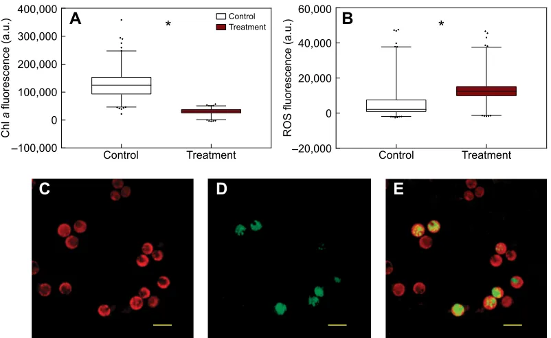
Using coral explants allowed us to investigate single-cell processes in endosymbionts in vivo. Fluorescence microscopy showed a significant decline of autofluorescence in heat-treated explants (67,902±1250 a.u., Mann–Whitney test, P<0.001) compared with the control (137,111±1498 a.u.; Fig. 2A,C), indicative of chlorophylladegradation in the chloroplasts (Coles and Jokiel, 1977; Warner et al., 1996). A decrease in the integrity of the photosynthetic apparatus is often accompanied by increases in the cellular concentration of ROS, as well as increases in enzymatic antioxidant defences in the host and/or in different strains of Symbiodinium(Tchernov et al., 2004). Indeed, our results show a concomitant increase in the fluorescence intensity of the general ROS dye (CM-H2DCFDA) in the heat-treated explants, from
16,943±355 a.u. in the control to 26,196±373 a.u. in the treatment (Mann–Whitney test, P<0.001; Fig. 2B). The increased fluorescence under heat stress would suggest net accumulation of
ROS fluorescence (a.u.)
Chl
a
fluorescence (a.u.)
Control Treatment
A
B
Control Treatment
–20,000 0 20,000 40,000 60,000
Control Treatment
–100,000 0 100,000 200,000 300,000 400,000
*
*
[image:3.612.107.508.56.302.2]C
D
E
Fig. 2. Fluorescence intensity measurements for chlorophyllaand reactive oxygen species (ROS)-stained cells.(A) Autofluorescence of chlorophylla, (B) CM-H2DCFDA dye (ROS) within theSymbiodiniumcells, for control (white) and heat treatment (red), (C) autofluorescence (red) ofSymbiodiniumwithin a coral
explant, (D) fluorescence ofSymbiodiniumstained with CM-H2DCFDA dye (green) after acute thermal stress and (E) overlay of chlorophyllaand CM-H2DCFDA
dye, where ROS is evident withinSymbiodiniumcells. Significant differences shown by asterisks (Mann–Whitney,P<0.001). Data represent means±s.e.m. (N=600 for control andN=521 for treatment). Scale bars, 10 µm.
Control Treatment Control Treatment Control Treatment
0 0.0001 0.0002 0.0003
0 0.0001 0.0002 0.0003
DMSP
(pmol
µ
m
–3)
DMSO (pmol
µ
m
–3)
DMSO:DMSP
ratio
A
B
C
1
0 2 3
*
*
[image:3.612.75.538.574.703.2]*
Fig. 3. Intracellular concentrations of sulphur compounds measured in coral explants.(A) Dimethylsulphoniopropionate (DMSP), (B) dimethylsulphoxide (DMSO) and (C) DMSO:DMSP ratio in control (white) and heat-treated (red) explants. Significant differences shown by asterisks (unpairedt-test,P<0.001). Data represent means±s.e.m. (N=10 for each group).
Journal
of
Experimental
hydrogen peroxide inSymbiodinium in response to acute thermal stress (Fig. 2D,E), which has been reported previously (Suggett et al., 2008). While it is generally believed that the primary trigger for coral bleaching during heat stress is ROS produced in the light (Downs et al., 2002; Lesser, 2011), bleaching can also be triggered in the dark (Tolleter et al., 2013), suggesting that photosynthetically derived ROS are not a pre-requisite for bleaching (Tolleter et al., 2013). The oxidative stress in our study is a measure of ROS generated during the 20 min dark incubation with the dye and is possibly an indication of mitochondrial-produced ROS, driven in part by negative effects of temperature on mitochondrial integrity (Dunn et al., 2012).
Thermal stress caused a significant decline in concentrations of intracellular DMSP (unpairedt-test, t19=3.67,P<0.001; Fig. 3A)
and DMSO (unpairedt-test,t19=3.47,P<0.01; Fig. 3B), resulting in
a significant decrease in the DMSO:DMSP ratio (unpaired t-test, t19=3.64,P<0.001; Fig. 3C), a measure often used as an indicator
for the conversion of DMSP to DMSO, potentially mediated by ROS. This decrease in DMSO:DMSP ratio under thermal stress indicates greater use (or faster rate of quenching) of DMSO than DMSP, highlighting the enhanced potency of DMSO as an ROS quencher (Sunda et al., 2002). Furthermore, DMSO can be oxidised to methanesulphonic acid (MSA) by reacting with and scavenging hydroxyl radicals (Lavoie et al., 2016); this additional oxidation step could account for the greater loss in the DMSO pool compared with DMSP.
Similar decreases in physiological health, increases in ROS (Franklin et al., 2004) and decreases in DMSP have been shown for Symbiodiniumcultures (Deschaseaux et al., 2014c), and previous work has found that changes in DMSO and DMSP often correlate (Deschaseaux et al., 2014a). The loss of DMSP/O in our study could be explained by an increase in DMSP lyase activity elicited by elevated temperatures, and thus the subsequent oxidation of DMS to DMSO. This has been described previously, where decreases in DMSP and DMS over 48 h in temperature-stressed Acropora intermedia(Fischer and Jones, 2012). Alternatively, because of the rapid increase in temperature, intracellular DMSP could have been used as an antioxidant (Sunda et al., 2002), with further oxidation of DMSO to MSA, reducing the available intracellular pools of both DMSP and DMSO well before anyde novosynthesis could take place. This is plausible, given that DMSP is energetically expensive to produce (Keller et al., 1999), and if coral cellular activity is compromised, as was indicated by the lowerFv/Fm, higher ROS and
degradation in chlorophyll a, the production of DMSP by the symbiont cells may have slowed or ceased under the acute temperature stress.
Here, we use coral explants as model organisms for host– symbiont cellular investigations. Coral explants were used for direct measurements of intracellular ROS in vivoduring a stress event, allowing for rapid and repeatable measurements of ROS activity in endosymbiotic Symbiodinium. While we did not investigate the oxidative stress response of the host, the same methodology could be applied to host cells, enabling assessment of both host and symbiont responses in an intact symbiosis. In this study we explored the links between intracellular ROS and DMSP dynamics in corals under acute thermal stress, a procedure previously made difficult owing to the complexity of measuring ROSin vivo, as the removal of tissue or symbionts from the coral skeleton is invasive and would likely influence the ROS activity being measured. Our results show increased ROS in endosymbionts under acute thermal stress and a decline in both DMSP and DMSO pools, supporting the involvement of sulphur compounds in the quenching of ROS in
corals. This study adds to the growing knowledge of the importance of dimethylated sulphur compounds in oxidative stress regulation by linking intracellular ROS with DMSP and DMSO dynamics under acute thermal stress. Investigating these biochemical links in corals at the cellular level will be helpful for future studies, as we uncover the site of DMSP production and its proximity to these short-lived oxygen radicals.
Acknowledgements
Thanks to Daniel A. Nielsen for assistance with microscopy and Elizabeth Deschaseaux for help with DMSO measurements.
Competing interests
The authors declare no competing or financial interests.
Author contributions
Conceived and designed the experiments: S.G.G. and K.P. Performed the experiments: S.G.G. and K.P. Analysed the data: S.G.G. and K.P. Wrote the paper: S.G.G. and K.P., with comments and suggestions from all authors.
Funding
This work was funded by Climate Change Cluster and School of Life Sciences, University of Technology Sydney and the Australian Research Council (DP140101045) awarded to Justin R. Seymour and Katherina Petrou.
Data availability
Data are available from Figshare: https://figshare.com/s/df0ae582b618fe710df8
References
Broadbent, A. D. and Jones, G. B.(2004). DMS and DMSP in mucus ropes, coral mucus, surface films and sediment pore waters from coral reefs in the Great Barrier Reef.Mar. Freshw. Res.55, 849-855.
Coles, S. L. and Jokiel, P. L.(1977). Effects of temperature on photosynthesis and respiration in hermatypic corals.Mar. Biol.43, 209-216.
Curran, M. A. J., Jones, G. B. and Burton, H.(1998). Spatial distribution of dimethylsulfide and dimethylsulfoniopropionate in the Australasian sector of the Southern Ocean.J. Geophys. Res.103, 16677-16689.
Deschaseaux, E. S. M., Jones, G. B., Deseo, M. A., Shepherd, K. M., Kiene, R. P., Swan, H. B., Harrison, P. L. and Eyre, B. D.(2014a). Effects of environmental factors on dimethylated sulfur compounds and their potential role in the antioxidant system of the coral holobiont.Limnol. Oceanogr.59, 758-768.
Deschaseaux, E. S. M., Kiene, R. P., Jones, G. B., Deseo, M. A., Swan, H. B., Oswald, L. and Eyre, B. D.(2014b). Dimethylsulphoxide (DMSO) in biological samples: a comparison of the TiCl3 and NaBH4 reduction methods using
headspace analysis.Mar. Chem.164, 9-15.
Deschaseaux, E. S. M., Beltran, V. H., Jones, G. B., Deseo, M. A., Swan, H. B., Harrison, P. L. and Eyre, B. D.(2014c). Comparative response of DMS and DMSP concentrations inSymbiodiniumclades C1 and D1 under thermal stress.
J. Exp. Mar. Biol. Ecol.459, 181-189.
Downs, C. A., Fauth, J. E., Halas, J. C., Dustan, P., Bemiss, J. and Woodley, C. M.(2002). Oxidative stress and seasonal coral bleaching.Free Radic. Biol. Med.33, 533-543.
Downs, C. A., Kramarsky-Winter, E., Woodley, C. M., Downs, A., Winters, G., Loya, Y. and Ostrander, G. K.(2009). Cellular pathology and histopathology of hypo-salinity exposure on the coralStylophora pistillata.Sci. Total Environ.407, 4838-4851.
Downs, C. A., McDougall, K. E., Woodley, C. M., Fauth, J. E., Richmond, R. H., Kushmaro, A., Gibb, S. W., Loya, Y., Ostrander, G. K. and Kramarsky-Winter, E.(2013). Heat-stress and light-stress induce different cellular pathologies in the symbiotic dinoflagellate during coral bleaching.PLoS ONE8, e77173.
Dunn, S. R., Pernice, M., Green, K., Hoegh-Guldberg, O. and Dove, S. G.(2012). Thermal stress promotes host mitochondrial degradation in symbiotic cnidarians: are the batteries of the reef going to run out?PLoS ONE7, e39024.
Falkowski, P. and Raven, J.(1997).Aquatic Photosynthesis. Oxford, UK: Blackwell Science.
Fischer, E. and Jones, G.(2012). Atmospheric dimethylsulphide production from corals in the Great Barrier Reef and links to solar radiation, climate and coral bleaching.Biogeochemistry110, 31-46.
Franklin, D. J., Hoegh-Guldberg, O., Jones, R. J. and Berges, J. A.(2004). Cell death and degeneration in the symbiotic dinoflagellates of the coralStylophora
pistillataduring bleaching.Mar. Ecol. Prog. Ser.272, 117-130.
Gardner, S. G., Nielsen, D. A., Petrou, K., Larkum, A. W. D. and Ralph, P. J.
(2015). Characterisation of coral explants: a model organism for cnidarian–
dinoflagellate studies.Coral Reefs34, 133-142.
Journal
of
Experimental
Gardner, S. G., Nielsen, D. A., Laczka, O., Shimmon, R., Beltran, V. H., Ralph, P. J. and Petrou, K.(2016). Dimethylsulfoniopropionate, superoxide dismutase and glutathione as stress response indicators in three corals under short-term hyposalinity stress.Proc. R. Soc. B Biol. Sci.283, 20152418.
Jones, G. and King, S. (2015). Dimethylsulphoniopropionate (DMSP) as an indicator of bleaching tolerance in scleractinian corals.J. Mar. Sci. Eng.3, 444-465.
Keller, M. D., Kiene, R. P., Matrai, P. A. and Bellows, W. K.(1999). Production of glycine betaine and dimethylsulfoniopropionate in marine phytoplankton. I. Batch cultures.Mar. Biol.135, 237-248.
Kiene, R. P. and Gerard, G. (1994). Determination of trace levels of dimethylsulfoxide (DMSO) in seawater and rainwater.Mar. Chem.47, 1-12.
Kiene, R. P., Linn, L. J., González, J., Moran, M. A. and Bruton, J. A.(1999). Dimethylsulfoniopropionate and methanethiol are important precursors of methionine and protein-sulfur in marine bacterioplankton. Appl. Environ.
Microbiol.65, 4549-4558.
Lavoie, M., Levasseur, M. and Sunda, W. G.(2016). A steady-state physiological model for intracellular dimethylsulfoxide in marine phytoplankton.Environ. Chem.
13, 212-219.
Lesser, M. P.(1997). Oxidative stress causes coral bleaching during exposure to elevated temperatures.Coral Reefs16, 187-192.
Lesser, M. P.(2011). Coral bleaching: causes and mechanisms. InCoral Reefs: an
Ecosystem in Transition (ed. Z. Dubinsky and N. Stambler), pp. 405-419.
New York: Springer.
Pernice, M., Meibom, A., Van Den Heuvel, A., Kopp, C., Domart-Coulon, I., Hoegh-Guldberg, O. and Dove, S. (2012). A single-cell view of ammonium assimilation in coral–dinoflagellate symbiosis.ISME J.6, 1314-1324.
Schneider, C. A., Rasband, W. S. and Eliceiri, K. W.(2012). NIH Image to ImageJ: 25 years of image analysis.Nature Methods9, 671-675.
Shapiro, O. H., Kramarsky-Winter, E., Gavish, A. R., Stocker, R. and Vardi, A.
(2016). A coral-on-a-chip microfluidic platform enabling live-imaging microscopy of reef-building corals.Nat. Commun.7, 10860.
Suggett, D. J., Warner, M. E., Smith, D. J., Davey, P., Hennige, S. and Baker, N. R. (2008). Photosynthesis and production of hydrogen peroxide by
Symbiodinium (Pyrrhophyta) phylotypes with different thermal tolerances.J.
Phycol.44, 948-956.
Sunda, W., Kieber, D. J., Kiene, R. P. and Huntsman, S.(2002). An antioxidant function for DMSP and DMS in marine algae.Nature418, 317-320.
Tchernov, D., Gorbunov, M. Y., de Vargas, C., Yadav, S. N., Milligan, A. J., Haggblom, M. and Falkowski, P. G.(2004). Membrane lipids of symbiotic algae are diagnostic of sensitivity to thermal bleaching in corals.Proc. Natl. Acad. Sci. USA101, 13531-13535.
Tchernov, D., Kvitt, H., Haramaty, L., Bibby, T. S., Gorbunov, M. Y., Rosenfeld, H. and Falkowski, P. G.(2011). Apoptosis and the selective survival of host animals following thermal bleaching in zooxanthellate corals.Proc. Natl. Acad.
Sci. USA108, 9905-9909.
Tolleter, D., Seneca, F. O., DeNofrio, J. C., Krediet, C. J., Palumbi, S. R., Pringle, J. R. and Grossman, A. R. (2013). Coral bleaching independent of photosynthetic activity.Curr. Biol.23, 1782-1786.
Warner, M. E., Fitt, W. K. and Schmidt, G. W.(1996). The effects of elevated temperature on the photosynthetic efficiency of zooxanthellaein hospitefrom four different species of reef coral: a novel approach.Plant Cell Environ.19, 291-299.