LEABHARLANN CHOLAISTE NA TRIONOIDE, BAILE ATHA CLIATH TRINITY COLLEGE LIBRARY DUBLIN OUscoil Atha Cliath The University of Dublin
Terms and Conditions of Use of Digitised Theses from Trinity College Library Dublin
Copyright statement
All material supplied by Trinity College Library is protected by copyright (under the Copyright and Related Rights Act, 2000 as amended) and other relevant Intellectual Property Rights. By accessing and using a Digitised Thesis from Trinity College Library you acknowledge that all Intellectual Property Rights in any Works supplied are the sole and exclusive property of the copyright and/or other I PR holder. Specific copyright holders may not be explicitly identified. Use of materials from other sources within a thesis should not be construed as a claim over them.
A non-exclusive, non-transferable licence is hereby granted to those using or reproducing, in whole or in part, the material for valid purposes, providing the copyright owners are acknowledged using the normal conventions. Where specific permission to use material is required, this is identified and such permission must be sought from the copyright holder or agency cited.
Liability statement
By using a Digitised Thesis, I accept that Trinity College Dublin bears no legal responsibility for the accuracy, legality or comprehensiveness of materials contained within the thesis, and that Trinity College Dublin accepts no liability for indirect, consequential, or incidental, damages or losses arising from use of the thesis for whatever reason. Information located in a thesis may be subject to specific use constraints, details of which may not be explicitly described. It is the responsibility of potential and actual users to be aware of such constraints and to abide by them. By making use of material from a digitised thesis, you accept these copyright and disclaimer provisions. Where it is brought to the attention of Trinity College Library that there may be a breach of copyright or other restraint, it is the policy to withdraw or take down access to a thesis while the issue is being resolved.
Access Agreement
By using a Digitised Thesis from Trinity College Library you are bound by the following Terms & Conditions. Please read them carefully.
Evaluation of polarising stimuli on bone
marrow-derived macrophage phenotypes
in two models of neuroinflammation
James Barrett
A thesis submitted to Trinity College Dublin for the degree of Doctor of
Philosophy.
Department of Physiology
Trinity College Institute of Neuroscience
f^TONITYCOLLEGE^
- < MA,'! 20!'.
5^ LI3RARY
D U B U J ^
I: Declaration of authorship
I declare that this thesis has not been submitted as an exercise for a degree at this or any
other university and it is entirely my own work, with the exception of certain results kindly
donated by Dr. Raasay Jones and Dr. Aedin Minogue and work carried out in conjunction
with Dr. Thelma Cowley. I agree to deposit this thesis in the University’s open access
institutional repository or allow the library to do so on my behalf, subject to Irish Copyright
Legislation and Trinity College Library conditions of use and acknowledgement.
II: Acknowledgements
Firstly I would sincerely like to thank my supervisor Professor Marina Lynch for all
of her encouragement, support, guidance and most of all, patience. You made sure I
didn’t feel sorry for myself when things didn't go to plan and no matter how busy you were,
your door was always open.
I would like to thank everyone in the MAL group past and present for making it
such a great place to work. I would like to particularly like to thank Aedin for all of her help
and support from the start of this PhD. I'd like to say thanks to Aedin and Raasay for being
great "work fronds” and making the lab a great place to be even on the bad days. Thanks
to Anto and Downer for all the chats, advice and introducing me to Beamish! Thanks to all
the other postgrads past and present: Donal, Rashers, Niamh, Colin, Rodrigo, Ronan,
Lorraine, Steph and Tara for the much needed laughs and cups of tea in the office.
A PhD is a long hard road, but it is made a lot easier having a friend beside you
who is going through the same process, so I would like to thank Jack for being such an
amazing friend and helping me along the way (I promise to repay all the proof reading). I
would also like to thank Ciaran for the free accommodation in Bordeaux, the breaks were
much appreciated! A big thanks to Simon for all the free tickets to gigs and making sure I
remembered to have fun along the way. People say it’s hard to live with a PhD student, so
I would like to thank my flatmate Aaron for putting up with Jack and I for the last three
years. HON THE LADS! Thanks to Murray for always being on the other end of the phone
and keeping me informed about the Championship!
I would also like to thank all the staff in the Department of Physiology for making it
an enjoyable place to study for the last 5 years, in particular Kieran, Aidan, Quentin and
Alice.
I would finally like to thank my family for all their never ending support and
Ill: Abstract
Inflammatory changes in the brain have been observed with aging and in
neurodegenerative disorders, and these are believed to be associated with the
accompanying decline in cognitive function. Recent evidence has suggested that
peripheral immune cells may play a role in the modulation of inflammatory events within
the central nervous system. The objective of this thesis was to investigate the responses
of macrophages prepared from aged rats and mice which overexpress amyloid precursor
protein and presenilin 1 (APP/PS1 mice) to stimuli which have been shown to polarise
macrophages to different activation states. Therefore, the effects of lipopolysaccharide
(LPS) and interferon-y (IFNy), which induce classical activation of macrophages, which is
also referred to as the M1 phenotype, and interleukin-4 (IL-4), which induces alternative
activation, also referred to as the M2 state, were compared in bone marrow-derived
macrophages (BMDMs) from young and aged rats and BMDMs from wildtype (WT) and
APP/PS1 mice.
The data demonstrate that macrophages infiltrate the brains of aged rats and
because of the age-related changes, it was important to investigate the responses of
these macrophages to inflammatory stimuli. The data showed that macrophages from
aged rats exhibited an enhanced response to LPS and IFNy compared with macrophages
from young rats, however there was no age-related change observed in response to IL-4.
It was also found that age was associated with increased hippocampal expression of the
TLR4 agonist, high mobility group box 1
(HMGB1), suggesting that infiltrating
macrophages responding to HMGB1 may exacerbate existing inflammation within the
brain.
It has been consistently reported that microglial activation is observed in animal
models of Alzheimer’s disease and the present data shows that there is an increase in
hippocampal concentration of IFNy in APP/PS1 mice which may trigger microglial
activation. The present data also demonstrate that macrophage infiltration was increased
in the brains of APP/PS1 mice and, significantly, BMDMs from APP/PS1 mice exhibit
enhanced responsiveness to IFNy. Therefore infiltrating macrophages may be further
An additional aim of this study was to evaluate the signalling events triggered by
IL-4 and IFNy. IL-4 increased activation of phosphorylated AKT (pAKT) in BMDMs from
wildtype mice; in contrast, IFNy treatment decreased pAKT activation. The evidence
indicates that activation of the PI3K/AKT pathway plays a role in the induction of the M2
markers Arginase-1, Chitinase-3-like-3 and found in inflammatory zone 1 and inhibition of
this pathway was associated with a dramatic decrease in IL-4-induced expression of these
markers. IFNy-induced tumour necrosis factor-a, major histocompatibility complex II and
intercellular cell adhesion molecule-1 was exaggerated in the presence of the PI3K
inhibitor Ly294002. Therefore it is concluded that manipulation of this pathway impacts on
the polarisation of macrophages.
IV: Table of contents
I: Declaration of Authorship
i
II: Acknowledgements
ii
III: Abstract
iii
IV: Table of
contents
v
V: List of figures
xiv
VI: List of Tables
xvii
VII: Abbreviations
xviii
Chapter 1
Introduction
1.1. Macrophages
2
1.2. Macrophage Activation
3
1.3. M1 activation and markers of the M1 activation state
6
1.3.1. TNFa and N0S2 are the archetypal markers of the M1 activation
7
state
1.3.1.1. TNFa
7
1.3.1.2. N0S2
8
1.3.2. Inflammatory Cytokines as markers of M l activation
9
1.3.2.2. IL-6
9
1.3.3.
Cellular proteins as markers of M l activation
10
1.3
.3.1
MHCII
10
1.3.3.2.
CD11b
10
1.3.3
.3.
CD40
10
1.3.3.4
.
CD68
11
1.3.3.5. N 0D 2
11
1.3.3.6
.
ICAM-1
12
1.4
.
M2 activation
12
1.4
.1.
Arginase 1
13
1.4
.2.
Mannose Receptor
13
1.4
.3.
Chitinase-3-like-3
14
1.4.4
.
Fizz1
16
1.5.
Macrophage signalling
16
1.5.1.
TLR4 signalling
16
1.5.2. The IFNy signalling pathway
17
1.5.3.
The IL-4 signalling Pathway
21
1.5.4.
PI3K/AKT Pathway
21
1.6.The blood-brain barrier
23
1.8 Alzheimer’s Disease
1.9 Aims and objectives
25
29
Chapter 2
Materials and methods
2.1 Animals
31
2.2. Cell Culture
32
2.2.1. Aseptic technique
32
2.2.2. Maintenance of L929 cell line
32
2.2.3. Cell treatments
33
2.2.4.
Generation
of bone
marrow-derived
macrophages 33
(BMDMs)
2.2.5. Cell counting
34
2.3. Mononuclear cell isolation
35
2.3.1. Preparation of Percoll gradients
35
2.3.2. Cell isolation
35
2.4.1. Assessment of phagocytosis
2.4.2. Cell surface staining
37
37
2.5. Western Immunoblotting
39
2.5.1. Preparation of cell lysates
39
2.5.2. Preparation of tissue
39
2.5.3. Gel electrophoresis
39
2.6. Analysis of mRNA by reverse transcriptase polymerase chain reaction (RT-
PCR)
41
2.6.1. Isolation of RNA
41
2.6.2. RNA quantification
41
2.6.3. cDNA synthesis
42
2.6.4. cDNA amplification by RT- PCR
42
2.6.5. RT-PCR quantification
42
2.7. Cytokine analysis
45
2.8. Mesoscale multiplex assays
47
2.8.1. Tissue preparation for Mesoscale
47
2.8.2. Mouse Pro-Inflammatory 7-Piex Assay
47
C hapter 3
The Im pact of aging on the activation state o f bone-m arrow derived m acrophages
3.1. Introduction 50
3.2. Methods
3.3. Results
3.3.1. Age-related increase in the number of macrophages present
in the brain
3.3.2. LPS induced CD11b and CD40 mRNA expression in
BMDMs
3.3.3. The effect of LPS on cytokine mRNA and supernatant
concentration in BMDMs from young and aged rats
3.3.4. The effect of IFNy on TNFa, and N 0 S 2 mRNA expression in
BMDMs from young and aged rats
3.3.5. The effect of IFNy on MHC I! and CD40 mRNA expression in
BMDMs from young and aged rats
3.3.6. The effect of IL-4 on MRC1 and ARG1 mRNA expression in
BMDMs from young and aged rats
3.3.7. Phagocytic activity of BMDMs from young and aged rats.
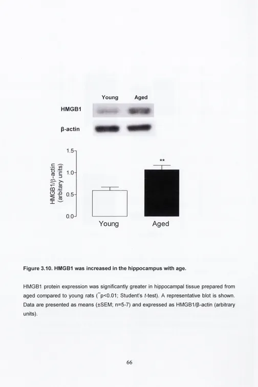
3.3.8. HMGB1 protein expression in the hippocampus of young
and aged rats
52
53
53
53
53
54
55
55
55
56
Chapter 4
Analysis o f the responsiveness of bone marrow-derived macrophages from WT and
APP/PS1 mice to inflam m atory stim uli
4.1. Introduction
4.2. Methods
4.3. Results
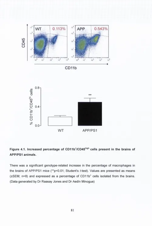
4.3.1. CD11b+/CD45high cells are present in the brains of
APP/PS1 animals.
4.3.2. The effect of IFNy on TNFa and N0S2 mRNA
expression in BMDMs from WT and APP/PS1 mice
4.3.3. The effect of IFNy on MHC II and N0D2 mRNA
expression in BMDMs from WT and APP/PS1 mice
4.3.4. The effect of LPS on CD11b and CD11b mRNA
expression in BMDMs from WT and APP/PS1 mice
4.3.5. LPS increased cytokine concentration in BMDMs in
BMDMs from WT and APP/PS1 mice
4.3.6. The effect of IL-4 on MRC1 and Arg1 mRNA expression
in BMDMs from WT and APP/PS1 mice
4.3.7. IFNyRI mRNA expression is increased in BMDMs from
APP/PS1 mice
73
76
77
77
77
77
78
78
78
4.3.8. IFNy concentration is increased in the hippocampus of
APP/PS1 mice
4.3.9. IFNy increases TNFa and N0S2 mRNA expression in
BMDMs from WT and CD200 '' mice
4.3.10. LPS increased TNFa and N0S2 mRNA expression in
BMDMs from WT and CD200 '" mice
4.3.11. IL-4 increased MRC1 and Arg1 mRNA expression in
BMDMs from WT and CD200 '’ mice
4.3.12. IL-4 increased MRC1 and Arg1 mRNA expression in
BMDMs from WT and CD200 '' mice
Chapter 5
Modulation of macrophage activation; a role for the PI3K/AKT pathway?
5.1. Introduction
99
5.2. Methods
101
5.3. Results
102
5.3.1. IL-4 induced the activation of pSTAT6 in BMDMs
102
5.3.2. The effect of IL-4 on PI3K/AKT pathway in BMDMs
102
5.3.3. IL-4 induced the M R C I.A rg I, ChiSliS and FIZZ1 in BMDMs
102
5.3.4. The effect of Ly294002 on IL-4-induced markers of M2
activation in BMDMs
5.3.5. The effect of Ly294002 on IL-4-induced STAT6 and
PI3K/AKT signalling in BMDMs
^^2
5.3.6. IFNy induced the activation of pSTATI in BMDMs
103
5.3.7. The effect of IFNy on PI3K/AKT activity in BMDMs
104
5.3.8. The effect of Ly294002 on IFNy-induced markers of Ml
activation in BMDMs
5.3.9. The effect of Ly294002 on IFNy-induced STATI and
PI3K/AKT signalling in BMDMs
Chapter 6
General discussion
6
.
1
.
General discussion
127
6.2
Limitations and future studies
136
References
138
V: L ist of figures
Figure 1.1 L-Arginine metabolism (Adapted from Varin & Gordon 2009)
15
Figure 1.2 TLR4 signalling pathway
19
Figure 1.3 IFNy induced classical activation
20
Figure 1.4 The IL-4 signalling pathway
22
Figure 2.1. Image of Rat BMDMs
34
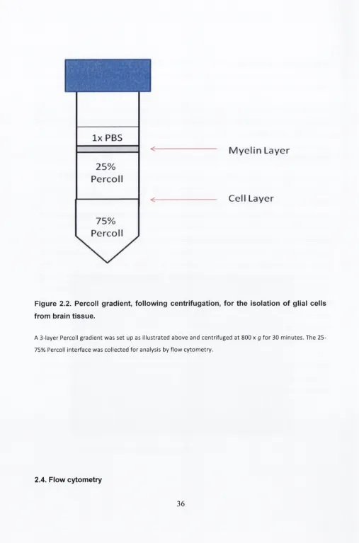
Figure 2.2. Percoll gradient, following centrifugation, for the isolation of glial
cells from brain tissue
36
Figure 3.1. Age was associated with an increase in CD11bVCD45^'®'' cells in
brain tissue
57
Figure 3.2. LPS induces CD11 b and CD40 mRNA expression in BMDMs from
young and aged rats
58
Figure
3.3. LPS increases
IL-1P mRNA
expression
and supernatant
concentration in BMDMs from
young and
aged rats
59
Figure
3.4. LPS increases TNFa mRNA
expression
and supernatant
concentration in BMDMs from
young and
aged rats
60
Figure
3.5. LPS increases
IL-6 mRNA
expression
and supernatant
concentration in BMDMs from
young and
aged rats
61
Figure 3.6. The effect of IFN-y on TNFa, and N0S2 mRNA expression in
BMDMs from young and aged rats
62
Figure 3.7. The effect of IFN-y on MHC II and CD40 mRNA expression in
BMDMs from young and aged animals
Figure 3.8. The effect of IL-4 on MRC1 and ARG1 mRNA expression in
BMDMs from young and aged rats
64
Figure 3.9. Phagocytic activity of BMDMs was increased with age
65
Figure 4.1. Increased percentage of CD11bVCD45^'®^ cells present in the
brains ofAPP/PS1 animals
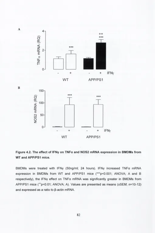
Figure 4.2. The effect of IFNy on TNFa and N0S2 mRNA expression in
BMDMs from WT and APP/PS1 mice
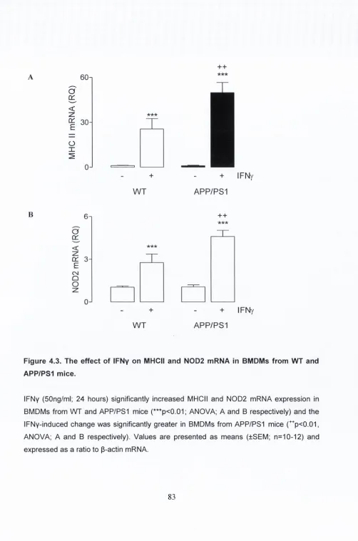
Figure 4.3. The effect of IFNy on MHC I! and N0D2 mRNA in BMDMs from
WT and APP/PS1 mice
Figure 4.4. The effect of LPS on CD40 and CD11b mRNA in BMDMs from
WT and APP/PS1 mice
Figure 4.5. The effect of LPS on TNFa and N0S2 mRNA in BMDMs from WT
and APP/PS1 mice
Figure 4.6. LPS increased TNFa and IL-6 in supernatant of BMDMs from WT
and APP/PS1 mice
Figure 4.7. The effect of IL-4 on MRC1 and Arg1 mRNA expression in
BMDMs from WT and APP/PS1 mice
Figure 4.8. IFNGR1 mRNA was increased in BMDMs from APP/PS1 mice
Figure 4.9. IFNy concentration was increased in hippocampus of APP/PS1,
compared with WT mice
Figure 4.10. The effect of IFNy on TNFa, and N0S2 mRNA expression in
BMDMs from WT and CD200 '’ mice
Figure 4.11. The effect of LPS on CD11b and CD40 mRNA in BMDMs from
WT and CD200 '' mice
Figure 4.12. The effect of IL-4 on MRC1 and Arg1 mRNA expression in
BMDMs from WT and CD200 '' mice
Figure 5.1. The effect of IL-4 on pSTAT6 expression in BMDMs
Figure 5.2. The effect of IL-4 on pPI3K and PI3K expression in BMDMs
107
Figure 5.3. The effect of IL-4 on pAKT and AKT expression in BMDMs
108
109
Figure 5.4. The effect of IL-4 on MRC1 and Argi mRNA expression in
BMDMs
Figure 5.5. The effect of IL-4 on Chi3li3 and Fizzi nnRNA expression in
BMDMs
110
Figure 5.6. The effect of Ly294002 on IL-4-induced MRC1 and Argi mRNA
expression in BMDMs
111
Figure 5.7. The effect of Ly294002 on IL-4-induced Fizzi and Chi3li3 mRNA
expression in BMDMs
112
Figure 5.8. The effect of Ly294002 on IL-4 signalling in BMDMs
113
Figure 5.9. The effect of IFNy on pSTAT 1 expression in BMDMs
114
Figure 5.10. The effect of IFNy on pPI3K and PI3K expression in BMDMs
115
Figure 5.11. The effect of IFNy on pAKT and AKT expression in BMDMs
116
Figure 5.12. The effect of Ly294002 on IFNy-induced TNFa and N0S2
mRNA in BMDMs
117
Figure 5.13. The effect of Ly294002 on IFNy-induced MHCII and ICAM1
mRNA in BMDMs
118
Figure 5.14. The effect of Ly294002 on IFNy-induced CD40 and N0D2
mRNA in BMDMs
119
Figure 5.15. The effect of Ly294002 on IFNy signalling in BMDMs
120
Figure 6.1. Proposed mechanism for AKT modulation on macrophage
VI: List o f tables
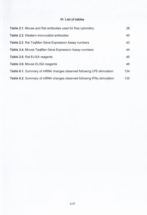
Table 2.1. Mouse and Rat antibodies used for flow cytometry
38
Table 2.2. Western immunoblot antibodies
40
Table 2.3. Rat TaqMan Gene Expression Assay numbers
43
Table 2.4. Mouse TaqMan Gene Expression Assay numbers
44
Table 2.5. Rat ELISA reagents
46
Table 2.6. Mouse ELISA reagents
46
VII: Abbreviations
AN OVA
Analysis of variance
APC
Antigen presenting cell
BBB
Blood-brain barrier
BCA
Bicinchoninic acid
BMDM
Bone marrow-derived macrophage
BSA
Bovine serum albumin
CD
Cluster of differentiation
cDNA
Complimentary deoxyribonucleic acid
CNS
Central nervous system
DAMP
Damage-associated molecular patterns
DMEM
Dulbecco’s modified Eagle’s medium
DNase
Deoxyribonuclease
dNTP
Deoxyribonucleotide triphosphate
EAE
Experimental Allergic Encephalomyelitis
ECM
Extracellular matrix
ELISA
Enzyme linked immunosorbent assay
ERK
Extracellular signal-regulated kinase
FBS
foetal bovine serum
Fizz1
Found in inflammatory zone 1
HMGB1
High mobility group protein B1
ICAM-1
Intracellular adhesion molecule 1
IFN
yInterferon-y
IKK
I
kB kinase
IL-10
Interleukin-10
IL -ip
Interleukin-1 p
IL-4
Interleukin-4
IL-6
Interleukin-6
IL-13
Interleukin-13
IRAK
Interleukin-1 receptor associated kinase
I
kB
Inhibitor of
kB
JAK
Janus tyrosine kinase
LBP
Lipopolysaccaride-binding protein
LPS
Lipopolysaccaride
LTA
Lipoteichoic acid
LTP
Long-term potentiation
MAL
MyDBB adaptor-like protein
MAPK
Ras/Mitogen-activated protein kinase
MHCII
Major histocompatibility complex class II
mRNA
Messenger RNA
MS
Multiple sclerosis
MyD88
Myeloid differentiation primary response gene 88
NEMO
NF-kappa-B essential modulator
NFkB
Nuclear factor kappa-light-chain-enhancer of activated B cells
NK cells
Natural Killer cells
N 0D 2
Nucleotide-binding oligomerization domain-containing protein 2
NOS
Nitric oxide synthase
PAMP
Pathogen-associated molecular pattern
PCR
Polymerase reaction chain
PI3K
phosphatidylinositol 3-kinases
PRR
Pattern recognition receptor
RasGAP
Ras GTPase activating protein
RNA
Ribonucleic acid
RNase
Ribonuclease
SEM
Standard error of mean
SHIP
Sh2-containing inositol phosphatase
SOCS
Suppressor of cytokine signaling
SphK
Spingosine phosphate kinase
STAT
Signal transducer and activator of transcription
Strep-HRP
Streptavidin-horseradish peroxidise linked
TGF-p
Transforming growth factor p
TLR
Toll-like receptor
Chapter 1
1.1 Macrophages
Elie Metchnikoff first described macrophages in 1905 (Metchnikoff, 1905), recognising their ability to eliminate pathogens and function in homeostasis. Macrophages form the first line of defence and, as part of the innate immune response, help mediate the immune response to microorganisms (Varin & Gordon, 2009). A primary role of macrophages is the phagocytosis of pathogens and the removal of debris resulting from apoptotic cells (Town et al., 2005).
Macrophage populations within tissue can be replenished from circulating blood monocytes; in fact as far back as 1939, Ebert and Florey observed that monocytes migrated from the blood and differentiated into macrophages in tissue (Gordon & Taylor, 2005). Other work has also described an increased number of monocytes recruited during an inflammatory event (Murray & Wynn, 2011). Blood monocytes originate in the bone marrow from hematopoietic stem cells, these cells then infiltrate tissue where they differentiate into mature macrophages and these tissue macrophages aid maintenance of homeostasis, with important roles in the clearance of cellular debris, tissue remodelling, repair and the initiation and resolution of inflammatory events. Macrophages exhibit great heterogeneity, as they adopt tissue specific functions which vary from osteoclasts that remodel bone and microglia that maintain homeostasis within the central nervous system (CNS) to liver macrophages, commonly referred to as Kupffer cells, which exhibit increased phagocytic activity as they remove foreign materials from the portal circulation. Alveolar macrophages are responsible for the removal of inhaled pathogens and, these macrophages can produce reactive oxygen species to aid this process. Perivacular macrophages are another subset of macrophages whose primary function is to remove debris from the perivascular space.
much debate as to the origin of microglia, but recent findings have suggested that these cells are derived from primitive macrophages in the yolk sac that migrate to the CNS during development (Saijo & Glass, 2011). However, it has also been proposed that bone marrow-derived cells have the ability to cross the blood-brain barrier (BBB) and differentiate into microglia (Hess et al., 2004; Malm et al., 2005; Simard et a i, 2006). Simard and colleagues transplanted stem cells expressing green fluorescent protein (GFP) into lethally-irradiated mice and later observed that cells expressing GFP had infiltrated the CNS. This has led to the theory that there may be two distinct populations within the CNS - resident microglia and bone marrow-derived microglia. The merits of use of irradiation have been called into question as other groups have demonstrated that irradiation can lead to disruption of the blood-brain barrier (BBB) and this may account for the appearance of bone marrow cells within the CNS.
Bone marrow-derived macrophages (BMDMs) are primary macrophage cells derived from bone marrow stem cells that can differentiate into mature macrophages in the presence of growth factors in vitro. The major benefit of using these cells is that, unlike peritoneal macrophages or alveolar macrophages, a high number of cells can be generated from a single animal. They respond to a variety of stimuli including interleukin (IL)-4, interferon-(IFN)Y and lipopolysaccharide (LPS) and, since they express the same genes as tissue macrophages, may prove to be a more relevant research tool than macrophage-like cell lines such as RAW 246.7 cells. It has been demonstrated that perivascular macrophages are bone marrow-derived, and that the turnover of these cells is approximately 30% over 2 months and, as mentioned above, it has been reported that bone marrow cells may migrate across the BBB and populate the CNS.
1.2 Macrophage Activation
The role of macrophages is to maintain tissue homeostasis and, to achieve, this they express cell surface proteins that allow them to respond to a wide range of stimuli. To enable them to respond to invading pathogens, macrophages are activated by pattern associated molecular patterns (PAMPs), which are structures expressed by pathogens (Olson & Miller, 2004). These cells express pathogen recognition receptors (PRRs), an example of which is the toll-like receptor (TLRs) family. There are over 10 TLRs in humans which are capable of interacting with a diverse range of PAMPs (Takeda & Akira, 2005). TLR4 recognises LPS, a component of the cell wall of gram-negative bacteria (Buchanan et al., 2010) and activation of TLR4 leads to the upregulation of pro-inflammatory mediators (Buchanan et al., 2010). Macrophages also respond to endogenous molecules associated with ‘danger’ and stress (Plowden et al., 2004). Release of damage-associated molecular patterns (DAMPs) into the microenvironment results in activation of nearby macrophages; adenosine triphosphate (ATP) is released from damaged or dying cells (Thomas & Salter, 2010) and induces the activation of the inflammasome both in macrophages (Mills, 2011) and microglia (Murphy & Lynch, 2012).
One of the main roles of macrophages is phagocytosis, which is defined as the uptake of particles greater than O.Spm. The primary function of this process is the removal of dying or damaged cells and pathogens to maintain homeostasis, and for example, it has been suggested that macrophages remove almost 2 x 1 0 ” erythrocytes each day (Mosser & Edwards, 2008). Phagocytosis is a crucial part of the immune response mediating the ingestion, destruction and processing of foreign materials and macrophages express receptors including TLRs and scavenger receptors (SRs) that mediate this process. Interaction of receptors on macrophages with ligands on the surface of the particle induces a signalling cascade that results in the plasma membrane surrounding the particle and internalising. The ingested material is then localised to the phagosome, which is a highly acidic environment equipped with peptides that degrade microbial components (Flannagan et al., 2009). Macrophages also function as antigen presenting cells by displaying antigens from digested pathogens on their cell surface; this allows interaction with T cells and initiates the adaptive immune response.
Upon activation, macrophages release inflammatory mediators, such as pro- inflammatory cytokines, however they also express cell surface markers which enable macrophages to communicate with other cells o f the immune system. Activated macrophages exhibit increased expression of MHCII which is involved in antigen presentation and allows communication between the innate and adaptive immune response. Increased expression of CD40 is a key feature of activated macrophages, CD40 receptor is expressed on macrophages while CD40 ligand is expressed on T cells, this allows further communication with the adaptive immune response.
In the last 100 years since the first description of macrophages, research has exposed the complexity of macrophage function. As stated by David Mosser ‘macrophages may be more heterogeneous group of cells than originally appreciated, with different physiologies and performing immunological functions' (Mosser, 2003). These groups of macrophages are broadly divided into M1 and M2 activated cells, which in the simplest possible terming perform pro and anti inflammatory functions respectively.
1.3 M1 activation and markers of the M1 activation state
As already described, macrophages become activated in response to changes within the tissue environment. M1 or classical activation of macrophages describes a shift in the cell phenotype from a quiescent state to a pro-inflammatory profile. The M1 state can be initiated by the Th1-derived cytokine IFNy, a PAMP such as LPS or DAMPs such as ATP or high mobility group box 1 (HMGB1). HMGB1 is primarily found within the nucleus and is released from necrotic cells but not apoptotic cells, which suggests that controlled cell death does not result in macrophage activation. HMGB1 has been shown to activate TLR4 signalling and results in the secretion of pro-inflammatory cytokines such as IL-1p, TNFa and IL-6. M1 macrophages are recruited to initiate the host response with the subsequent release of pro-inflammatory mediators.
Activation of macrophages through PRR has been extensively studied; LPS, a component of gram negative bacteria, is frequently used to induce inflammation in experimental models (Lund et al., 2006). LPS mediates its effects through TLR4, and induces a robust activation of macrophages, leading to the release of cytokines and expression of cell surface markers. The activation of TLRs is crucial to mount an immune response and macrophages from TLR4-deficient mice fail to mount a response to LPS treatment. While activation of TLRs is crucial in activating the immune system to mounting an effective immune response, it is also implicated in the pathogenesis of autoimmune, chronic inflammatory and infectious diseases
1.3.1. TNFa and N 0 S 2 a re th e archetypal markers of the M1 activation state
1.3.1.1.TN Fa
TNFa was first identified nearly 40 years ago as a product of macrophages (Carswell et al., 1975). TNFa inherited its name from its ability to induce cell death especially in tumour cells, though it also has the ability to activate an effective immune response to combat infection when necessary (Walczak, 2011). TNFa can exist in two forms, as a membrane-associated protein and also in a soluble form (Locksley et al., 2001). The membrane-bound TNFa forms heterodimers (Walczak, 2011) which the metalloprotease, TNFa converting enzyme (TACE), can cleave to release the soluble cytokine (Black et al., 1997). Release of pro-inflammatory cytokines such as TNFa from microglia/macrophages is an indication of increased activation of these cells (Lynch, 2009).
been implicated in the chronic activation of microglia observed in neurodegenerative diseases possibly as a result of its ability to act as an autocrine/paracrine signal for microglia (Mir et al., 2008).
1.3.1.2. N0S2
Like TNFa upregulation, an increase in the expression of the enzyme nitric oxide synthase (NOS) 2 is considered to be a marker of M1 activation. NOS can exist in 3 isoforms N0S1, N 0S 3 and N0S2 (iNOS). The N0S1 and N 0S 3 isoforms (also known as nNOS and eNOS) are constitutively expressed, while the expression of N 0S 2 is induced during an immune response (MacMaking et al., 1997). NOS activity ultimately leads to the production of NO through an enzymatic pathway involving the substrate arginine from which NO and citrulline are produced (Colton, 2009).
Activation of microglia/macrophages is associated with an increase in the production of NO (Kim et al., 2004; Mir et al., 2008). The production of NO promotes the death of bacteria and it can mediate cell death in a number of ways. Firstly, it can induce the phosphorylation of p53 which is involved in triggering apoptosis (Messmer et al., 1994). It can also inactivate cytochrome oxidase in the mitochondria and, as a result, disrupt cellular respiration (Colton, 2009). Thirdly, NO can mediate the oxidative and nitrosative damage to proteins, through the production of superoxides (MacMaking et al., 1997). Excessive activity of N0S2 can have severe detrimental effects, and NO has been postulated to have a critical role in mediating neurotoxicity associated with neuroinflammation (Dawson & Dawson, 1998). N0S2 immunoreactivity has been reported to be increased in the spinal cord of ALS patients (Sasaki et al., 2000), while LPS-induced production of NO by microglia has been associated with motor neuron injury (Zhao et al., 2004).
1.3.2 Inflammatory Cytokines as markers of M1 activation
1.3.2.1. IL -lp
The increased expression of inflammatory cytokines like IL-1(3 and IL-6 are also considered markers of the M1 activation state. IL-1P is considered to be the classic pro- inflammatory cytokine, and was first described in the 1980’s. It is produced by activated immune cells including macrophages and microglia. Systemic administration of IL-1P has been shown to induce sickness behaviour, and increased expression of other pro- inflammatory cytokines in the CNS. It has been consistently shown that IL-1P impairs the induction of long-term potentiation (LTP) and its concentration in the hippocampus is increased in the brains of aged rats in which a deficit in LTP has been shown (Minogue et a i, 2007). In the brain, microglia are considered to be the primary source of IL-ip . Exposure of microglia to Ap has been shown to result in increased expression of IL-1P; consequently it is not surprising that increased expression of IL -ip has also been observed in the brain of individuals with Alzheimer’s disease (AD) (Griffin et al., 1989).
1.3.2.2. IL-6
IL-6 is described as a pleiotropic cytokine, which can have both pro-inflammatory and anti-inflammatory effects. The effect of IL-6 on cells is mediated through the IL-6 receptor (IL-6R) and gp130 which is a signal transducer utilised by cytokines related to IL- 6; this leads to the activation of the JAK/STAT and mitogen-activated protein kinase (MAPK) cascade. The increased expression of acute phase proteins during an inflammatory response is chiefly mediated by IL-6 activity and it is thought that IL-6 plays a role in the recruitment of leukocytes to the site of infection. The role of IL-6 in inflammation is complex as conflicting reports have suggested beneficial and detrimental effects of this cytokine.
brain. However, it has also been shown that IL-6-deficient mice exhibit slower recovery following traumatic brain injury (Spooren et al., 2011), identifying a reparative role. Work from this laboratory has suggested that in LPS-stimulated rat glia, IL-6 regulates IL-1(3 and TNFa production (Minogue etal., 2012)
1.3.3. Cellular proteins as markers of M1 activation
1.3.3.1. MHCII
MHCII is expressed on antigen presenting cells (APC) which includes macrophages and microglia, and its upregulation is a hallmark of activated cells. It has been proposed that expression of MHCII promotes phagocytosis of pathogens and cellular debris which contributes to increased release of pro-inflammatory mediators (Lynch, 2009). Its expression on macrophages/microglia, together with expression of co stimulatory molecules CD80 and CD86, allows interaction with T cells (Frank et al., 2006). It has been suggested that MHCII upregulation is an indicator of the M l activation state in macrophages and microglia (Colton & Wilcock, 2010).
1.3.3.2. CD11b
1.3.3.3. CD40
CD40 is a member of tiie TNF receptor superfamily and is expressed on many immune cells such as macrophages and dendritic cells (Nguyen & Benveniste, 2002). Microglia express CD40 within the CNS, although expression is low under resting conditions though it is increased with age and following inflammatory stimuli (Lynch 2009). LPS treatment of macrophages or co-treatment of macrophages with Th1 cells leads to an increase in CD40 expression in vitro (Aloisi et a!., 1998; Aloisi et al., 2000; Nguyen & Benveniste, 2000; Lynch, 2009). CD40 ligand (CD40L), also known as CD154 is expressed on T cells (Henn et al., 1998). CD40-CD40L interaction is involved in the development of many immune responses and this is a key mechanism in enabling the interactions between APC and T cells (Aloisi, 2001). Binding of CD40 on microglial cells with its ligand CD40L leads to an increased production of pro-inflammatory mediators such as TNF-a, IL-12 and NO (Chen et al., 2006). Activated macrophages/microglia and CD40L-expressing T cells have been identified in MS and EAE (Gerritse et al., 1996) and this may contribute to the inflammation observed in these conditions. Indeed upregulation of CD40 accompanies upregulation of other markers, including MHCII in M l activated microglia (Colton & Wilcock, 2010). CD40 has also been implicated in a number of disorders that involve hyperactive immune response such as rheumatoid arthritis (RA) and MS (Aloisi, 2001). Blocking the CD40-CD40L interaction has been demonstrated to be beneficial in an animal model of MS (Gerritse et al., 1996)
1.3.3.4. CD68
1.3.3.5. N0D2
NOD-like receptors (NLRs) are a family of PRR, which are localised intracellularly and involved in the recognition of peptidoglycan moieties from bacteria. N0D2 activation is induced by muranyl dipeptide (MDP) which is a peptidoglycan expressed by both gram- positive and gram-negative bacteria. It is suggested that MDP treatment in combination with TLR activation can induce the production of pro-inflammatory mediators (Moreira & Zamboni, 2012), though it has been argued by others that this is only. N 0D2 mutations have been associated with susceptibility to infectious diseases such as leprosy and tuberculosis. Interestingly BMDMs from N0D2-deficient mice show no response to MDP stimulation (Pauleau & Murray, 2003), and a separate study reported that N0D2-deficient mice exhibit increased bacterial burden following oral administration of Listeria (Kobayashi et al., 2005). N 0D 2 activation may also be involved in the recruitment of other immune cells to the site of infection, as N 0D 2 agonists have been shown to induce the production of RANTES (CCL5) by macrophages (Werts et a!., 2007).
1.3.3.6. ICAM-1
Intracellular adhesion molecule 1 (ICAM-1), also known as CD54, is an adhesion molecule that is important in the cell-cell interaction of endothelial cells and leukocytes. It is also expressed on other cells including macrophages and microglia. ICAM-1 interacts with LFA-1, a receptor found on leukocytes, and it this interaction that allows binding of leukocytes to endothelial cells and migration into the tissue. Expression on macrophages and microglia is low under resting conditions, however LPS and IFNy stimulation have been shown to upregulate ICAM-1 expression. It is expressed on a number of cells and has been suggested that it mediates the infiltration of leukocytes.
1.4. M2 activation
morphology from classically-activated macrophages. In addition to MRC1 which is involved in endocytosis, M2 macrophages are characterised by increased expression of arginase which is involved in NO homeostasis, and the molecules chitinase-3-like-3 (Chi3li3) and found in inflammatory zone 1 (Fizz1), both of which are involved in the rebuilding of the extracellular matrix (Colton, 2009). The M2 state can also be induced by the anti-inflammatory cytokines IL-5 and IL-13 (Varin & Gordon, 2009).
M2 macrophages prevent excessive inflammation, mediating tissue repair and restoration of homeostasis (Gordon & Martinez, 2010) and this is thought to be initiated by the release of trophic factors. Although responses of M2 activated macrophages are beneficial in the resolution of the inflammatory response, it dampens the host immune response and it has been demonstrated that M2 macrophages promote Leishmania infection by inhibiting the Th1 response and macrophage-killing activity (Holscher et al., 2006).
1.4.1. Arginase 1
Arginase can exist in two isoforms, arginase 1 (A rg i) and arginase 2 (Arg2) that share approximately 60% amino acid sequence homology (Durante et al., 2007). Arg1 is an inducible isoform that is upregulated in M2 macrophages, while Arg2 is a mitochondrial- associated protein constitutively expressed in many cells throughout the body (Colton, 2009). A rg i is expressed at high levels within areas of the brain such as cerebellum, though expression of Arg2 appears to be low throughout the brain (Yu et al., 2003). The role of A rg i remains to be fully elucidated however a key function is considered to be in the regulation of NO production.
microglial cell line showed that treatment with IL-4/IL-13 attenuated the IFNy increase in N0S2 and TNFa mRNA (Colton, 2009).
The role of arginase in alternative activation may not be limited to decreasing the levels of NO during inflammatory events as it has been suggested that arginase may also be involved in the production of ornithine-derived proline a precursor in the production of collagen (Munder et al., 1999) which may be used in the rebuilding of the ECM.
1.4.2. Mannose Receptor
MRC1 is a member of the C-type lectins receptor (CLRs) family (Underhill & Ozinsky, 2002) and obtains its name from its ability to recognise sugars terminating in mannose, fucose or N-acteyl glucosamine (Taylor et al., 2005). MRC1 mediates the phagocytosis of bacteria/yeasts (Burudi & Regnier-Vigouroux, 2001) and is highly expressed on alternatively-activated macrophages/microglia as well as dendritic cells.
MRC1 exists in a membrane-bound or soluble form (Burudi & Regnier-Vigouroux, 2001) and cleavage of the MRC1 from the cell membrane leads to release of the soluble form (Martinez-Pomares et al., 1998). Engagement of the macrophage-bound MRC1 with its ligand initiates an anti-inflammatory signalling cascade which results in the production of IL-10 and IL-1 Ra and a decrease in IL-12 and TNFa mRNA and protein (Colton, 2009).
1.4.3. C hitinase-3-like-3
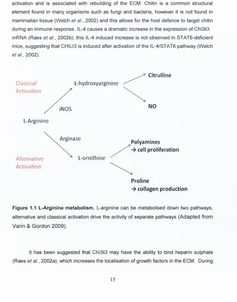
Also known as YM1, chitinase-3-like-3 (Chl3ll3) is another marker of alternative activation and is associated with rebuilding of the ECM. Chitin is a common structural element found in many organisms such as fungi and bacteria, however it is not found in mammalian tissue (Welch et al., 2002) and this allows for the host defence to target chitin during an immune response. IL-4 causes a dramatic increase in the expression of Chi3li3 mRNA (Raes et al., 2002b); this IL-4 induced increase is not observed in STAT6-deficient mice, suggesting that CHILI3 is induced after activation of the IL-4/STAT6 pathway (Welch et al., 2002).
Citrulline
Classical
Activation
L-hydroxyargininc
iNOS
NO
L-Arglninc
Arginasc
Poiyamines
ceil proliferation
Alternative
Activation
L-ornithine
Prollne
-> collagen production
Figure 1.1 L -A rg inlne m etabolism . L-arginine can be metabolised down two pathways,
alternative and classical activation drive the activity of separate pathways (A dapted from
V arin & G ordon 2009).
[image:37.530.27.516.167.791.2]an inflammatory response the levels of heparin are decreased and binding of Chi3li3 to heparin is believed to decrease the loss of growth factors from the ECM which ultimately allows rebuilding of the ECM after an inflammatory event (Colton, 2009). Others have suggested that Chi3li3 may have a role in the recruitment of eosinophils (Owhashi et al., 2000), suggesting a possible mechanism for macrophage and eosinophil communication. In further support of this function, it was demonstrated that blocking IL-4 and IL-13 activity or deletion of STAT6 prevents eosinophil recruitment in murine models of allergy (Welch et al., 2002).
1.4.4. Fizz1
Fizz1 also known as RELM-a, a member of the family of resistin-like molecules (RELM) is also upregulated in alternatively-activated macrophages/microglia. Fizz1 was first identified in the alveolar lavage of mice with experimentally-induced asthma (Nair et al., 2003) and it has been suggested that Fizz1 may act in an anti-inflammatory manner during lung allergy and asthma (Nair et al., 2009). Similar to YM1, Fizz1 has been implicated in the reconstruction of the extracellular matrix (ECM). Fizz1 upregulation increases the expression of collagen (Liu et al., 2004b), which is integral to the integrity of the ECM.
1.5. Macrophage signalling
1.5.1. TLR4 signalling
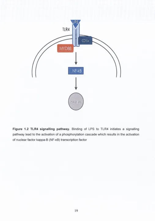
Signalling through TLR4 is mediated through two different pathways, the MyD88-
dependent and TRIF-associated (MyD88 independent) pathways (Figure 1.2). The
MyD88-dependent pathway is conserved throughout most of the TLR family; only TLR4
and TLR3 employ an additional pathway (Akira, 2006). Signalling through the MyD88-
dependent pathway is initiated through the interaction of the Toll/interleukin-1 receptor
(TIR) domains of TLR4 and MyD88 (Horng
et al.,
2002). MyD88 interacts with the
interleukin-1 receptor-associated kinase (IRAK) family; the interaction of the death
domains of MyD88 and IRAK4 allows the propagation of the signalling event (Kawai &
Akira, 2007). Both pathways lead to the activation of a phosphorylation cascade which
results in the activation of nuclear factor kappa-B (NF-kB) transcription factor. The TRIF-
dependent pathway is referred to as the ‘late phase’ of NF-kB activation, as NF-kB
activation occurs much earlier through the MyD88-dependent pathway which is aptly
called ‘early phase’ (Buchanan
et al.,
2010). NF-kB controls the expression of several
genes that are involved in many processes within the CNS including synaptic plasticity
and neurogenesis (Sarnico
et al.,
2009a). Although NF-kB activation can ultimately be
neuroprotective, it can also contribute to inflammatory processes and lead to apoptosis of
cells after injury (Caso
et al.,
2007; Sarnico
et al.,
2009b).
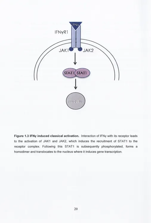
1.5.2. The IFNy signalling pathway
The IFN family comprises the, type I IFNs (such as IFN3) and type II IFN of which
IFNy is the only member. While IFNy differs from the type I IFNs its inclusion within the
IFN family occurred because of its ability to ‘interfere’ with microbial infections (Platanias,
2005). IFNy binding to its receptor induces a signalling cascade that utilises the Janus-
activated kinases/signal transducer of transcription (JAK/STAT) pathway, ultimately
inducing gene transcription (Saha
et al.,
2010) (Figure 1.3).
IFNy to its receptor leads to the activation of JAK1 and JAK2 resulting in the recruitment of STAT1 to the receptor complex. STAT1 is subsequently phosphorylated, forms a homodimer and translocates to the nucleus where it induces upregulation of the transcription of IFNy-responsive genes (Platanias, 2005).
IFNyRl
JAK2
STATU STATl
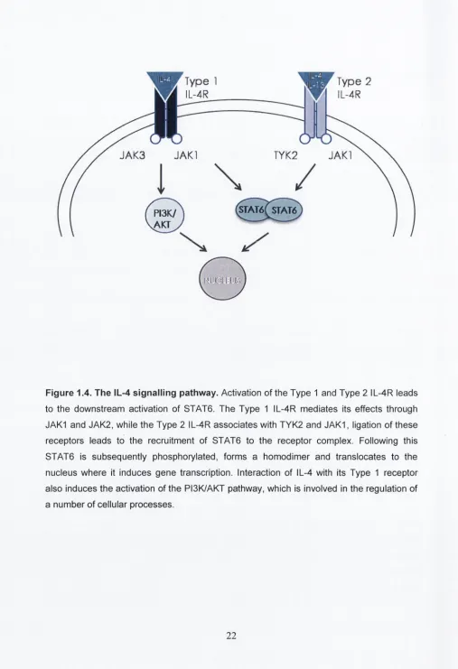
[image:42.530.11.517.39.788.2]1.5.3. The IL-4 signalling Pathway
IL-4 and IL-13 signal transduction is mediated through the JAK/STAT pathway. IL- 4 can signal through two receptors, type I IL-4 receptor (composed of two subunits, IL-4Ra and IL-2Ry) and type II receptor (IL-4Ra and IL-13Ra1). IL-13 can only signal through the type II receptor. IL-13 has a high affinity for the IL-13Ra2, though it has been suggested that this may act as a decoy receptor (Lupardus et a!., 2010). Type I IL-4 receptor is expressed predominantly on haemopoietic cells while type II IL-4R is found on non- haemopoietic cells, however some cells such as macrophages express both types (Gordon & Martinez, 2010).
Ligation of IL-4 to type I IL-4R induces the heterodimerisation of the IL-4Ra and IL- 2Ry subunits leading to activation of JAK1 and JAK3. JAK1 phosphorylates the tail of the receptor which acts as a docking site for STAT6 where it is phosphorylated. STAT6 forms a homodimer with other phosphorylated STAT6 molecules and translocates to the nucleus where it causes the induction of IL-4-responsive genes (Varin & Gordon, 2009) (Figure 1.4). Activation of type II IL-4R also results in the homodimerisation of STAT6 though this is mediated through the activation of JAK1 and tyrosine kinase 2 (Tyk2). Unlike type I IL- 4R, type II IL-4R can be activated by either IL-4 or IL-13. The type I IL-4R also activates IRS-1/2 which results in activation PI3K and RAS-MAPK and induces cell proliferation (Gordon & Martinez, 2010). IL-4 treatment also decreases the expression of Sh2- containing inositol phosphatase (SHIP), a negative regulator of the PI3K/AKT pathway, and this leads to increased activation of the PI3K/AKT pathway.
1.5.4. PI3K/AKT Pathway
Type 2
IL-4R
JAK3
JAK
[image:44.530.9.516.36.777.2]/
STAT6( STAT6/threonine kinase and recent reports suggest that AKT act on as many as 40 substrates. Although this pathway is involved often crucial to the survival and proliferation of cells, hyperactivation of this pathway has been implicated in some human cancers.
The PI3K/AKT pathway is activated by molecules including growth factors and cytokines, leading to the activation of receptor tyrosine kinases (RTKs) on the cell surface. Activation of RTKs leads to interaction of the p85 catalytic subunit of PI3K with the tyrosine kinase domain; binding leads to the liberation of the p110 subunit and this leads to full activation of PI3K. IL-4 signalling triggers activation of IRS1/2, which contains phosphotyrosine residues that interact with the Src homology 2 (Sh2) domain of p85 subunit of RISK which activates p110 activity. The p110 subunit converts phosphatidylinositol-4,5-biphosphate 2 (PIP2) to PIPS, which acts as a docking site which allows the interaction of PDK1 and AKT. This results in phosphorylation of AKT. AKT mediates its effects through which phosphorylation of several proteins (Minichiello, 2009).
In recent years, focus has turned to the possible role the PI3K/AKT pathway plays in modulating inflammation. Activation of the PI3K/AKT pathway by IL-4 has been shown to be important in the induction of the M2 state, and the evidence indicates that IL-4- induced Arg1, Chi3li3 and Fizz1 is significantly attenuated when this pathway is inhibited. Other groups have also demonstrated that TLR-induced cytokine production is negatively regulated by the PI3K/AKT pathway (Luyendyk et ai, 2008). Consequently, it has been demonstrated that LPS induced TNFa production is exaggerated in the THP-1 monocytic cell line following PI3K inhibition (Guha & Mackman, 2002).
1.6. The blood-brain barrier
dom ains o f occluded intercellular clefts and the tw o tight junction proteins most extensively studied are occludin and claudins. Occludin is thought to be involved in the regulation of paracellular perm eability and cell adhesion, w hile it has been suggested that claudins establish the barrier properties and may m ediate paracellular ion permeability.
Recent evidence from this laboratory has dem onstrated an increase in the perm eability o f the BBB in aged rats and m ouse m odels in which neuroinflam m ation were marked (Blau et a!., 2012; Denieffe et a i, 2013; Kelly et al., 2013) and this w as associated with increased infiltration of peripheral im m une cells including T cells. Although the concentration of IFNy in the brain is negligible under normal conditions, increased concentration o f the T h i cytokine IFNy has also been observed in the brains o f these anim als. Predictably increased IFNy w as accom panied with evidence o f increased m icroglial activation and this is consistent with the suggestion that the infiltration of peripheral cells into the CNS is the source o f IFNy (Benveniste, 1998). The origin o f IFNy within the CNS is still undeterm ined and a great deal o f evidence suggests its expression in resident cells is very low. How ever studies have detected IFNy m RNA expression in neurons, astrocytes and m icroglia (N eum ann et al., 1997; De Sim one et al., 1998) and although De Sim one and colleagues showed that isolated cultures o f m icroglia and astrocytes expressed IFNy mRNA; no detectable levels of IFNy protein were dem onstrated. Overall, it seem s likely th a t w heneve r IFNy concentrations in the brain are detected, the sources is infiltrating cells.
1.7. Aging
M any studies have dem onstrated an age-related increase in inflam m ation in the brain (M aher et al., 2005; Nolan et al., 2005; G riffin et al., 2006; Lyons et al., 2009), and this is characterised by increased levels o f pro-inflam m atory cytokines such as IL -ip , IL-6 and TN Fa (G odbout et al., 2005; M aher et al., 2005). These increases in are also seen in neurodegenerative disorders, especially A lzh e im e r’s disease (M rak & Griffin, 2005; Griffin, 2006) (Stout & Suttles, 2005)
(Lyons et a!., 2007a; Lyons et al., 2009). Microglia express the receptors for both of these molecules and their activation helps to maintain microglia in a quiescent state. A decrease in CD200 with age was accompanied by an age-related increase in major histocampatibility complex II (MHCII), a marker of microglial activation (Lyons et al., 2007a) and this increased in MHCII was reversed by injection of CD200 fusion protein. Similarly age-related increases in CD40, IL-1P and MHC I! mRNA were associated with a decrease in fractalkine protein and mRNA (Lyons et al., 2009), Evidence has also revealed that a decrease in the expression of anti-inflammatory cytokines, such as IL-4 and IL-10 occurs with age (Frank et al., 2006) (Ye & Johnson, 2001; Maher et al., 2005; Nolan et al., 2005).
Another possible explanation for the inflammatory environment within the aged brain is the age-related changes observed in BBB permeability (Pakulski et al., 2000; Pelegri et al., 2007; Blau et al., 2012) and this is often accompanied by infiltration of cells (Lynch, 2013). Entry of peripheral immune cells may exacerbate any inflammatory activity that already exists in the brain and Browne and colleagues showed that injection T h i cells markedly increased inflammation (Browne et al., 2013a). A detailed study carried out by Allan and colleagues demonstrated that macrophages induced cell death in organotypic hippocampal slices (Girard et al., 2013), W hereas the infiltration of macrophages in C D 200'' mice increased inflammation (Denieffe et al., 2013). Previous studies have reported an increase in the concentration of IFNy in the CNS of aged animals (Maher et al., 2005; Clarke et al., 2008), and this has been attributed to the infiltration of peripheral immune cells.
which contributes to the susceptibility of aged mice to streptococcal infection (Goldmann
et al., 2010). It has also been reported that LPS-induced TNFa production was increased in macrophages from aged compared to young rats (Tang et al., 2000) and several studies showed that LPS-induced IL-6 and IL-8 production was significantly increased in monocytes isolated from aged, compared with young, human subjects (Plowden et al.,
2004). Kohut and colleagues showed that age-related responses in macrophages were variable depending upon the nature of the stimulus and the origin of the macrophages (Kohut et al., 2004). They demonstrated that splenic and alveolar macrophages from aged animals exhibited an enhanced response to a combined LPS and IFNy stimulation, although peritoneal macrophages from aged animals showed an impaired response.
1.8. Alzheimer’s Disease
Alzheimer’s disease (AD) is a progressive neurodegenerative disorder and the most common form of dementia affecting over 35.6 million people worldwide. Auguste D was the first documented case and was described in 1906 by Alois Alzheimer; she suffered from progressive cognitive decline and disorientation. Postmortem analysis of the AD brain reveals extensive neuronal loss, the presence of Ap plaques and neurofibrillary tangles. The accumulation of aberrant proteins is thought to contribute to the progressive neuronal loss that leads to subsequent symptoms.
Similar to other neurodegenerative disorders, evidence of increased inflammation is observed in the parenchyma of the AD brain. Post-mortem analysis of AD brains revealed enhanced expression of MHCII, a classic marker of microglial activation (Luber- Narod & Rogers, 1988). Increased expression of the pro-inflammatory cytokines IL -ip and IL-6 has also been observed in the AD brain (Wood et al., 1993) and activated microglia have been shown to surround A[3 plaques (Combs, 2009). In experimental systems it is well documented that Ap activates microglia in vivo and in vitro (Clarke et al., 2007; Lyons
The presence o f A[3 plaques is one o f the hallm arks of AD pathology. Ap is produced by th e enzym atic cleavage of the type 1 transm em brane glycoprotein, am yloid precursor protein (APP). The prim ary function of A P P rem ains largely unknown although it is has been suggested that it may regulate neurite outgrowth. It has also been dem onstrated that APP deficient mice exhibit im paired LTP as well as im paired perform ance in spatial learning (Seabrook e t al., 1999; Ring e t al., 2007). APP cleavage can o ccu r through tw o distinct pathways, the am yloidogenic and the non-am yloidogenic pathways, this proteolytic action is carried out by three enzym es, a-,(3- and y-secretases. The am yloidogenic pathway results in the production o f A(3, (3-secretase cleaves APP resulting in the production o f sAPPP and a m em brane bound C -term inal fragm ent which is further cleaved by y-secretase resulting in the production of A p peptide, y-secretase may cleave this fragm ent at several sites, resulting in the production of a num ber o f A p species varying in size (39-43 am ino acids). APi^o is the m ost com m on product com pared to the less abundant A p, ^ 2 In the non-am yloidogenic pathway, APP is cleaved by a-secretase which results in the production o f sAPPa and a short m em brane bound C-term inal fragm ent. H ow ever since a-secretase cleaves A P P w ithin the Ap dom ain subsequent cleavage by y-secretase does not result in A p generation.
In the last decade evidence is mounting that peripheral immune cells may play a role in the pathology of AD. An increase in BBB permeability has been observed in AD patients (Desai et a i, 2007), and evidence from this laboratory has also demonstrated that integrity of the BBB is compromised in a mouse model of AD (Kelly e ta i., 2013). Attention has now turned to the possible roles infiltrating peripheral immune cells may play in AD pathology and specifically the infiltration of IFNy-produdng cells has been proposed to increase inflammation within the CNS. Recent evidence from this laboratory has shown that intrahippocampal injection of IFNy induced microglial activation in mice (Denieffe et a/., 2013).
1.10 Aims
The present studies were prompted by considering how macrophages which
infiltrate a brain in which inflammatory changes are ongoing, specifically the brains of aged
animals and APP/PS1 mice, might respond to the inflammatory Stimuli. The specific aims
of the study are:
1. To investigate whether macrophages infiltrate the brains of young or middle-aged
rats. To examine the phenotype of BMDMs cultured from young and aged rats, and
their responses to inflammatory stimuli.
2. To investigate the phenotype of BMDMs cultured from WT and APP/PS1 mice, and
their responses to inflammatory stimuli and to assess the impact of CD200
expression on BMDM phenotype.
3. To investigate the differential effects of IL-4 and IFNy on PI3K/AKT activity in
BMDMs and to determine the role of the PI3K/AKT pathway in the induction of M2
Chapter 2
2.1. Animals
All animals were maintained in a specific pathogen-free environment under constant veterinary supervision at the Bioresources Unit of Trinity College Dublin. Animals were housed in groups of 2 or 3 per cage, at 22-23°C with a 12-hour light-dark cycle. All animals used in these studies had free access to food and water and were fed a standard laboratory diet. Experiments were carried out under licence obtained from the Department of Health and Children (Ireland) under the Cruelty to Animals Act 1876, the European Community Directive, 86/609/EEC, and in agreement with experimental procedures laid down by the local ethics committee. During all studies, every effort was made to minimise stress to the animals. All animals were sacrificed by decapitation whilst under anaesthesia induced by urethane (1.5 g/kg; Sigma Aldrich; IRE).
Infiltration of monocytes into the brain was assessed in groups of young (4 months; n=4) and middle-aged (16 months; n=4) male rats. The bone marrow of femurs and tibias for preparation of Bone marrow-derived macrophages (BMDMs) were obtained from young (3-5 months)