Original Article
Decreased expression of let-7f associates with
the poor prognosis of glioma patients
Jianxin Zhu1,2, Yilei Xiao2, Li Li2, Baojun Fang2, Fuhua Yu2, Shubao Zhang2, Ming Sun2, Qingju Zhao2, Gang Li1
1Department of Neurosurgery, Qilu Hospital, Shandong University, Jinan 250012, Shandong, China; 2Department
of Neurosurgery, Liaocheng People’s Hospital, Liaocheng 252000, Shandong, China
Received November 2, 2015; Accepted January 5, 2016; Epub September 1, 2016; Published September 15, 2016
Abstract: Objectives: Let-7f has been reported to play regulatory roles in several types of cancer and could act as a prognostic biomarker for cancer patients. The purpose of this study was to evaluate the prognostic value of let-7f in glioma patients. Methods: The relative expression levels of let-7f in tumor tissues and corresponding normal tissues of 108 glioma patients were detected by qRT-PCR. Chi-square test was used for analyzing the association between the expression level of let-7f and the clinical features. Besides, Kaplan-Meier method was performed to estimate the overall survival of glioma patients. The prognostic significance of let-7f was evaluated by Cox regres-sion analysis. Results: The relative expresregres-sion level of let-7f in tumor tissues was lower than in normal tissues (0.45 vs 1.09). Chi-square test indicated that let-7f expression was associated with tumor size (P=0.004) and WHO grade (P=0.000). The results of Kaplan-Meier method suggested that patients with low expression level of let-7f
had shorter overall survival time than those with high expression level (24.1 months vs 34.5 months, log rank test,
P=0.000). Moreover, let-7f could act as an independent biomarker for glioma prognosis (HR=1.971, 95% CI=1.381-2.813, P=0.000). Conclusion: The decreased expression of let-7f is detected in glioma tissues and the expression levels are associated with tumor progression. Let-7f may be a potential indicator for prognosis of glioma patients.
Keywords:Let-7f, glioma, prognosis
Introduction
Glioma is one of the most common brain tumors, accounting for about 30%-40% of all intracranial tumors [1]. The prognosis of pa-
tients with glioma is poor, with a five-year mor -tality over 95% despite multiple therapies [2]. The high death rate of glioma is associated with its rapid growth, angiogenesis, and inva-sion throughout the brain [3]. Although great progresses have been made in the advanced treatments, the results are still disappointing [4]. Recently, glioma epidemiology has focused on identifying factors that are associated with tumor progression and prognosis. The relevant factors may provide valuable information about the features of the disease and therapy targets [1, 5].
MicroRNAs (miRNAs) are short non-coding RNAs, which play important regulatory roles in organism by interfering with the functions of target mRNAs [6, 7]. The fundamental roles of miRNAs are regulating basic cell cycle including
The purpose of this study was to evaluate the
clinical significance of let-7f in glioma. We detected the expression levels of let-7f in tumor tissues and corresponding normal tissues of 108 glioma patients. The associations between the gene expression and clinical features of glioma patients were analyzed and the prog-nostic value of let-7f was also estimated in the study.
Materials and methods Patients and tissue specimens
108 glioma patients confirmed by pathological
and clinical diagnoses in Liaocheng People’s Hospital from December 2011 to February 2014 were enrolled in this study. The
pathologi-UV absorbance (A260/A280) and 1% agarose gel electrophoresis was used for testing the quality of the total RNA.
PrimeScript RT reagent kit (Takara, China) was used to obtain the complementary DNA (cDNA)
from the total RNA. Moreover, fluorescence
quantitative real-time PCR (qRT-PCR) was per-formed using SYBR Green assay (Takara, Ch- ina). U6 was emerged as the internal control. The data analysis was calculated by 2-ΔΔCt
meth-od. The sequences of the primers used in this study were listed in Table 1. Each sample was in triplicate.
Statistical analysis
[image:2.612.89.383.85.152.2]The relative expression levels of let-7f in the Table 1. The sequences of the primers used in this study
Name Sequences
Let-7f Forward 5’-GCCGTGAGGTAGTAGATTGTAT-3’ Reverse 5’-GTGCAGGGTCCGAGGT-3’
U6 Forward 5’-CGCTTCGGCAGCACATATAC-3’
Reverse 5’-TTCACGAATTTGCGTGTCAT-3’
Table 2. The association between let-7f expression and clinical fea-tures of glioma patients
Characteristics Total num-ber (n) Let-7f expression x2 P value
High (n) Low (n)
Gender 0.160 0.689
Male 56 28 28
Female 52 24 28
Age 0.060 0.807
≥ 55 61 30 31
< 55 47 22 25
Tumor location 0.252 0.616
Supratentorial 68 34 34
Subtentorial 40 18 22
Tumor size 8.306 0.004
> 3 cm 55 19 36
≤ 3 cm 53 33 20
Karnofsy performance status 0.572 0.449
≥ 80 56 25 31
< 80 52 27 25
WHO grade 13.588 0.000
I+II 57 37 20
III+IV 51 15 36
Relapse 0.250 0.617
Yes 67 31 36
No 41 21 20
mal tissue samples were col-lected from the patients and the samples were put in liq-uid nitrogen immediately th- en stored at -80°C for use. None of the patients had received chemotherapy or radiotherapy before the sur-gery. The study was approved by the Ethic Committee of the hospital and all the par-ticipators had signed written informed consents in adva- nce.
The follow-up investigation was about 5 years and all of the patients were participat-ed in. The investigation infor-mation and the clinical fea-tures of the glioma patients were collected to estimate
the clinical significance of
let-7f in glioma patients.
RNA extraction and qRT-PCR
[image:2.612.91.380.200.503.2]tissues were analyzed by T test and the results were shown as mean ± SD. The correlation between expression level of the gene and clini-cal features of glioma patients was estimated
[image:3.612.92.382.73.277.2]according to their average expression level. The results of Chi-square test indicated that the expression level of let-7f was associated with tumor size (P=0.004) and WHO grade (P=
Figure 1. Relative expression of let-7f in tumor tissues and corresponding nor-mal tissues of glioma patients (U6 as nornor-malized control).
by Chi-square test. And the overall survival analysis was assessed using Kaplan-Me- ier method. In addition, Cox regression analysis was us- ed to evaluate the prognostic
significance of let-7f. All the data analyses were per-formed in SPSS 18.0 ware and Sigma Plot soft-ware was used for drawing. P < 0.05 was considered as
statistical significance.
Results
Relative expression of let-7f in glioma patients
The 108 glioma patients col-lected in this study included 56 males and 52 females and their average age was 56.3 years. The clinical infor-mation of the glioma patients was summarized in Table 2. QRT-PCR was used to test the relative expression of let-7f in pathological tissues and corresponding normal tissues of glioma patients. The relative expression level in tumor tissues was 0.45± 0.203, while that in the cor-responding normal tissues was 1.09±0.211. There was
a significant difference
bet-ween them (Figure 1, P= 0.000).
The relationship between let-7f expression and clinical features
In order to analyze the con-nection between let-7f exp- ression and the clinical fea-tures, the glioma patients were divided into two groups
[image:3.612.93.381.324.609.2]0.000). However, it had nothing to do with age, gender, tumor location, Karnofsy performance status or relapse (P > 0.05).
Association between let-7f expression and the overall survival of glioma patients
Kaplan-Meier method was used for overall sur-vival analysis. The patients with low expression level of the let-7f had shorter overall survival time than those with high expression level (24.1 months vs 34.5 months, Figure 2). There was a remarkable difference between the two groups (Log rank test, P=0.000).
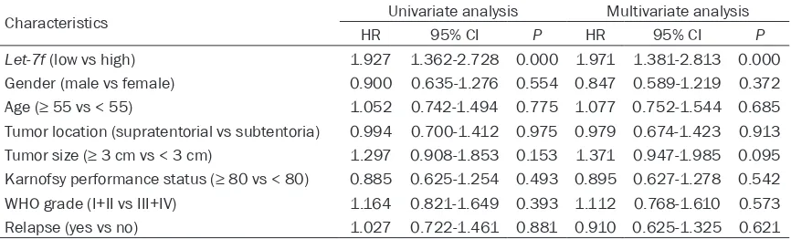
Univariate and multivariate Cox regression an- alyses were used for analyzing prognostic fac-tors in glioma patients. The results indicated that let-7f was associated the glioma progno- sis and could act as an independent biomark- er for prognosis of glioma patients (Table 3, HR=1.971, 95% CI=1.381-2.813, P=0.000). Discussion
Recent years, glioma as a typical type of brain tumor has attracted more and more attentions
but the therapeutic efficacy for glioma patients
are not satisfactory. The prognosis of glioma is poor despite multiple advance treatments, due to excessive proliferation, relentless invasion,
and angiogenesis [20]. The identification of
new biomarkers for glioma therapy target and prognosis may improve the clinical outcomes of the patients. Recently, with the development of the molecular biological techniques, the genes which could alter during the tumor progression are proved to improve the outcomes of the can-cer patients. Guo et al. had reported that FRAT1 was an important factor in the tumorigenesis and progression of glioma and could be ex- plored as an indicator for pathological
diagno-sis, therapy target and prognosis [21]. CDKN2A (p16) mRNA was proved to be a prognostic marker for malignant high-grade glioma [22]. Wang et al. indicated that transmembrane-4-L-six-family-1 was a potential predictor for prog-nosis in human glioma [23]. These studies sug-gested that the endogenous biomarkers might improve the clinical outcomes of patients with glioma.
The let-7 family is a high conserved family of miRNAs, including fourteen members [24]. Currently, many members of let-7 family were reported to be associated with cancer tumo- rigenesis and progression. Let-7a had been reported to inhibit growth and migration of breast cancer cells [25]. Han et al. demonstrat-ed that let-7b could suppress invasion and migration of gastric cancer cells through direct-ly targeting the tumor metastasis-associated gene ING1 [26]. And let-7f was also proved to play regulatory roles in several types of cancer, such as gastric cancer, breast cancer, hepato-cellular carcinoma and so on [15-17]. Then weather let-7f participates in the development of glioma or affects the clinical outcome re- quires further investigation.
In this study, we detected the expression level of let-7f in pathological tissues and correspond-ing normal tissues of patients with glioma. The results suggested that the expression level of let-7f was decreased in tumor tissues and a fur-ther analysis indicated that it was associated with tumor size and WHO grade. These results might suggest that the abnormal expression of let-7f in tumor tissues was associated with the
[image:4.612.90.523.86.217.2]cancer development. The findings were accor -dance with previous studies, such as Yan et al., had reported that let-7f overexpression inhibit-ed glioma cell proliferation, migration, and inva-sion via targeting periostin [19]. Nahid et al. Table 3. Univariate and multivariate analyses for prognostic factors in glioma patients
Characteristics Univariate analysis Multivariate analysis
HR 95% CI P HR 95% CI P
Let-7f (low vs high) 1.927 1.362-2.728 0.000 1.971 1.381-2.813 0.000 Gender (male vs female) 0.900 0.635-1.276 0.554 0.847 0.589-1.219 0.372 Age (≥ 55 vs < 55) 1.052 0.742-1.494 0.775 1.077 0.752-1.544 0.685 Tumor location (supratentorial vs subtentoria) 0.994 0.700-1.412 0.975 0.979 0.674-1.423 0.913 Tumor size (≥ 3 cm vs < 3 cm) 1.297 0.908-1.853 0.153 1.371 0.947-1.985 0.095 Karnofsy performance status (≥ 80 vs < 80) 0.885 0.625-1.254 0.493 0.895 0.627-1.278 0.542 WHO grade (I+II vs III+IV) 1.164 0.821-1.649 0.393 1.112 0.768-1.610 0.573
had proved that let-7f expression was de- creased during hepatic differentiation [27]. Decreased expression of let-7f was also detect-ed in metastasis gastric cancer tissues and the over-expression of the gene could inhibit inva-sion and migration of gastric cancer cells th- rough directly targeting the tumor metastasis-associated gene MYH9 [28]. In a word, all of the results indicated that let-7f might be a potential tumor suppressor.
In the previous studies, many miRNAs were proved to act as prognostic biomarkers and as non-invasive biomarkers, they could provide more credible outcomes for patients [29, 30]. For example, miRNA-124 had been reported to be associated with prognosis of lung cancer and could act as a potential biomarker for
fur-ther risk stratification in the treatment of lung
cancer [31]. Yuan et al. reported that miRNA-940 could inhibit the growth of hepatocellular carcinoma and might be a indicator for progno-sis [32]. Over-expression of miRNA-155 was proved to correlate with a poor outcome for patients with bladder cancer [33]. In this study, we evaluated the prognostic value of miRNA let-7f in glioma. The results suggested that pa- tients with low expression levels of the gene had lower survival rate and the expression lev-els of let-7f was associated with glioma progno-sis. Let-7f could act as an independent predic-tor in patients with glioma.
In conclusion, the decreased expression of let-7f is found in tumor tissues of glioma patients. And the gene is correlated with tumor size and WHO grade. Besides, the expression levels of let-7f are associated with survival rate of patients and let-7f may be a potential biomark-er for prognosis and thbiomark-erapy target for glioma. Disclosure of conflict of interest
None.
Address correspondence to: Dr. Gang Li, Department of Neurosurgery, Qilu Hospital of Shandong University, Jinan 250012, Shandong, China. E-mail: zhaone373h@sina.com
References
[1] Zeng T, Cui D and Gao L. Glioma: an overview of current classifications, characteristics, mo -lecular biology and target therapies. Front Biosci (Landmark Ed) 2015; 20: 1104-1115.
[2] McLendon RE and Halperin EC. Is the long-term survival of patients with intracranial glioblastoma multiforme overstated? Cancer 2003; 98: 1745-1748.
[3] Furnari FB, Fenton T, Bachoo RM, Mukasa A, Stommel JM, Stegh A, Hahn WC, Ligon KL, Lou-is DN, Brennan C, Chin L, DePinho RA and Cavenee WK. Malignant astrocytic glioma: ge-netics, biology, and paths to treatment. Genes Dev 2007; 21: 2683-2710.
[4] Shen J, Li G, Liu Q, He Q, Gu J, Shi Y and Lou H. Marchantin C: a potential anti-invasion agent in glioma cells. Cancer Biol Ther 2010; 9: 33-39.
[5] Schwartzbaum JA, Fisher JL, Aldape KD and Wrensch M. Epidemiology and molecular pa-thology of glioma. Nat Clin Pract Neurol 2006; 2: 494-503; quiz 491 p following 516.
[6] Bartel DP. MicroRNAs: genomics, biogenesis, mechanism, and function. Cell 2004; 116: 281-297.
[7] Hwang HW and Mendell JT. MicroRNAs in cell proliferation, cell death, and tumorigenesis. Br J Cancer 2007; 96 Suppl: R40-44.
[8] Bueno MJ and Malumbres M. MicroRNAs and the cell cycle. Biochim Biophys Acta 2011; 1812: 592-601.
[9] Carleton M, Cleary MA and Linsley PS. MicroR-NAs and cell cycle regulation. Cell Cycle 2007; 6: 2127-2132.
[10] Qin J and Luo M. MicroRNA-221 promotes colorectal cancer cell invasion and metastasis by targeting RECK. FEBS Lett 2014; 588: 99-104.
[11] Petrovic N, Mandusic V, Stanojevic B, Lukic S, Todorovic L, Roganovic J and Dimitrijevic B. The difference in miR-21 expression levels be-tween invasive and non-invasive breast can-cers emphasizes its role in breast cancer inva-sion. Med Oncol 2014; 31: 867.
[12] Gong J, Cui Z, Li L, Ma Q, Wang Q, Gao Y and Sun H. MicroRNA-25 promotes gastric cancer proliferation, invasion, and migration by direct-ly targeting F-box and WD-40 Domain Protein 7, FBXW7. Tumour Biol 2015; 36: 7831-40. [13] Lee YS and Dutta A. MicroRNAs in cancer.
Annu Rev Pathol 2009; 4: 199-227.
[14] Kuehbacher A, Urbich C, Zeiher AM and Dim-meler S. Role of Dicer and Drosha for endothe-lial microRNA expression and angiogenesis. Circ Res 2007; 101: 59-68.
[15] Tao WY, Liang XS, Liu Y, Wang CY and Pang D. Decrease of let-7f in low-dose metronomic Pa-clitaxel chemotherapy contributed to upregula-tion of thrombospondin-1 in breast cancer. Int J Biol Sci 2015; 11: 48-58.
in gastric cancer and precancerous disease. Tumour Biol 2015; 36: 3337-3343.
[17] Ge W, Yu DC, Li QG, Chen X, Zhang CY and Ding YT. Expression of serum miR-16, let-7f, and miR-21 in patients with hepatocellular carci-noma and their clinical significances. Clin Lab 2014; 60: 427-434.
[18] Geraldo MV, Fuziwara CS, Friguglieti CU, Costa RB, Kulcsar MA, Yamashita AS and Kimura ET. MicroRNAs miR-146-5p and let-7f as prognos-tic tools for aggressive papillary thyroid carci-noma: a case report. Arq Bras Endocrinol Me-tabol 2012; 56: 552-557.
[19] Yan S, Han X, Xue H, Zhang P, Guo X, Li T, Yuan G, Deng L and Li G. Let-7f Inhibits Glioma Cell Proliferation, Migration, and Invasion by Tar-geting Periostin. J Cell Biochem 2015; 116: 1680-92.
[20] Moller HG, Rasmussen AP, Andersen HH, John-sen KB, HenrikJohn-sen M and Duroux M. A system-atic review of microRNA in glioblastoma multi-forme: micro-modulators in the mesenchymal mode of migration and invasion. Mol Neurobiol 2013; 47: 131-144.
[21] Guo G, Zhong CL, Liu Y, Mao XG, Zhang Z, Jin J, Liu J, Yang L, Mao JM, Guo YH and Zhao YL. Overexpression of FRAT1 is associated with malignant phenotype and poor prognosis in human gliomas. Dis Markers 2015; 2015: 289750.
[22] Sibin MK, Bhat DI, Narasingarao KV, Lavanya C and Chetan GK. CDKN2A (p16) mRNA de-creased expression is a marker of poor progno-sis in malignant high-grade glioma. Tumour Biol 2015; 36: 7607-14.
[23] Wang P, Bao W, Zhang G, Cui H and Shi G. Transmembrane-4-L-six-family-1, a potential predictor for poor prognosis, overexpressed in human glioma. Neuroreport 2015; 26: 455-461.
[24] Pasquinelli AE, Reinhart BJ, Slack F, Martin-dale MQ, Kuroda MI, Maller B, Hayward DC, Ball EE, Degnan B, Muller P, Spring J, Sriniva-san A, Fishman M, Finnerty J, Corbo J, Levine M, Leahy P, Davidson E and Ruvkun G. Conser-vation of the sequence and temporal expres-sion of let-7 heterochronic regulatory RNA. Na-ture 2000; 408: 86-89.
[25] Liu K, Zhang C, Li T, Ding Y, Tu T, Zhou F, Qi W, Chen H and Sun X. Let-7a inhibits growth and migration of breast cancer cells by targeting HMGA1. Int J Oncol 2015; 46: 2526-2534. [26] Han X, Chen Y, Yao N, Liu H and Wang Z.
Mi-croRNA let-7b suppresses human gastric can-cer malignancy by targeting ING1. Cancan-cer Gene Ther 2015; 22: 122-129.
[27] Davoodian N, Lotfi AS, Soleimani M, Mola SJ and Arjmand S. Let-7f microRNA negatively regulates hepatic differentiation of human adi-pose tissue-derived stem cells. J Physiol Bio-chem 2014; 70: 781-789.
[28] Liang S, He L, Zhao X, Miao Y, Gu Y, Guo C, Xue Z, Dou W, Hu F, Wu K, Nie Y and Fan D. Mi-croRNA let-7f inhibits tumor invasion and me-tastasis by targeting MYH9 in human gastric cancer. PLoS One 2011; 6: e18409.
[29] Fabbri M. miRNAs as molecular biomarkers of cancer. Expert Rev Mol Diagn 2010; 10: 435-444.
[30] Wang F, Lou JF, Cao Y, Shi XH, Wang P, Xu J, Xie EF, Xu T, Sun RH, Rao JY, Huang PW, Pan SY and Wang H. miR-638 is a new biomarker for outcome prediction of non-small cell lung can-cer patients receiving chemotherapy. Exp Mol Med 2015; 47: e162.
[31] Zhang Y, Li H and Han J. Down-regulation of microRNA-124 is correlated with tumor metas-tasis and poor prognosis in patients with lung cancer. Int J Clin Exp Pathol 2015; 8: 1967-1972.
[32] Yuan B, Liang Y, Wang D and Luo F. MiR-940 inhibits hepatocellular carcinoma growth and correlates with prognosis of hepatocellular car-cinoma patients. Cancer Sci 2015; 106: 819-24.