Original Article
Substance P induces monocyte chemoattractant
protein-1 production in skin fibroblasts
from genetically-diabetic mice via
activation of NF-κB
Tao Ni1, Zhigang Jia4, Fenghou Gao1, Yong Fang1, Weirong Yu1, Xiong Zhang1,2, Min Yao1,3
1Department of Burns and Plastic Surgery, No. 9 People’s Hospital, and Institute of Traumatic Medicine; School
of Medicine, Shanghai Jiao Tong University, Shanghai, China; 2Department of Burns and Plastic Surgery, Ruijin
Hospital, School of Medicine, Shanghai Jiao Tong University, Shanghai, China; 3Wellman Center for
Photomedi-cine, Department of Dermatology, Massachusetts General Hospital, Harvard Medical School, Boston, MA, USA;
4Department of Burns and Plastic Surgery, Wuxi No. 3 People’s Hospital, Wuxi, China
Received February 15, 2016; Accepted May 20, 2016; Epub July 1, 2016; Published July 15, 2016
Abstract: Substance P (SP) is a prominent neuropeptide that function in the active communication between the nervous system and immune response and pathological conditions such as inflammatory disorders and chronic wound healing. This functionality has been attributed to modulation of SP on production of inflammatory media-tors, including chemokine and cytokines by binding to tachykinin receptors. In the present study, the effects of SP-induced production of chemokine MCP was examined and the activation of the proinflammatory transcription factor NF-κB was investigate as a molecular mechanism by which SP stimulate inflammatory process underlying impaired wound healing. Using genetically diabetic mice skin fibroblasts, we showed that SP up-regulates mRNA and protein level of MCP-1 in a dose- and time-dependent manner via its receptor neurokinin-1. Moreover, this stimulatory ef-fect requires activation of transcription factor, NF-κB, which contributed to MCP-1 gene transcription. Furthermore, we found that MG132, a NF-κB inhibitor, inhibits the phosphorylation and degradation of IκB and MCP-1 production by SP management in diabetic skin fibroblasts. Identification of the functionality exhibited by SP and its molecular mechanisms has important implications for modulation of impaired inflammation in chronic wounds and inflamma-tory disorders.
Keywords: Neuropeptide, tachykinin receptor, MCP-1, NF-κB activation, chronic wound
Introduction
Substance P (SP), an 11 -amino acid neuropep-tide member of the tachykinin family, is wide- ly distributed (found) throughout the central and peripheral nervous systems and released by unmyelinated sensory nerve endings as well as immune cells [1, 2]. SP elicit biological responses in effector cells by binding to at least one of three distinct G protein-coupled recep-tors, the neurokinin (NK)-1, NK-2, and NK-3 receptors on the surface of endothelial cells, epithelial cells, and fibroblast [3]. Several stud-ies also confirm a major pro-inflammatory role for SP and the NK-1R in the pathogenesis and progression of inflammation of different etiolo-gies, which is associated with pathopsychology
of impaired wound healing, such as diabetic wounds [1, 4-10]. It has been reported that the pro-inflammatory responses of SP involve up-regulation both gene expression and pro- tein synthesis of monocyte chemoattractant protein-1 (MCP-1), which is a putative chemo-kine for recruiting inflammatory cells into the site of wound during wound healing. However, the mechanisms for SP inducing the product- ion of MCP-1 still reminds unclear.
wounds site and activating these cells to ful- fill their functions. Therefore, MCP-1 plays an important regulation role in normal inflam- matory process and in many inflammatory disorders.
Diabetes-related impaired wounds remain a profound clinical problem with increasing prev-alence and a high-risk for amputation. Previous studies showed that the characteristics includ-ed inflammatory response insufficiency at early stage and delayed wound healing, eventually [11, 12]. Importantly, the protein level of SP in the wound-healing impairment associated with diabetes was decreased by recent reports [13-15]. In addition, a study has found that MCP-1, the main chemokine in diabetic wounds, pre-sented a low level in early stage, which indicat-ing that SP and MCP-1 are critical in inflamma-tion response during impaired wound-healing in diabetes [16, 17]. The external application of SP promotes wound healing by improving early infiltration of inflammatory cells in diabet-ic wounds and inflammatory response disor-ders [18-20]. The date suggests possible co-regulation of neuropeptide (SP) and chemo- kine MCP-1 in wound healing processes, par-ticularly in abnormal inflammations and diabet-ic wounds.
We hypothesed that SP may modulate the pro-duction of MCP-1 on genetically diabetic mice skin fibroblasts and that increased MCP-1 might then be responsible for improving im- paired wound healing in diabetes.In the pres-ent study, we sought to investigate the effect of inflammatory mediators, substance P, on MCP-1 production in skin fibroblasts from ge- netically diabetic mice, as well as its possible mechanisms.
Materials and methods
Animals
All procedures were subject to prior institu- tional approval by the Subcommittee for Re- search Animal Care at the Shanghai No. 3 Peoples’ Hospital. Four female genetically dia-betic C57BL/KsJ Lepdb mice weighting 20-30 g (referred to as db+/db+ mice) were obtained from Charles River Laboratories (Wilmington, USA). They were acclimatized to caged labora-tory conditions for three days prior to research, then the back skin were harvested from these mice after they were sacrificed.
Materials
Trypsin, DMEM culture solution and fetal bo- vine serum were purchased from the Gibco Inc. (Langley, USA). SP was obtained from Sigma (St. Louis, US). CCK-8 cell proliferation kit, mod-erate protein lysate, NA-Red, ponceau staining solution were purchased from the Beyotime Institute of Biotechnology (Shanghai, China); Trizol reagent was purchased from the Invitro- gen (Carlsbad, USA); Takara RT-PCR amplifica-tion kit was obtained from the Sangon (Dalian, China); rabbit anti mouse MCP-1 primary anti-body was purchased from the Abcam (Cam- bridge, USA); Nk-1R inhibitor L703606, NF-κB inhibitor MG132 were purchased from Santa Cruz Biotechnology (Santa Cruz, USA); Anti- bodies of phospho-NF-κBp65 (Ser276), Phos- pho-IκB-α (Ser32), IΚB-α were purchased from the Cell Signaling Technology (Danvers, USA).
Mouse skin fibroblasts culture
After hair removal, the back skin specimens from genetically diabetic C57BL/KsJ Lepdb mice were cut into pieces sized 0.5×0.5 cm. We put them onto the bottom of culture dish and col-lected the fibroblasts for primary culture. Then the primary fibroblasts were cultivated in DMEM medium containing 10% fetal bovine serum. The fourth generation cells were seed-ed in Petri dishes. A total of 10 ml of DMEM medium containing 10% fetal bovine serum was added into each Petri dish, and then the cells was placed in an incubator (Heal Force, China) in 37 degree and at 5% CO2.
Reverse transcription-polymerase chain reac-tion (RT-PCR) analysis
MCP-1 consisted of an initial denaturation at 94°C for 2 min, 30 cycles of 94°C for 30 s, 61°C for 30 s and 72°C for 1 min. The follow- ing specific primer pairs (Shanghai Sangon Bio-tech, China) were used: β-actin, sense 5’TGA- CAGGATGCAGAAGGAGA3’ and antisense 5’GC- TGGAAGGTGGACAGTGAG3’, MCP-1, sense 5’C- CCCAGTCACCTGCTGTTAT3’ and antisense 5’T- CCTGAACCCACTTCTGCTT3’. PCR products were analyzed using 1% agarose gel containing 0.05 mg/100 ml ethidium bromide. The result was analyzed using Mig gel analysis system, and expressed as the corrected ratio of absorbance to the β-actin absorbance in each group. The experiments were conducted independently three times.
Enzyme-linked immunosorbent assay (ELISA)
The protein levels of MCP-1 were examined using ELISA. Briefly, for SP-treated experiment, the primary cultured fibroblasts were added high glucose DMEM media (containing 0.5% FBS) for 2 h and treated with various concen-trations of SP (0.1 nM, 1 nM, 10 nM, 100 nM and 1 μM) and different times (2, 4, 8, 12, 24 and 48 h). No treatment was used as control. The supernatant was collected and centrifuged for 10 min at 2000 rpm. For MG132-treated ex- periment, the cells were cultured with DMEM containing 10% FBS in an incubator with 5% CO2 at 37°C. The cells were treated with SP alone, SP plus MG132 with indicated concen-tration (0.5, 1, 1.25, 1.5 and 2 μM), and with-out treatment. The supernatant was collected for measuring MCP-1 concentration by ELISA assay according to ELISA kit manufacturer’s instruction. Absorbance was measured at 450 nm using an automatic microplate reader. The experiments were performed in duplicate, three times independently.
Western blot analysis
Western blot analysis was performed to deter-mine the protein level of MCP-1 in the primary fibroblasts, or in the cells treated with SP, or the inhibitor MG132 plus SP, or NK-1R inhi- bitor L703606 plus SP. Briefly, the cells were lysed with RIPA buffer containing 1% NP-40, 0.5% deoxycholate and 0.1% SDS, PMSF. Total protein concentrations were determined by the BCA protein assay. Samples with 50 μg of total protein were loaded into 15% SDS-poly- acrylamide gel and electrophoretically
trans-ferred to PVDF membrane. Non-specific bind- ing was blocked by incubating the membranes with 5% nonfat milk in PBST (0.05% Tween 20 in PBS). The membranes were then incubated overnight at 4°C with the primary antibo- dies (1:1,000) in buffer containing 2.5% nonfat dry milk, After three times wash with PBS for 10 min each, the membranes were incubated with goat anti-rabbit HRP-conjugated second-ary antibody (1:4,000) for 2 h at room tempera-ture. The blots were developed for visualiza- tion using an enhanced chemiluminescence (ECL) detection kit (Pierce, Rockford, IL). Se- mi-quantitative analysis was performed using an image analyzer with GAPDH as internal con-trol. The experiment was repeated three times independently.
Immunofluorescence staining
Mice skin fibroblasts were inoculated in a six-well plate at 2×10³ cells/well and treated with SP, SP plus MG132, and without treatment. In SP plus MG132 management, the cells were pretreated with MG132 for 30 min, and then treated with SP for 60 min. The cells were washed three times with PBS and then fixed for 15 min with 4% paraformaldehyde. Triton was added into the cells for 30 min at room tem-perature. The cells were blocked with 6% fetal bovine serum for 30 min, and then incubated with primary antibody or PBS (negative control) over night at 4°C. After washing, the samples were treated with Cy3 labeled goat anti rabbit secondary antibody for 2 hours in dark room. The slide was sealed with glycerol and analyzed under a fluorescence microscope.
Electrophoretic mobility shift assay (EMSA)
chemiluminescent EMSA kit following the man-ufacturer’s instructions. Briefly, DNA binding reactions were set up by incubating 10 μg nuclear extract and 20 fmol of biotin-labeled probe together with 1 μg poly (dI-dC) for 20 min at room temperature in 20 μl of binding buffer (10 mM Tris, pH 7.5, 50 mM KCl, and 1 mM DTT). Samples were fractionated on native 6% polyacrylamide gels in 0.5 Tris-borate-EDTA buffer and then transferred onto Zeta-Probe cationized nylon membranes. After being cross-linked by a UV-light cross-linker (Spectronics), the biotin-labeled DNA on the membranes was detected by chemiluminescence. Band shifts were visualized by exposure to X-ray films.
Statistics
The statistical analysis was performed using SPSS17.0. Student t test was used to evaluate difference between the study groups. Data was expressed as mean ± s. Statistical significance was set at P < 0.05.
Results
SP increases mRNA expression and protein level of MCP-1 in diabetic fibroblasts
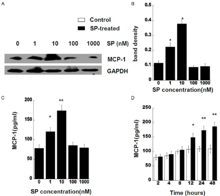
Because SP has been proposed as a factor in the modulation of the inflammation. We exam-ined whether this neuropeptide can modulate inflammatory MCP-1 production in skin fibro-blasts from genetically diabetic C57BL/KsJ Lepdb mice. SP significantly up-regulated mRNA expression of MCP-1 from 1 nM to 10 nM after 24 h, compared with control subject at 24 h
(Figure 1A and 1B). At 10 nM treatment, SP stimulation of MCP-1 gene expression reached a peak level (P < 0.01).
In addition to MCP-1 gene expression, we also determine the effect of SP on the MCP-1 pro-tein production. Diabetic skin fibroblasts were treated with various SP concentrations (from 1 nM to 1000 nM) for 24 h. Subsequently, MCP-1 protein levels were analyzed by Western blot or ELISA (Figure 2A-C). Chemokine MCP-1 level in the fibroblasts treated with 1 nM and 10 nM SP were significantly higher than that in untreated cells at 24 h (P < 0.05). The data was coinci-dent with the MCP-1 mRNA expression results. Moreover, we observed the changes of MCP-1 by SP in different time points. SP stimulation of MCP-1 protein levels in the fibroblasts were much higher than that in non-treated cells at 12 h (P < 0.05), 24 h, and 48 h (P < 0.01) after treatment. Our data indicated that ap- plication of SP with 10 nM concentration, for 24 h substantially increases production of MCP-1 in both mRNA and protein levels. Based on these results, we decided to use 10 nM SP and incubation time of 24 h for all sub- sequent experiments.
NK-1 inhibitor abolishes SP stimulation of MCP-1 in diabetic fibroblasts
We next examined whether SP stimulates MCP-1 through its neurokinin-1 receptor (NK-1 receptor) [21, 22] in the genetically diabetic fibroblasts. Using pre-treatment of L703606 (a specific NK-1 receptor inhibitor) at 20 nmol for 10 min. Our data revealed that L703606 effi-Figure 1. SP stimulates MCP-1 gene expression. A. Skin
fibro-blasts from C57BL/KsJ Lepdb mice were treated with SP as
[image:4.612.338.518.73.263.2]ciently inhibited the protein level of MCP-1 that stimulated by SP in the pre-treated fibroblasts (Figure 3). The results demonstrated the spe-cific NK-1 receptor inhibitors almost completely blocked SP-induced MCP-1 production, sug-gesting that MCP-1 augmentation was a result of direct activation by SP through its receptor NK-1 in genetically diabetic murine fibroblasts.
SP stimulates activation of NF-κB through NK-1 receptor in diabetic fibroblasts
To delineate the molecular mechanisms of SP-stimulated MCP-1 production, the diabetic fibroblasts were treated using 10 nM SP for
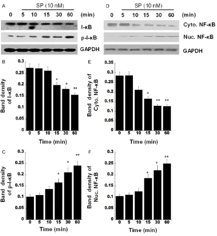
[image:5.612.90.522.71.455.2]dif-ferent times (from 5 min up to 60 min), and then the activations of transcription factor (NF-κB) were analyzed. Because activation of NF- κB mainly occurs via IkappaB kinase (IKK)-me- diated phosphorylation of inhibitory protein- I-κB, the phosphorylation and degradation of I-κB is one of hallmarks for NF-κB activation. The Western blot results showed that the amount of I-κB decreased notably after 15, 30, and 60 min after 10 nM SP stimulation (Figure 4A, upper row). In contrast, with time exten-sion, the level of phospho-IκB increased signifi-cantly as early as 15 min, further up to 60 min, following SP stimulation (Figure 4A, mid row). The densities of blotting were indicated on Figure 2. SP induces MCP-1 production. A. Skin fibroblasts from C57BL/KsJ Lepdb mice were treated with SP
Figure 4B, 4C. Our data suggested that stimu-lation of SP causes degradation of I-κB and increases of I-κB phosphorylation, which sub-sequently leads to NF-κB nuclear translocation. Additionally, a specific NK-1 receptor inhibitor, L703606, was used to pre-treat the cells be- fore SP treatment. The cell lysates were sub-jected to Western Blotting with I-κB or anti-phospho-IκB. As shown on Figure 5, L703606 inhibited distinctly the degradation of I-κB and phosphorylation of I-κB that stimulated by SP. It indicated that SP activates NF-κB via its re- ceptor, NK-1.
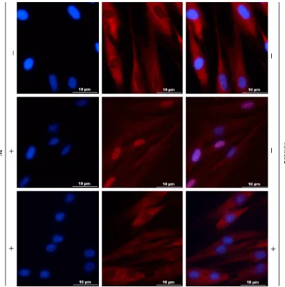
To ensure SP can activate NF-κB in diabetic fibroblasts, we further analyzed translocation of NF-κB by determining the amount of nuclear factor (NF)-kappaB p65 in cytoplasm or in nuclear, respectively. As shown in Figure 4D-F, the amount of nuclear p65 raised greatly at 15, 30, and 60 min after SP treatment. However, the amount of p65 in cytoplasm declined obvi-ously from 13 min to 60 min after treatment. Furthermore, an immunofluorescent staining result presented that RelA content indicated by red fluorescence was mainly found in nuclei (Figure 6, mid panel) after SP treatment, not in cytoplasm, compared to that in untreated cells (Figure 6, top panel). We used an inhibitor of NF-κB activation, MG132, to pre-treated the cells. RelA staining was mostly seen in cyto-plasm after SP treatment (Figure 6, bottom panel), suggesting MG132 significantly inhibit-ed NF-κB activation that inducinhibit-ed by SP. These
results revealed that SP treatment leads acti-vation of NF-κB in diabetic fibroblasts.
NF-κB activation is critical for SP-induced MCP-1 in diabetic fibroblasts
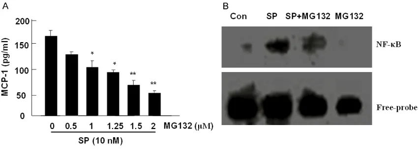
To determine whether the activation of NF-κB could be required for SP-induced MCP-1 pro-duction, MG132, a proteasome inhibitor for inhibiting NF-κB activation was used to pre-treat cells and the amount of MCP-1 was mea-sured by ELISA. The pre-treatment of MG132 significantly decreased the production of MCP-1, which was induced by SP, in a dose-depen-dent manner (Figure 7A). The above data dem-onstrated that NF-κB activation involved in SP-induced MCP-1 production in diabetic fibro-blasts. To verify the importance of NF-κB acti-vation for regulating MCP-1 production induced by SP, EMSA was performed in cells treated with or without MG132 plus SP. The data showed that SP activates the DNA binding activity of NF-κB (Figure 7B, lane 2), however, MG132 inhibits DNA binding activity of NF-κB induced by SP (Figure 7B, lane 3). The results suggested that NF-κB activation plays a key role in regulating SP-induced MCP-1.
Discussion
In this study, we investigated the molecular mechanisms by which Substance P stimulated the production of inflammatory mediator MCP-1 in skin fibroblasts from genetically diabetic Figure 3. SP-induced MCP-1 production is abrogated by
inhibi-tion of SP receptor. A. Skin fibroblasts from C57BL/KsJ Lepdb
mice. We found that Substance P stimulates MCP-1 production in a dose- and time-depen-dent manner via its receptor neurokinin-1 (NK-1) in diabetic skin fibroblasts. Additionally, this stimulation required activation of transcription factor, NF-κB, which contributed to MCP-1 gene transcription. These results suggest SP is not only a potent neuropeptide involved in
[image:7.612.93.521.70.536.2]neuro-genic response, but is also a peptide activated transcription factor and modulated inflamma-tory mediators. To our knowledge, this is the first demonstration of SP-induced NF-κB acti- vation leading to chenomkine gene transcrip-tion activatranscrip-tion in diabetic skin fibroblasts. Our findings contribute to the understanding of the molecular mechanisms underlying chronic in- Figure 4. SP induces activation of NF-κB. A. Skin fibroblasts from C57BL/KsJ Lepdb mice were treated with 10 nM SP
Figure 5. SP induces activation of NF-κB via NK-1. A. Skin fibroblasts from C57BL/KsJ Lepdb mice were pre-treated
with 20 nmol L703606 for 10 min, and then were treated by 10 nM SP for 5, 15, and 30 min. The cell lysates were subjected to western blotting for determining I-κB or phospho- IκB. GAPDH was used to verify equal loading. B, C. Bar graphs show the average band density of I-κB or of phospho- IκB. Mean ± SD, n = 3.
Figure 6. SP stiumlats nuclear translocation of NF-κB. Diabitc skin fibroblasts from C57BL/KsJ Lepdb mice were
[image:8.612.98.510.257.672.2]flammation and impaired wound healing such as diabetic ulcers, which may be beneficial for developing future therapeutic approaches. Diabetic wound is known as an impaired heal-ing model with inadequate inflammatory response in the early stage, which is required to achieve wound healing. Several studies report-ed that the diabetic wound impairment is asso-ciated with insufficient inflammatory cells acti-vation and cytokine and/ or growth factors secretion in the early stage [23-25]. Similar to the results, in our previous study, we observed a decreased cutaneous inflammatory cells in- filtration, a lower level of inflammatory media-tors, such as cytokines or chemokines, and de- layed wound healing in a diabetic animal with deep partial-thickness burn injury model. More- over, the external applications of SP improve impaired healing by enhancing inflammatory cells infiltration and inflammatory mediators secretion in diabetic injury or inflammatory re- sponse disorders [26, 27]. These results sug-gest that SP plays an important role in chronic and impaired wounds. In the present study, we utilized cultivated skin fibroblasts from geneti-cally diabetic mice as a clinigeneti-cally relevant model that can serve to understand the molecular mechanisms underlying diabetic wound heal- ing impairment. Our data reveal that SP up-reg-ulates the gene expression of MCP-1 and in- creases MCP-1 protein level. MCP-1, as a che-mokine, can recruit adequate inflammatory cells infiltration by chemoattracting neutrophils, ma-
crophages, and T cells to wound (ulcer) site, which result in the improvement of impaired wound healing.
SP binding to its high-affinity neurokinin-1 (one of three tachykinin receptors) mediates a vari-ety of inflammatory processes, including up-regulation of lung inflammation, microvascular leakage, and mucosal permeability [17, 19]. To examine whether SP-induced MCP-1 produc-tion is a direct effect via binding to NK-1, we used a selective nonpeptide NK1 tachykinin receptor antagonist, L703606, toblock NK-1. As shown in this study, inhibition of NK-1 recep-tor by L703606 resulted in abrogation of MCP-1 production in diabetic skin fibroblasts. Our data suggest that interaction of SP and NK-1R medi-ate the process necessary for SP-induced MCP-1 production, which may therefore accel-erate diabetic wound healing.
[image:9.612.93.519.74.228.2]Given the function of transcription factor NF-κB in the regulation gene expression of many in- flammatory mediators, it is not surprising that this transcription factor can be responsible for SP-induced MCP-1 production. SP has been reported to activate NF-κB and stimulate pro-duction of cytokines in a variety of cell types, including mast cells, macrophages, and epithe-lial cells [28-30].Three mechanisms of NF-κB activation have been described, the classic pathway dependent on NF-κB inhibitory pro- tein IκB degradation and two atypical path-ways, one through the processing of p100 and Figure 7. SP induces MCP-1 production via NF-κB activation. MG-132 could block the NF-kB activation and MCP-1 production induced by SP. A. Skin fibroblasts from C57BL/KsJ Lepdb mice were pre-treated with MG132 as indicated
release of p52/RelB into the nucleus and the other through the phosphorylation of p65 at multiple serine sites by protein kinases [31, 32].Our results have identified that activation of NF-κB is involved in the SP-induced MCP-1 production. SP activated the classic NF-κB pathway as evidenced by the phosphorylation and subsequent degradation of I-κB as well as nuclear translocation of phosphorylated NF-κB. Moreover, the transcriptional dependence of MCP-1 on NF-κB by SP was confirmed with an inhibitor of NF-κB activation, MG132. Because MG132 selectively prevents I-κB phosphoryla-tion and disrupts NF-κB funcphosphoryla-tion by sparing I-κB from proteasomal degradation, thereby inacti-vate NF-κB. With the present study, we have demonstrated that the treatment of MG132 effectively blocked NF-κB activation and sig- nificantly inhibited MCP-1 production, which stimulated by SP in diabetic skin fibroblasts. Further studies will be done to elucidate the in- tracellular signaling pathways between down-stream of NK-1 and updown-stream of NF-κB by SP stimulation.
Conclusion
In conclusion, this study suggests substance P plays an important role in impaired wound healing, such as diabetic wounds, by up-re- gulating chemokine MCP-1 production, via ac- tivation of NF-κB. Therefore, our findings may be beneficial for developing new strategies to treat chronic wounds and impaired healing disorders.
Acknowledgements
We sincerely appreciate Yin-bo Peng and Shi- ting Wang for technical support. This study was supported by a grant from Science and Technology Commission of Shanghai Muni- cipality (134119b1300) and the National Natural Science Fund for Young Scholars of China (No. 81401585).
Disclosure of conflict of interest
None.
Address correspondence to: Dr. Min Yao, Depart- ment of Burns and Plastic Surgery, No. 3 People’s Hospital, Shanghai Jiao Tong University, School of Medicine, Shanghai, China. Tel: 86-21-66792005; Fax: 86-21-56691662; E-mail: my058@vip.sina.com
References
[1] O’Connor TM, O’Connell J, O’Brien DI, Goode T, Bredin CP, Shanahan F. The role of substance P in inflammatory disease. J Cell Physiol 2004; 201: 167-180.
[2] Brain SD, Cox HM. Neuropeptides and their re-ceptors: innovative science providing novel therapeutic targets. Br J Pharmacol 2006; 147 Suppl 1: S202-11.
[3] Zhang Y, Berger A, Milne CD, Paige CJ. Tachy- kinins in the immune system. Curr Drug Tar- gets 2006; 7: 1011-20.
[4] Maggi CA. The effects of tachykinins on inflam-matory and immune cells. Regul Pept 1997; 70: 75-90.
[5] Ramnath RD, Bhatia M. Substance P treat-ment stimulates chemokine synthesis in pan-creatic acinar cells via the activation of NF-kappaB. Am J Physiol Gastrointest Liver Physiol 2006; 291: G1113-9.
[6] Sun J, Ramnath RD, Zhi L, Tamizhselvi R, Bhatia M. Substance P enhances NF-kappaB transactivation and chemokine response in murine macrophages via ERK1/2 and p38 MAPK signaling pathways. Am J Physiol Cell Physiol 2008; 294: C1586-96.
[7] Tokuda M, Miyamoto R, Sakuta T, Nagaoka S, Torii M. Substance P activates p38 mitogen-activated protein kinase to promote IL-6 induc-tion in human dental pulp fibroblasts. Connect Tissue Res 2005; 46: 153-8.
[8] Zampetaki A, Mitsialis SA, Pfeilschifter J, Kourembanas S. Hypoxia induces macrophage inflammatory protein-2 (MIP-2) gene expres-sion in murine macrophages via NF-kappaB: the prominent role of p42/ p44 and PI3 kinase pathways. FASEB J 2004; 18: 1090-2.
[9] Karin M, Ben-Neriah Y. Phosphorylation meets ubiquitination: the control of NF-[kappa]B ac-tivity. Annu Rev Immunol 2000; 18: 621-63. [10] Guo S, Dipietro LA. Factors affecting wound
healing. J Dent Res 2010; 89: 219-29. [11] Brem H, Tomic-Canic M. Cellular and
molecu-lar basis of wound healing in diabetes. J Clin Invest 2007; 117: 1219-22.
[12] Pierce GF. Inflammation in nonhealing diabetic wounds: the space-time continuum does mat-ter. Am J Pathol 2001; 159: 399-403.
[13] Gibran NS, Jang YC, Isik FF, Greenhalgh DG, Muffley LA, Underwood RA, Usui ML, Larsen J, Smith DG, Bunnett N, Ansel JC, Olerud JE. Diminished neuropeptide levels contribute to the impaired cutaneous healing response as-sociated with diabetes mellitus. J Surg Res 2002; 108: 122-8.
[15] Pradhan Nabzdyk L, Kuchibhotla S, Guthrie P, Chun M, Auster ME, Nabzdyk C, Deso S, Andersen N, Gnardellis C, LoGerfo FW, Veves A. Expression of neuropeptides and cytokines in a rabbit model of diabetic neuroischemic wound healing. J Vasc Surg 2013; 58: 766-775 e12.
[16] Fang Y, Shen J, Yao M, Beagley KW, Hambly BD, Bao S. Granulocyte-macrophage colony-stimulating factor enhances wound healing in diabetes via upregulation of proinflammatory cytokines. Br J Dermatol 2010; 162: 478-86. [17] Pradhan L, Cai X, Wu S, Andersen ND, Martin
M, Malek J, Guthrie P, Veves A, Logerfo FW. Gene expression of pro-inflammatory cyto-kines and neuropeptides in diabetic wound healing. J Surg Res 2011; 167: 336-42. [18] Delgado AV, McManus AT, Chambers JP.
Exogenous administration of Substance P en-hances wound healing in a novel skin-injury model. Exp Biol Med (Maywood) 2005; 230: 271-80.
[19] Scott JR, Tamura RN, Muangman P, Isik FF, Xie C, Gibran NS. Topical substance P increases inflammatory cell density in genetically diabet-ic murine wounds. Wound Repair Regen 2008; 16: 529-33.
[20] Kant V, Gopal A, Kumar D, Bag S, Kurade NP, Kumar A, Tandan SK, Kumar D. Topically ap-plied substance P enhanced healing of open excision wound in rats. Eur J Pharmacol 2013; 715: 345-53.
[21] Killough SA, Lundy FT, Irwin CR. Substance P expression by human dental pulp fibroblasts: a potential role in neurogenic inflammation. J Endod 2009; 35: 73-7.
[22] Luo YL, Guo HM, Zhang YL, Chen PX, Zhu YX, Huang JH, Zhou WL. Cellular mechanism un-derlying formaldehyde-stimulated Cl- secretion in rat airway epithelium. PLoS One 2013; 8: e54494.
[23] Pierce GF. Inflammation in nonhealing diabetic wounds: the space-time continuum does mat-ter. Am J Pathol 2001; 159: 399-403. [24] Health Quality O. Management of chronic
pres-sure ulcers: an evidence-based analysis. Ont Health Technol Assess Ser 2009; 9: 1-203. [25] Scott JR, Muangman P, Gibran NS. Making
sense of hypertrophic scar: a role for nerves. Wound Repair Regen 2007; 15 Suppl 1: S27-31.
[26] Delgado AV, McManus AT, Chambers JP. Exogenous administration of Substance P en-hances wound healing in a novel skin-injury model. Exp Biol Med 2005; 230: 271-280. [27] Scott JR, Tamura RN, Muangman P, Isik FF, Xie
C, Gibran NS. Topical substance P increases inflammatory cell density in genetically diabet-ic murine wounds. Wound Repair Regen 2008; 16: 529-533.
[28] Sipos G, Sipos P, Altdorfer K, Pongor E, Feher E. Correlation and immunolocalization of sub-stance P nerve fibers and activated immune cells in human chronic gastritis. Anat Rec (Hoboken) 2008; 291: 1140-8.
[29] Azzolina A, Bongiovanni A, Lampiasi N. Sub- stance P induces TNF-alpha and IL-6 produc-tion through NF kappa B in peritoneal mast cells. Biochim Biophys Acta 2003; 1643: 75-83.
[30] Williams R, Zou X, Hoyle GW. Tachykinin-1 re-ceptor stimulates proinflammatory gene ex-pression in lung epithelial cells through activa-tion of NF-kappaB via a G(q)-dependent path-way. Am J Physiol Lung Cell Mol Physiol 2007; 292: L430-7.
[31] Moynagh PN. The NF-kappaB pathway. J Cell Sci 2005; 118: 4589-4592.