Original Article
Fentanyl inhibits proliferation and invasion of colorectal
cancer via β-catenin
Xiu-Lai Zhang, Min-Li Chen, Sheng-Li Zhou
Department of Anesthesiology and Surgery, Department of International Health Care Center, Second Affiliated
Hospital, School of Medicine, Zhejiang University, Hangzhou 310009, Zhejiang, China
Received August 24, 2014; Accepted September 15, 2014; Epub January 1, 2015; Published January 15, 2015
Abstract: Background and aim: Fentanyl is widely used for relieving pain and narcotizing in cancer patients. However, there are few published reports regarding the effects of fentanyl on tumor control and treatment. Here we inves-tigated the effects of fentanyl on tumor growth and cell invasion in the human colorectal carcinoma (HCT116) cells. Methods: Nude mice xenografts of HCT116 cells were established to assess the inhibition effect on tumor growth by fentanyl. MTT and Transwell were employed to determine the cell survival rate and cell invasion,
respec-tively. MicroRNAs and mRNAs expression were quantified by real-time PCR. β-catenin and matrix metalloprotein
-ases (MMP-2 and MMP-9) expression were assayed by western blotting. β-Catenin-specific small interfering RNA (Si-β-catenin) and miR-182 mimics were transfected in cells to investigate the mechanism underlying the effects of fentanyl on the colorectal tumor and HCT116 cells. Results: Treatment with fentanyl inhibited the tumor growth
and HCT116 cells invasion. Fentanyl also downregulated the expression of β-catenin and miR-182 in both xenograft
tumors and HCT116 cells, and decreased the protein level of MMP-9 in HCT116 cells. Downregulation of β-Catenin
resulted in the decrease of miR-182 expression in colorectal cells. In addition, the overexpression of miR-182 re-versed the effect of fentanyl on MMP-9 expression and cell invasion of HCT116 cells. Conclusions: The current study demonstrated that the inhibition of tumor growth and cell invasion in colorectal cancer by fentanyl is probably due
to downregulation of miR-182 and MMP-9 expression by β-catenin. Keywords: Colorectal cancer, fentanyl, β-catenin, miR-182, HCT116 cells
Introduction
Colorectal cancer is one of the most common cancers and the leading cause of cancer death in developed country [1]. Colorectal cancers are largely due to lifestyle factors and increas-ing age with a minority of individuals due to genetic basis of inherited [2, 3]. The treatment of colorectal cancer depends mostly on pa- tient’s health and stage of the tumor [4]. Fentanyl, is an opioid agonist, has been exten-sively applied to breakthrough pain manage-ment and acts as a pain reliever as well as a narcotic [5, 6]. It has been reported that fen-tanyl could suppress cell growth and prolifera-tion and interfere with the cell cycle in gastric carcinoma cells, which suggests a potential therapeutic role in gastric cancer treatment [7]. However, few reports about fentanyl on cancer treatment or chemotherapy are known to date.
Therefore, whether fentanyl inhibits cell inva-sion and metastasis in colorectal cancer is still unknown.
MicroRNAs (miRNAs), a new class of endoge-nous and non-coding RNAs, are implicated in regulation of target genes expression [8]. Currently, more than several hundred miRNAs are reported to be involved in tumorigenesis or tumor suppressors [9], suggesting a target strategy to prevent a variety of cancers. In colorectal cancer, four hundred miRNAs includ-ing miR-182, miR-150, miR-146a, miR-120 and miR-122 are in a deregulation [10, 11], takes part in promoting the proliferation and metasta-sis of colorectal cancer.
complex with regulating the cell-cell adhesion and gene transcription. Abnormal expression of contributes to tumor formation and partici-pates in the mechanism of the cancer disease, including hepatocellular carcinoma, lung can-cer, breast tumors, endometrial cancer and colorectal cancer [13]. In addition, it is found that in hepatocellular carcinoma cells, the increase of β-catenin upregulates the matrix metalloproteinases (MMPs) expression, which results in the regulation of hepatocellular carci-noma invasion and metastasis [14].
Our present studies investigated the effects of fentanyl on colorectal tumor growth and cell invasion in the human colorectal carcinoma (HCT116) cells and its underlying mechanism. Materials and methods
Xenograft in nude mice
5 × 10⁶ human colorectal carcinoma (HCT116) cells(Nanjing, China) were suspended in 100 μL PBS and subcutaneously injected into the flanks of 4-week-old BALB/c athymic nude mice (Gene-Cell, China). Five days after inoculation, 24 animals were randomly divided into four groups (injected with 0 mg/kg, 0.05 mg/kg, 0.1 mg/kg and 0.2 mg/kg fentanyl in tumor for every two days respectively). The mice were sacrificed and tumors were removed and weighted after the three weeks of fentanyl treatment. Tumor size was calculated accord-ing to the followaccord-ing formula: π×length×width2/6 [15].
All experiments with animals were approved by a local animal committee for ethics.
Cell culture
HCT116 cells were cultured in DMEM medium (contained 10% fetal bovine serum, 100 IU/mL penicillin, 100 IU/mL penicillin) at 37°C in humidified atmosphere of 5% CO2. Cells cul-tured in fresh medium containing fentanyl at clinically relevant concentrations: 0 ng/mL, 0.5 ng/mL and 2 ng/mL in DMEM medium. After 24 h, an MTT assay (Beyotime, China) was employed to determine the cell inhibitory effect of fentanyl (104 cells/ml was used).
Invasive assay
Transwell chambers with Matrigel (BD Bio- science, USA) were used to evaluate the cell
invasion. Briefly: HCT116cells were incubated in the upper chambers of a Transwell plate (Corning, USA) with serum-free medium. Lower chambers with polycarbonate membranes, received 10% FBS-containing medium, served as the attractant. After 24 h, the cells in upper chambers were removed; migrated cells on lower side were observed after being fixed with 4% paraformaldehyde and stained with crystal violet under a microscope. Migrating cells in five fields on each chamber were counted to calculate the average.
Quantitative real-time PCR
Total RNA and miRNAs from xenografts or cells were extracted using the TRIzol reagent (Invitrogen, USA) and 2 µg of RNA was used to reverse transcription-PCR with ImProm-II™ (Promega, USA) following the manufacturer’s instructions. The mRNA and miRNAs levels were quantified by real-time PCR using TransStartTM SYBR Green qPCR Supermix (TransGen Biotech, China), and with β-actin and U6 small nuclear RNA served as an internal nor-malized reference respectively.
Western blotting
In the logarithmic phase of cell growth, cells were harvested and prepared to do western blotting. Proteins were loaded onto the sodium dodecyl sulfate -polyacrylamide gels for elec-trophoresis and transferred to PVDF mem-branes, which were then blocked with 5% BSA prior to incubation with the indicated primary antibodies and the secondary antibodies (Cell signaling, USA). Immunoreactivity was deter-mined using enhanced chemiluminescence (Millipore, USA) and observed using autoradiog-raphy (Protein Simple, USA). β-actin was served as a control of the amount of protein loading.
β-catenin by RNA interference and over-expression of miR-182
HCT116 cells were transiently transfected with siRNA using the Lipofectamine 2000 reagent (Invitrogen, USA) according to the manufactur-er’s instructions. β-catenin-specific small interfering RNA (Si-β-catenin) and control siRNA were synthesized by RIBOBIO (Ribobio, China).
ChIP-Quantitative real-time PCR analysis
Cells were prepared as above described. ChIP assays were performed with cells using chro-matin immunoprecipitation assay kit (Upstate, USA) following the manufacturer’s instructions. Quantitative real-time PCR was used to analyze binding to the miR-182 enhancer.
Statistical analysis
All data were presented as means ± SD. Each experiment in vitro was performed at least in triplicate. Variance (ANOVA) or Student’s t-test
by SPSS 17.0 software was performed to statis-tical analysis. P value < 0.05 was regarded as statistically significant.
Results
Effect of fentanyl on growth of tumor and
ex-pression of β-catenin and miR-182
[image:3.612.93.521.75.492.2]and weight of tumor in a dose-dependent man-ner, and 0.05 mg/kg fentanyl had no effect on the decrease of tumor growth. In addition, fen-tanyl at 0.1 mg/kg and 0.2 mg/kg concentra-tion downregulated the expression of β-catenin protein by western blotting (Figure 1C), and reduced the level of miR-182 expression by quantitative real-time PCR (Figure 1D) in xeno-graft tumors. However, no changes of the expression of miR-150, miR-146a, miR-210 and miR-122 were observed in tumors treated with different concentrations fentanyl (Figure 1D).
Fentanyl inhibites the survival and invasion of HCT116 cells
To identify the mechanisms underlying the effects of fentanyl on the colorectal tumor, MTT
in 0.5 ng/ml fentanyl group (Figure 3A, 3B). Therefore, the 2 ng/ml fentanyl was used in cell treatment to determine the binding of β-catenin on miR-182 promoter region. The ChIP data showed that 2 ng/mL fentanyl decreased the combination of β-catenin and miR-182 promot -er in HCT116 cells.
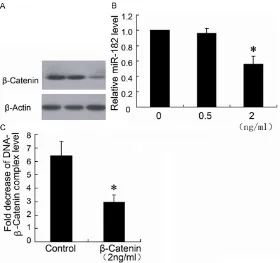
Knockdown of β-catenin protein decreased miR-182 expression of HCT116 cells
To evaluate the role of β-catenin in the effects on miR-182 expression, we used Si-β-catenin to downregulate the β-catenin expression. As shown in Figure 4A, the Si-β-catenin effectively silenced the β-catenin expression in HCT116 cells, which resulted in significant down regula -tion of the miR-182 level.
Figure 2. Fentanyl inhibites the proliferation and invasion of HCT116 cells. HCT116 cells were treated with different concentrations (0 ng/ml, 0.5 ng/ ml, 2 ng/ml, 5 ng/ml and 10 ng/ml) of fentanyl for 24 hours. 104 cells per ml was used to determine the cell inhibition rate by MTT assays (A). The cell invasion was evaluated with Transwell (B). *P < 0.05 by Student’s t-test,
indi-cates a significant difference from the group without fentanyl treatment. All
experiments were repeated three times.
assay and Transwell were per-formed to detect the inhibition rate and cell invasion in HCT116 cells. Cells treated with different concentrations (0.5 ng/ml, 2 ng/ml, 5 ng/ml and 10 ng/mL) of fentanyl resulted in a dose-dependent increase in inhibition rate of cell (Figure 2A), but 0.5 ng/ml and 2 ng/ml fentanyl treat-ment had no significant increase in inhibition rate of cell growth. However, 2 ng/mL fentanyl decreased the cell invasion to 62.3 ± 8.2% (Figure 2B).
Fentanyl inhibites the
expres-sion of β-catenin protein and miR-182 of HCT116 cells
Effect of fentanyl on MMP-2 and MMP-9 ex-ffect of fentanyl on MMP-2 and MMP-9 ex-pression of HCT116 cells
[image:5.612.93.373.71.334.2]The real-time PCR data showed that MMP-2 and MMP-9 expression levels in HCT116 cells
Figure 3. Fentanyl inhibits the expression of β-catenin protein and miR-182
of HCT116 cells. HCT116 cells were treated with different concentrations (0
ng/ml, 0.5 ng/ml and 2 ng/ml) of fentanyl for 24 hours. β-catenin expression
was detected by Western blotting (A) and miR-182 was detected by real-time
PCR (B). ChIP assay was performed to determine the binding of β-catenin on
miR-182 promoter region (C). *P < 0.05 by Student’s t-test, indicates a signifi -cant difference from the group without fentanyl treatment. All experiments were repeated three times.
Figure 4. Knockdown of β-catenin expression decreased miR-182 expres
-sion of HCT116 cells. The HCT116 cells were transfected with Si-β-catenin (Si-β-catenin) or control siRNA (NC). Western blotting (A) was used to de
-termine β-catenin expression and real-time PCR (B) was used to de-termine
miR-182 expression. *P < 0.05 by Student’s t-test, indicates a significant difference from control (NC). All experiments were repeated three times.
treated with different concen-trations of fentanyl had no dif-ference with the group with-out fentanyl treatment (Figure 5A, 5B). However, 2 ng/ml fentanyl downregulated the MMP-9 expression in protein level, but had no effect on MMP-2 expression (Figure 5C).
Overexpression of miR-182 reversed the effect of fen-tanyl on HCT116 cells
To investigate whether miR-182 is involved in the inhibi-tion effect of fentanyl on cell invasion. Cells treated by fen-tanyl transfected with miR-182 mimic to upregulate the miR-182 expression. The resu- lts demonstrated that overex-pression of miR-182 signifi -cantly attenuated fentanyl-induced downregulation of M- MP-9 expression (Figure 6A) and inhibition of cell invasion (Figure 6B).
Discussion
Fentanyl acts as a potent analgesic, clinically used to relieve pain and narcotize in preoperative and postopera-tive. Qin et al has found that fentanyl inhibited gastric can-cer progression in vitro experi-ment [7]. Colorectal cancer is one of the most common malignant tumors in the world, with high rate of recurrence and metastasis [16]. However, there has been very few reports on fentanyl in colorec-tal tumor inhibition. Current study demonstrated that fen-tanyl inhibits the colorectal tumor growth and HCT116 cell invasion via downregulating β-Catenin and miR-182 expression.
[image:5.612.93.374.444.591.2]inhibited the invasion of HCT116 cells in this study. In vitro experiment, HCT116 cells treated with different concentrations of fentanyl result-ed in a dose-dependent increase in cell inhibi-tion rate and decrease in cell invasion. However, 2 ng/mL fentanyl had no effect on the inhibi-tion rate of HCT116 cells, which was corre-spond with the report of literature [17]. This is probably concerned with the low fentanyl con-centration in medium. We also illustrated that both xenograft tumor and HCT116 cells treated with fentanyl had a decrease in β-catenin protein expression by western blotting. The pro-sion by western blotting. The pro-posed mechanism of inhibition of HCT116 cells invasion by fentanyl was correlated with down regulation of β-catenin.
β-catenin, mainly located in cell membrane and plays a critical role in cell-cell communications, is identified as a multifunctional protein [12]. It also acts as a key transcriptional factor in Wnt pathway, which participates widely in regulation of cell proliferation and migration [18]. In many cancers, β-catenin plays pivotal role in malignant transformation of cells [13]. It has been reported that β-catenin mediated
expression of matrix metalloproteinases (MMPs) enhance epithelial-mesenchymal tran-sition in tumor development [19]. The key point of tumor invasion and metastasis is degrading extracellular matrix. MMPs take part in the deg-radation of extracellular matrix, so it was relat-ed closely with the tumor occurrence and devel-opment [20]. Western blotting results showed that fentanyl (2 ng/ml) downregulated the expression of β-catenin as well as MMP-9 pro -tein in HCT116 cells. The research by Tutton et al concluded that MMP-2 and MMP-9 levels in plasma are potential indicators of invasion or metastasis in colorectal tumor [21]. The de- creased expression of MMP-9 confirmed the fentanyl inhibition effect on human colorectal cancer cell invasion.
[image:6.612.93.521.73.387.2]PCR was used to measure the expression of miR-150, miR-146a, miR-210 and miR-122 and miR-182 in colorectal cancer xenograft tumor, and the results showed that only miR-182 expression was downregulated by fentanyl injection. miR-182 is involved in carcinogenesis by targeting the tumor suppressor gene [22], and the up-regulation of miR-182 was signifi -cantly correlated with large tumor size, positive regional lymph node metastasis, and advanced TNM stage. It is also reported that miR-182 is upregulated in breast cancer [23], prostate Cancer [24] and colorectal cancer [10]. In vitro experiments, downregulation of miR-182 expression by fentanyl treatment was also observed in HCT116 cells, which indicated the mechanism of fentanyl on tumor inhibition. To evaluate the role of β-catenin in the effects of miR-182 expression in HCT116 cells, we used Si-β-catenin to silence the β-catenin expression. As expected, miR-182 was down-regulated by silence of β-catenin. The similar results were observed in breast cancer
report-ed [25]. In addition, Our ChIP data demonstrat-ed that binding of β-catenin on miR-182 promoter region was significantly decreased by fentanyl treatment, implying fentanyl inhibits invasion of colorectal cancer via down-regula-tion of miR-182 by β-catenin. On the other hand, overexpression of miR-182 attenuated the fentanyl-induced decrease in cell invasion and MMP-9 expression. Therefore, it was sup-posed that miR-182 participated in the effect of fentanyl on HCT116 cell invasion.
In conclusion, we revealed in the present study that fentanyl exerts the inhibition of colorectal tumor growth and invasion in human colorectal carcinoma cells. Our results also suggest that the inhibition effects associated with fentanyl treatment are due to downregulation of β-catenin and miR-182 expression. The role of fentanyl in the inhibition of colorectal tumor and more cancers should be further studied. Disclosure of conflict of interest
[image:7.612.93.333.77.420.2]None.
Figure 6. The overexpression of miR-182 reversed the effect of fentanyl in HCT116 cells. HCT116 cells treated by fentanyl were transfected with or without the miR-182 mimic. MMP-9 expression were detected by Western blotting (A). The cell invasion was evaluated with Transwell chamber (B). *P < 0.05 by Student’s t-test, indicates a
Address correspondence to: Dr. Sheng-Li Zhou, Department of Anesthesiology and Surgery, Department of International Health Care Center,
Second Affiiated Hospital, School of Medicine,
Zhejiang University, Hangzhou 310009, Zhejiang, China. E-mail: sheli_zhou0811@163.com
References
[1] Siegel R, Desantis C and Jemal A. Colorectal cancer statistics, 2014. CA Cancer J Clin 2014; 64: 104-117.
[2] Johnson CM, Wei C, Ensor JE, Smolenski DJ, Amos CI and Levin B. Meta-analyses of colorec-tal cancer risk factors. Cancer Causes Control 2013; 24: 1207-1222.
[3] Yacoub G, Nagalla S and Aklilu M. Oncologic management of hereditary colorectal cancer. Clin Colon Rectal Surg 2012; 25: 118-122. [4] Stein A, Atanackovic D and Bokemeyer C.
Current standards and new trends in the pri-mary treatment of colorectal cancer. Eur J Cancer 2011; 47: S312-314.
[5] Davis MP. Fentanyl for breakthrough pain: a systematic review. Expert Rev Neurother 2011; 11: 1197-1216.
[6] Kitamura T, Fujiwara H, Nagata O, Usui H, Suzuki T and Ogawa M. [Anesthetic manage-ment using propofol and fentanyl for transrec-tal ultrasound-guided prostatic biopsy]. Masui 2001; 50: 1209-1212.
[7] Qin Y, Li L, Chen J, Tang X, Liao C and Xie Y. Fentanyl inhibits progression of human gastric cancer MGC-803 cells by NF-kappaB down-regulation and PTEN updown-regulation in vitro. Oncol Res 2012; 20: 61-69.
[8] Felekkis K, Touvana E, Stefanou C and Deltas C. microRNAs: a newly described class of en-coded molecules that play a role in health and disease. Hippokratia 2010; 14: 236-240. [9] Hogan NM, Joyce MR and Kerin MJ. MicroRNA
expression in colorectal cancer. Cancer Biomark 2012; 11: 239-243.
[10] Liu H, Du L, Wen Z, Yang Y, Li J and Wang L. Up-regulation of miR-182 expression in colorectal cancer tissues and its prognostic value. Int J Colorectal Dis 2013; 28: 697-703. [11] Pizzini S, Bisognin A, Mandruzzato S, Biasiolo
M, Facciolli A and Perilli L. Impact of microR-NAs on regulatory networks and pathways in human colorectal carcinogenesis and develop-ment of metastasis. BMC Genomics 2013; 14: 589.
[12] Li X, Zhang X, Liu X, Tan Z, Yang C and Ding X. Caudatin induces cell apoptosis in gastric can-cer cells through modulation of Wnt/beta-catenin signaling. Oncology Rep 2013; 30: 677-684.
[13] Fu Y, Zheng S, An N, Athanasopoulos T,
Popplewell L and Liang A. β-catenin as a poten -tial key target for tumor suppression. Int J Cancer 2011; 129: 1541-1551.
[14] Qu B, Liu BR, Du YJ, Chen J, Cheng YQ and Xu W. Wnt/beta-catenin signaling pathway may regulate the expression of angiogenic growth factors in hepatocellular carcinoma. Oncol Lett 2014; 7: 1175-1178.
[15] Cao Q, Mani RS, Ateeq B, Dhanasekaran SM, Asangani IA, Prensner JR, Kim JH, Brenner JC, Jing X, Cao X, Wang R, Li Y, Dahiya A, Wang L, Pandhi M, Lonigro RJ, Wu YM, Tomlins SA, Palanisamy N, Qin Z, Yu J, Maher CA, Varambally S, Chinnaiyan AM. Coordinated regulation of polycomb group complexes through microRNAs in cancer. Cancer Cell 2011; 20: 187-199.
[16] Kin C1, Kidess E, Poultsides GA, Visser BC, Jeffrey SS. Colorectal cancer diagnostics: bio-markers, cell-free DNA, circulating tumor cells
and defining heterogeneous populations by
single-cell analysis. Expert Rev Mol Diagn 2013; 13: 581-599.
[17] Nomura Y, Kawaraguchi Y, Sugimoto H, Furuya H and Kawaguchi M. Effects of morphine and
fentanyl on 5-fluorouracil sensitivity in human
colon cancer HCT116 cells. J Anesth 2014; 28: 298-301.
[18] Kim W, Kim M and Jho EH. Wnt/beta-catenin signalling: from plasma membrane to nucleus. Biochem J 2013; 450: 9-21.
[19] Orlichenko LS and Radisky DC. Matrix metal-loproteinases stimulate epithelial-mesenchy-mal transition during tumor development. Clin Exp Metastasis 2008; 25: 593-600.
[20] Fagan-Solis KD, Schneider SS, Pentecost BT, Bentley BA, Otis CN and Gierthy JF. The RhoA pathway mediates MMP-2 and MMP-9-independent invasive behavior in a triple-nega-tive breast cancer cell line. J Cell Biochem 2013; 114: 1385-1394.
[21] Tutton MG, George ML, Eccles SA, Burton S,
Swift RI and Abulafi AM. Use of plasma MMP-2
and MMP-9 levels as a surrogate for tumour ex-pression in colorectal cancer patients. Int J Cancer 2003; 107: 541-550.
[22] Guo Y, Liao Y, Jia C, Ren J, Wang J and Li T. MicroRNA-182 promotes tumor cell growth by targeting transcription elongation factor A-like 7 in endometrial carcinoma. Cell Physiol Biochem 2013; 32: 581-590.
[23] Guttilla IK and White BA. Coordinate regulation of FOXO1 by miR-27a, miR-96, and miR-182 in breast cancer cells. J Biol Chem 2009; 284: 23204-23216.
[24] Casanova-Salas I, Rubio-Briones J, Calatrava A, Mancarella C, Masia E and Casanova J.
-markers of Early Diagnosis and Prognosis in Patients with Prostate Cancer Treated with Radical Prostatectomy. J Urol 2014; [Epub ahead of print].
[25] Chiang CH, Hou MF and Hung WC. Up-regulation of miR-182 by beta-catenin in