Original Article
Downregulation of ring-finger protein 43 in glioma
associates with poor prognosis
Shaoyan Xi1,2*, Xinke Zhang1,2*, Huadong Chen3, Zhihai Zhong3, Jiabin Lu1,2, Wanming Hu1,2, Qiuliang Wu1,2, Jing Zeng1,2
1State Key Laboratory of Oncology in South China, Collaborative Innovation Center of Cancer Medicine, Sun
Yat-Sen University Cancer Center, Guangzhou, China; 2Department of Pathology, Sun Yat-Sen University Cancer
Center, Guangzhou, China; 3Department of Pediatric Surgery, The First Affiliated Hospital, Sun Yat-sen University,
Guangzhou, China. *Equal contributors.
Received October 25, 2014; Accepted December 22, 2014; Epub January 1, 2015; Published January 15, 2015
Abstract: Ring-finger protein 43 (RNF43) has been identified as a RING-type E3 ubiquitin ligase. The overexpres
-sion of RNF43 results in a significant enhancement of cell growth, and knockdown of the expres-sion of RNF43 by siRNAs exerts a growth-suppressing effect. RNF43 has been proposed as a tumor suppressor in colorectal cancers. However, its expression significance in glioma has not been well studied. In the present study, we found that the decreased expression of RNF43 was associated with poor prognosis by the Kaplan-Meier survival analysis (P <
0.001). Importantly, multivariate analysis suggested RN43 as an independent predictor of overall survival ([hazard
ratio (HR) 0.547, 95% confidence interval (95% CI) 0.382-0.784, P = 0.001]). Collectively, our study demonstrated that RNF43 could be served as a potential prognostic marker for patients with this deadly disease.
Keywords: Ring-finger protein 43 (RNF43), glioma, survival, prognostic marker
Introduction
Glioma is the most common human primary central nervous tumor, and astrocytic glioma composes the largest subgroup of gliomas [1-3]. Radiation exposure and certain genetic syndromes are well-known and well-defined risk factors for glioma [4]. The World Health Organization (WHO) grade is an estimate of the malignancy of a glioma [5]. This grading has been systematically evaluated and successful-ly applied to a spectrum of diffusesuccessful-ly infiltrative astrocytic tumors that range from grade I to IV.
The survival time of glioma patients is known to be variable, and this variability might be related to the WHO grade, tumor location (i.e., supra-tentorial or subsupra-tentorial), surgical treatment, postoperative course, radiotherapy, chemo-therapy, patient age, glioblastoma multiforme (GM) recurrence, imaging characteristics, serum, molecular markers and other factors. However, few factors are likely to be good pre-dictors of long-term survival. More than two decades of research has identified several
important molecular events, such as the over-activation of the epidermal growth factor recep-tor (EGFR) and phosphatidylinositide 3 kinase (PI3K)/Akt pathways and inactivation of the p53/Rb pathways, as important contributors to glioma development. These alterations facili -tate cell survival, proliferation, and migration and are thus crucial for tumorigenesis [6]. Another important factor that has recently emerged as a significant determinant of glioma initiation and progression is the canonical Wnt/ β-catenin signaling pathway, and this contribu -tion is independent of the direct survival advan-tage conferred by Wnt signaling to the tumor cells [7].
Ring-finger protein 43 in glioma
carcinogenesis [8]. Moreover, RNF43 has also been found to be an important tumor suppres-sor gene in mucinous ovarian tumours [9]. However, the role and mechanism of RNF43 in the progression of carcinogenesis have not been fully elucidated. The present study is the first study of the role of RNF43 in glioma. Methods
Patients and tissue specimens
In the present study, paraffin-embedded patho -logic specimens from 229 patients with glioma were obtained from the archives of the Department of Pathology, Sun Yat-sen Uni- versity Cancer Center, China, between November 2000 and December 2008. The cases were selected based on the distinctive pathologic diagnoses of gliomas of patients who had undergone primary and curative resec-tions for tumors without preoperative chemo-therapy or radiochemo-therapy, the availability of resection tissue, and the follow-up data. The glioma cases included 125 men and 104 women, and the mean age of the patients was 58.8 years. The average follow-up time was 52.46 months (median, 45.25 months; range, 4-116 months). Patients whose cause of death was unknown were excluded from the study. The clinicopathologic characteristics of these patients, including age, sex, tumor location,
WHO grade and relapse, are detailed in Table 1. WHO grade was defined according to the 4th edition of WHO Classification of Tumors of the Central Nervous System [10]. The institute research medical ethics committee of Sun Yat-sen University Cancer Center granted approval for this study. All patients provided signed con-sent forms indicating that they agreed to donate the tissue for the medical study when they were hospitalized. Therefore, our ethics committee waived the need for consent.
Tissue microarray (TMA) construction
Tissue microarray was constructed for the study. Briefly, formalin-fixed, paraffin-embed -ded tissue blocks and the corresponding Hematoxylin and Eosin -Stained slides were overlaid for TMA sampling. A senior pathologist (J. Z) reviewed the slides to determine and mark out representative tumor areas. Duplicates of 1 mm diameter cylinders were punched from rep-resentative tumor areas of individual donor tis-sue block and re-embedded into a recipient paraffin block at defined position, using a tis -sue arraying instrument. (Beecher Instruments, Silver Spring, MD).
Immunohistochemical (IHC) staining
[image:2.612.92.523.84.350.2]The TMA slides were dried overnight at 37°C, deparaffinized in xylene, rehydrated through Table 1. Relationship between the expression of RNF43 and clinicopathologic characteristics
Variable RNF43
No. Low expression No. (%) High expression No. (%) P value
Age 0.055
< 50 y 172 60 (34.9) 112 (65.1)
≥ 50 y 57 28 (49.1) 29 (50.9%)
Sex 0.279
Male 125 52 (41.6) 73 (58.4)
Female 104 36 (34.6) 68 (65.4)
Tumor location 0.309
Supratentorial 199 79 (39.7) 120 (60.3) Subtentorial 30 9 (30) 21 (70)
WHO grade 0.631
Low grade (grade I to II) 93 34 (36.6) 59 (63.4) High grade (grade III to IV) 136 54 (39.7) 82 (60.3)
Status 0.002
Live 105 29 (27.6) 76 (72.4) Death 124 59 (47.6) 65 (52.4)
Relapse 0.778
graded alcohol, immersed in 3% hydrogen oxide for 10 minutes to block endogenous per-oxidase activity, and antigen retrieved by pres-sure cooking for 3 minutes in EDTA buffer (pH 8.0). The slides then were preincubated with 10% normal goat serum at room temperature for 30 minutes to reduce nonspecific reaction. Subsequently, the slides were incubated with mouse polyclonal anti-RNF43 (1:50dilution, ab31218; Abcam, Cambridge, UK) at a concen -tration of 3 ng/mL for overnight at 4°C. The slides were incubated with a secondary anti-body (Envision; Dako, Glostrup, Denmark) for 1 hour at room temperature and stained with DAB (3,3-diaminobenzidine). Finally, the sec -tions were counterstained with Mayer hema -toxylin, dehydrated, and mounted. A negative control was obtained by replacing the primary antibody with a normal murine immunoglobulin G. Known immunostaining positive slides were used as positive controls.
Evaluation of IHC staining
The IHC stained samples were assessed as fol -lows: each TMA spot was assigned an intensity score from 0-3 (I0, I1-3), and the proportion of tumor cells for that intensity over the total num-ber of tumor cells was recorded in 5% incre-ments over the range of 0-100 (P0, P1-3); a final H score (range 0-300) was calculated by adding the sum of the scores obtained for each intensity and the proportion of area stained (H score = I1 × P1 + I2 × P2 + I3 × P3); and the expression of RNF43 was scored by two inde -pendent pathologists (S.X. and J.Z.) who were blinded to the clinicopathologic data.
Statistical analysis
[image:3.612.91.523.70.398.2]Statistical analysis was performed by using the SPSS statistical software package (standard version 16.0; SPSS, Chicago, IL). The correla -tion between the expression patterns of RNF43 Figure 1. The expression patterns of RNF43 immunostaining in Glioma tissue. A. The high expression of RNF43 in a glioma case, showing the immunoreactivity as a block mass (upper panel, ×100; lower panel, ×200). B. The low expression of RNF43 in a glioma case, showing the immunoreactivity as a block mass (upper panel, ×100; lower
Ring-finger protein 43 in glioma
and clinicopathologic features of glioma patient was evaluated by χ2-test. Univariate and multivariate survival analyses were performed using the Cox proportional hazards regression model. Survival curves were obtained with the Kaplan-Meier method. Differences were con -sidered significant if the P-value from a two-tailed test was < 0.05.
Results
Immunochemical expression of RNF43 in glioma
To investigate the expression pattern of RNF43 in glioma tissues, we performed IHC staining on 229 cases of glioma tissues. Positive staining of RNF43 protein was observed in most of glio -ma cases. The positive for the immunochemi -cal staining of RNF43 was cytoplasmic stain -ing. As the median score of the expression was 100.5, we classified the expression score that less than and equal to 100 was low expression, otherwise was high expression (As shown in Figure 1).
Relationship between the expression of RNF43 and clinicopathologic characteristics
The relationship between the expression of RNF43 and the clinicopathological characteris
-tics such as age, sex, tumor location, WHO grade, status or relapse were analyzed by the correlation analysis. We found that the expres-sion of RNF43 was significantly related to the status of patients (As shown in Table 2).
Relationship of RNF43 expression and glioma patients’ survival: univariate survival analysis
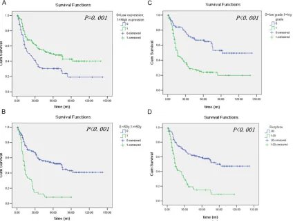
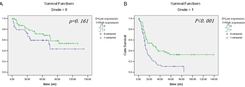
Kaplan-Meier survival analysis was used to demonstrate the prognostic factors of glioma patients’ survival. Such as age (<50 y or ≥ 50 y), WHO grade [low grade (grade I and II) or high grade (grade III and IV)], tumor location (supra -tentorial or sub-tentorial) and the high or low expression of RNF43. Patients with a high RNF43 expression were likely to have a signifi -cantly longer survival (P = 0.001). As our result showed, age, WHO grade, tumor location and relapse were also significant prognostic factors in glioma by the analysis of Kaplan-Meier sur -vival analysis (As shown in Table 2 and Figure 2). Stratified survival analysis was further per -formed with regard to the RNF43 expression in subsets of glioma patients with different WHO grade and age. As shown in the Figures 3 and
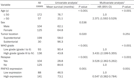
[image:4.612.90.523.95.350.2]4, in high grade glioma, high expression of RNF43 indicated the better survival (P < 0.001). In the younger patients, high expression of RNF43 indicated the better survival (P = 0.009). Table 2. Univariate and multivariate analysis of different prognostic factors in 229 patients with glioma
Variable casesAll Univariate analysis* Multivariate analysis†
Mean survival (months) P value HR (95% CI) P value
Age (y) < 0.001 < 0.001
≤ 50 172 76.7 1.0
> 50 57 21.1 2.371 (1.592-3.529)
Sex 0.536
Male 104 62.1
Female 125 64.8
Tumor location 0.020 0.251
Supratentorial 199 58.0 Subtentorial 30 96.3
WHO grade < 0.001 < 0.001
Low grade (grade I to II) 93 90.4 1.0 High grade (grade III to IV) 136 43.8 3.431 (2.199-5.355)
Relapse < 0.001 < 0.001
Yes 104 28.8 3.526 (2.362-5.262)
No 125 84.6 1.0
RNF43 expression 0.001 0.001
Low expression 88 46.5 1.0 High expression 141 73.1 0.547 (0.382-0.784)
Independent prognostic factors of glioma: mul-tivariate Cox regression analysis
Since variables observed to have prognostic significance by univariate analysis might covari -ate, the expression of RNF43 as well as other clinicopathological features (WHO grade, age, tumor location and relapse) that were signifi -cant in univariate analysis was examined in multivariate analysis (Table 2). Our results showed that high expression of RNF43 was an independent prognostic factor for patient sur-vival [hazard ratio (HR) 0.547, 95% confidence interval (95% CI) 0.382-0.784, P = 0.001] (Table 2). Of the clinicopathological features, WHO grade (P < 0.001), age (P < 0.001) and relapse (P < 0.001) were also evaluated as good independent prognostic factors.
Discussion
Glioma was the most common tumor in the central nervous system. RING finger proteins
act by mediating diverse protein-protein inter-actions. The main biological function of RING finger proteins is the catalysis of ubiquitylation as an ubiquitin-protein isopeptide ligase [11, 12]. The relevance of ubiquitination to cancer might arise from oncogenic mutations that dis-rupt ubiquitination of the proteins that control cell growth and death [13]. RNF43 has fre -quently been found to be up-regulated in human colorectal cancer. In this study, to eluci-date the clinical role of RNF43 in glioma, we applied TMA and IHC to examine its expression in a cohort of Chinese patients. As our knowl-edge, this is the first study to analyze RNF43 expression in glioma using TMA-based IHC method.
[image:5.612.93.517.76.398.2]In results of Kaplan-Meier survival analysis, we found that patients with high RNF43 expres -sion had longer survival. This might be explained by the findings that high RNF43 expression positively correlated with a higher proliferation
Ring-finger protein 43 in glioma
index [14]. The expression of RNF43 has been shown to be regulated by the Wnt-signaling pathway [15-17]. RNF43 is also a direct target gene of Tcf4/b-catenin complex, it functions as a negative feedback regulator that is modulat-ed in the Wnt-signaling pathway by the Tcf4/b-catenin complex, which acted as a significant determinant of glioma initiation and progres-sion [7]. Furthermore, Cox regresprogres-sion analysis suggested RNF43 as an independent prognos -tic factor, indicating that RNF43 may be a piv -otal modulator involved in cancer development. This may be explained by the role of RNF43 in Wnt-signaling pathway, and the reactivation of Wnt/β-catenin signaling could cause an increase in the proliferation of glioma cells [18].
Stratified survival analysis of glioma, according to age, tumor location and WHO grade,
evalu-ated RNF43 expression to be closely correlevalu-ated with survival of glioma patients with younger patients and grade III-IV. This suggested that downregulation of RNF43 in glioma could be of clinical use for distinguishing a set of patients with poor prognosis. Age and WHO grade are the most commonly agreed survival indices for affecting the prognosis of glioma patients [19, 20]. In this study, our results indicated RNF43 expression evaluated by IHC could be used as an additional tool in identifying those patients at risk of glioma progression.
[image:6.612.94.522.71.222.2]Taken together, our study provides compelling clinical evidence that RNF43 can be served as an independent prognostic marker for survival in glioma. Although the current results which are based on a cohort of Chinese patients should be further confirmed in other glioma
Figure 3. Kaplan-Meier survival analysis of RNF43 expressed in a subset of Glioma patients with different age (log-rank test). A. The survival of the different RNF43 expression in glioma patients at not more than 50 years old: low expression, n = 60; high expression, n = 112. B. The survival of the different RNF43 expression in glioma patients at more than 50 years old: low expression, n = 28; high expression, n = 29.
Figure 4. Kaplan-Meier survival analysis of RNF43 expressed in a subset of Glioma patients with different WHO grade (log-rank test). A. The survival of the different RNF43 expression in glioma patients at low grade: low expres
[image:6.612.96.521.295.447.2]cohorts, our findings suggest RNF43 as a new and promising prognostic biomarker for glioma progression and prognosis.
Acknowledgements
This study was supported by Science and Technology Planning Project of Guangdong Province, China (No. 2011B031800159).
Disclosure of conflict of interest
None.
Addresscorrespondence to: Dr. Zeng Jing, Depart- ment of Pathology, Sun Yat-sen University Cancer
Center, 651 Dongfeng Road East, Guangzhou
510060, China. Tel: 20-87342270; Fax:
+86-20-87343807; E-mail: zengjing1320@163.com
References
[1] Jemal A, Siegel R, Ward E, Murray T, Xu J and Thun MJ. (2007)Cancer statistics. CA Cancer J
Clin 2007; 57: 43-66.
[2] Jemal A, Bray F, Center M, Ferlay J, Ward E and Forman D. Global cancer statistics. CA Cancer
J Clin 2011; 61: 69-90.
[3] Siegel R, Naishadham D and Jemal A. Cancer statistics, 2012. CA Cancer J Clin 2012; 62: 10-29.
[4] Omuro A and Deangelis L. Glioblastoma and
other malignant gliomas: a clinical review.
JAMA 2013; 310: 1842-1850.
[5] Kleihues P, Louis D, Scheithauer B, Rorke L, Reifenberger G, Burger P and Cavenee W. The WHO classification of tumors of the nervous
system. J Neuropathol Exp Neurol 2002; 61: 215-225.
[6] Mclendon RF, Bigner D. Comprehensive ge
-nomic characterization defines human glio -blastoma genes and core pathways. Nature 2008; 455: 1061-1068.
[7] Anastas J and Moon R. WNT signalling path -ways as therapeutic targets in cancer. Nat Rev Cancer 2013; 13: 11-26.
[8] Shinada K, Tsukiyama T, Sho T, Okumura F, Asaka M and Hatakeyama S. RNF43 interacts
with NEDL1 and regulates p53-mediated tran-scription. Biochem Biophys Res Commun 2010; 404: 143-147.
[9] Ryland G, Hunter S, Doyle M, Rowley S, Christie M, Allan P, Bowtell D, Gorringe K and Campbell IG. RNF43 is a tumour suppressor gene mu -tated in mucinous tumours of the ovary. J Pathol 2012; 229: 469-476.
[10] Nakazato Y. [The 4th Edition of WHO Classification of Tumours of the Central Nervous System published in 2007]. No Shinkei Geka 2008; 36: 473-491.
[11] Cadwell K and Coscoy L. Ubiquitination on non
-lysine residues by a viral E3 ubiquitin ligase.
Science 2005; 309: 127-130.
[12] Pickart, C. Mechanisms underlying ubiquitina -tion. Annu Rev Biochem 2001; 70: 503-533.
[13] Ding F, Xiao H, Wang M, Xie X and Hu F. The role of the ubiquitin-proteasome pathway in cancer development and treatment. Front
Biosci (Landmark Ed) 2014; 19: 886-895.
[14] Arshad H, Ahmad Z and Hasan S. Gliomas: cor
-relation of histologic grade, Ki67 and p53 ex -pression with patient survival. Asian Pac J Cancer Prev 2010; 11: 1637-1640.
[15] Takahashi N, Yamaguchi K, Ikenoue T, Fujii T and Furukawa Y. Identification of two Wnt-responsive elements in the intron of RING fin
-ger protein 43 (RNF43) gene. PLoS One 2014;
9: e86582.
[16] Leung J, Kolligs F, Wu R, Zhai Y, Kuick R, Hanash S, Cho K and Fearon E. Activation of AXIN2 expression by beta-catenin-T cell factor.
A feedback repressor pathway regulating Wnt signaling. J Biol Chem 2002; 277: 21657-21665.
[17] Niida A, Hiroko T, Kasai M, Furukawa Y,
Nakamura Y, Suzuki Y, Sugano S and Akiyama
T. DKK1, a negative regulator of Wnt signaling, is a target of the beta-catenin/TCF pathway.
Oncogene 2004; 23: 8520-8526.
[18] Yan Z, Wang J, Wang C, Jiao Y, Qi W and Che S. miR-96/HBP1/Wnt/beta-catenin regulatory ci-
rcuitry promotes glioma growth. FEBS 2014;
588: 3038-46.
[19] Lv S, Dai C, Liu Y, Shi R, Tang Z, Han M, Bian R, Sun B and Wang R. The Impact of Survivin on Prognosis and Clinicopathology of Glioma Patients: A Systematic Meta-Analysis. Mol Neurobiol 2014; [Epub ahead of print]. [20] Arshad H, Ahmad Z and Hasan S. Gliomas: cor


![2 [N (4 Methoxyphenyl)acetamido] 1,3 thiazol 4 yl acetate](data:image/gif;base64,R0lGODlhAQABAIAAAP///wAAACH5BAEAAAAALAAAAAABAAEAAAICRAEAOw==)