Original Article
MicroRNA-27a
acts as a
novel biomarker in the diagnosis of
patients with laryngeal squamous cell carcinoma
Bin Zhou1, Lin-Lin Dai1, Ping Xu1, Shuo Wu1, Ting-Ting Li1, Zhong-Tao Cui1, Yong Yang2
1Department of Otolaryngology, The Fourth Hospital of Harbin Medical University, Harbin 150001, Heilongjiang,
China; 2Department of Otolaryngology, The People’s Hospital of Jixi, Jixi 158000, Heilongjiang, China
Received August 21, 2015; Accepted September 25, 2015; Epub February 1, 2016; Published February 15, 2016
Abstract: Background: Increased evidences suggest that the microRNA-23a/24-2/27a (miR-23a/24-2/27a) cluster may play a crucial role in carcinogenesis and act as a novel oncogene. Among these members, miR-27a has been reported to promote proliferation and suppresses apoptosis in laryngeal carcinoma Hep2 cell lines. In this study,
we examined the serum expression of miR-27a and its clinical significance in laryngeal squamous cell carcinoma
(LSCC) patients. Methods: Serum miR-27a expression in 107 patients with LSCC and 104 healthy volunteers were
detected using reverse transcription quantitative real-time polymerase chain reaction (qRT-PCR). Then the relation -ship between its expression and clinical factors was analyzed. Finally, receiver operating characteristic (ROC) curve was established to estimate the diagnostic value of miR-27a in LSCC. Results: Serum miR-27a expression was
higher in LSCC patients than in healthy volunteers (P<0.05). And the high miR-27a expression was significantly
associated with clinical stage (P=0.036). However, there was no association between miR-27a expression and
patients’ age, gender, tumor site, cell differentiation, T classification and N classification (P>0.05). An AUC of 0.914 corresponding with a sensitivity of 86.0% and a specificity of 85.6% were obtained according to ROC curve. Besides,
the optimal cutoff value was 3.155. These revealed miR-27a played an important role in the diagnosis of LSCC. Conclusion: Serum miR-27a expression was increased in LSCC patients compared to healthy controls. Moreover, it could be a potential diagnostic marker in LSCC.
Keywords:MicroRNA-27a, diagnosis, laryngeal squamous cell carcinoma
Introduction
Laryngeal squamous cell carcinoma (LSCC) is
one of the most common malignancies in the head and neck region [1]. Most LSCC originate
in glottis (>60%) and supraglottis, with the sub -glottis representing the minority of patients (<5%) [2]. The diagnostic methods for laryngeal cancer including physical examination, ultra-sound, and computer tomography, but they are
often insufficient for early detection [3].
Moreover, there are few typical symptoms in the early stages of LSCC and the lack of
sensi-tive and specific bio-markers results in a delay
of diagnosis [1]. Therefore, an accurate and reli-able method for early diagnosis of LSCC was necessary in LSCC.
MicroRNAs (miRNAs) are an abundant class of endogenous non-coding small RNAs with a length of 18-25 nucleotides. They are highly
conserved in the genomes of most species, including plants, animals and DNA viruses [4]. These miRNAs play important regulatory roles
by sequence-specific base pairing on the 3’ untranslated region (3’-UTR) of target messen -ger RNAs (mRNAs), in promoting mRNA degra-dation or inhibiting translation [5]. Growing evi-dences showed that miRNAs play an important role in a number of biological processes, includ-ing cell differentiation, proliferation, apoptosis and metabolism processes [6]. microRNAs
have been functionally classified as proto-onco -genes or tumor suppressors and aberrantly expressed in different cancers [7]. More impor-tantly, miRNAs have also been detected in human serum and plasma in remarkably stable
forms, which makes the unique plasma/serum
verte-supermix (TransScript, Invitrogen) following the manufacturer’s instructions. Real-time PCR was performed on an ABI 7500 Real-Time PCR instrument (Applied Biosystems, Inc). Primers of miR-27a and U6 were obtained from Ribobio (Guangzhou, People’s Republic of China). Each sample was examined in triplicate and the expression of miR-27a was normalized with U6 using the 2-ΔΔCT method.
Statistical analysis
All statistical analysis was carried out using Origin pro 9.0. All experiments were repeated at least three times, and the data were presented as means ± SD. The difference between two groups was analyzed using students’ t tests. The relationship between serum miR-27a expression and clinicopathological factors were assessed using x2 tests or Fisher’s exact tests. Receiver operating characteristic (ROC) analy-sis was established to determine the diagnos-tic performance of serum miR-27a in patients with LSCC. P<0.05 were considered to be
sta-tistically significant.
brate genomes [9]. It has been demonstrated that members of this cluster play an important role in mammary tumorigenesis and act as a novel class of oncogenes [10, 11]. The aberrant expression of miR-27a has been observed in various human cancer types including larynge-al carcinoma [12]. However, the role of serum miR-27a and its clinical significance in LSCC
patients remains unclear.
In this study, we examined the serum expres-sion levels of miR-27a using qRT-PCR analysis
and investigated the relationship between serum miR-27a expression and clinicopatho-logical characteristics of LSCC patients. Moreover, we evaluated the potential role of miR-27a as a noninvasive bio-marker in the diagnosis of LSCC.
Materials and methods
Patients and samples collection
107 patients with pathologically diagnosed LSCC at the Fourth Hospital of Harbin Medical
University during March 2006 until
March 2007. None of the patients enrolled in this study had received any chemotherapy or radiotherapy before sampling. Besides, 104 healthy volun-teers from with tumor-free and had no history of oncological diseases were recruited as healthy controls. The study was permitted by the Ethnic Committee of the hospital and all participant had signed written inform consent in advance.
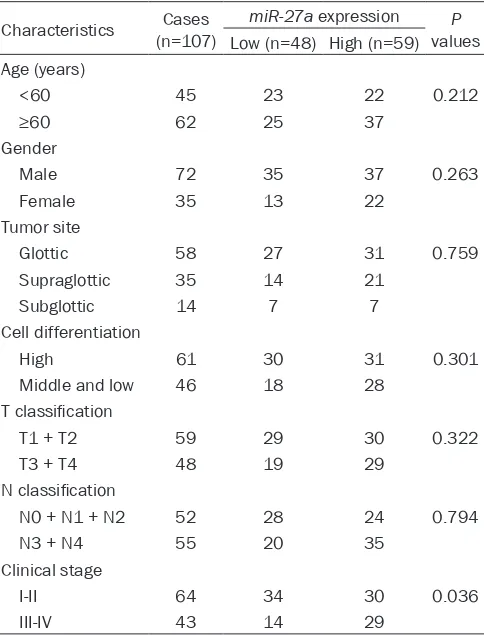
All blood specimens from patients and tumor-free volunteers were collected. The n the samples were separated by centrifuging the blood at 1200× g for 15 min at 4°C. Finally, the obtained serum samples were stored in 1.5 mL RNase free tubes at -80°C for RNA extraction. The clinicopathological char-acteristics of the LSCC patients are summarized in Table 1.
RNA extraction and qRT-PCR analysis
[image:2.612.92.334.96.414.2]Total RNA was isolated from serum of patients with LSCC and healthy controls with TRIzol reagent (Invitrogen). RNA was reverse transcribed into cDNA with TransScript First-Strand cDNA synthesis
Table 1. The relationship between miR-27a expression and clinicopathological features of LSCC patients
Characteristics (n=107)Cases miR-27a expression valuesP Low (n=48) High (n=59) Age (years)
<60 45 23 22 0.212
≥60 62 25 37
Gender
Male 72 35 37 0.263 Female 35 13 22
Tumor site
Glottic 58 27 31 0.759 Supraglottic 35 14 21
Subglottic 14 7 7 Cell differentiation
High 61 30 31 0.301 Middle and low 46 18 28
T classification
T1 + T2 59 29 30 0.322 T3 + T4 48 19 29
N classification
N0 + N1 + N2 52 28 24 0.794 N3 + N4 55 20 35
Clinical stage
Results
Expression level of miR-27a in LSCC patients
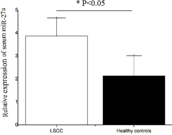
QRT-PCR was used to evaluate serum miR-27a expression level in 107 LSCC patients, and 104 healthy volunteers. As shown in Figure 1, the
serum miR-27a expression
was significantly lower in
patients with LSCC than that in healthy controls (P<0.05). Relationship between miR-27a expression and clinico-pathological characteristics of patients with LSCC
To further investigate whether the expression level of miR-27a was correlated with the development of LSCC, we further associated the rela-tionship between miR-27a expression and the clinico-pathological factors. It was found that miR-27a expres-sion was tightly associated with clinical stage (P=0.036). However, there was no asso-ciation between miR-27a expression and patients’ age, gender, tumor site, cell di-
[image:3.612.92.372.77.297.2]fferentiation, T classification and N classification (P>0.05;
Table 1).
Diagnostic value of miR-27a for LSCC
ROC curves showed that the miR-27a expression had a high diagnostic value with an
AUC of 0.914. Moreover, it
had a high sensitivity (86.0%)
and specificity (85.6%) (Figure 2). The ideal cutoff value of miR-27a expression level was 3.155. Therefore, miR-27a might be an independent diagnostic marker in LSCC.
Discussion
LSCC is one of the most fatal human carcinomas that often occurs in larynx which is a Figure 1. Serum miR-27a expression level in patients with LSCC and healthy
controls. It was increased in patients with LSCC compared to healthy controls (P<0.05).
Figure 2. ROC curve for evaluating the diagnostic value of serum miR-27a.
The AUC was 0.914 combing with a sensitivity of 86.0% and a specificity of
85.6%. The ideal cutoff value of miR-27a expression level was 3.155.
specialized area that is meaningful for breath-ing, sound production, and protecting the tra-chea against food aspiration in human [13]. A conventional biopsy from the primary tumor act
as an accurate technique for the diagnosis of
[image:3.612.94.376.364.576.2]anesthesia with physical or psychological dis-comfort. Therefore, it is particularly necessary
to identify novel and efficient bio-markers for
improving the strategies for the diagnosis of patients with LSCC.
In previous studies, many molecules had been
confirmed to be related to the diagnosis of
LSCC. For instance, serum HMGB1 was over-expression in LSCC and could serve as an inde-pendent diagnostic and prognostic marker [14].
Cyclin D1 was confirmed to be a highly sensi -tive marker in differentiating LSCC from LD or HLM via the study of Jovanovic et al. [15]. FGF3 and p21 protein expression were detected in LSCC and Wang et al., found the combination expression of serum exosomal miR-21 and
HOTAIR had a high diagnostic value with a AUC
of 0.876 as well as a good prognostic value in LSCC [16]. Besides, the expression of miR-21, miR-375 and miR-106b were also proved to be important diagnostic marker in LSCC according to stimulated studies [17, 18].
miR-27a has been reported to have both onco-genic and tumor suppressive functions in dif-ferent cell lines and human cancer tissues. Tang et al. reported that miR-27a expression might be elevated and have clinical potentials as a non-invasive diagnostic bio-marker in osteosarcoma patients [19]. It was revealed that oncogenic miR-27a could play an impor-tant role in the cell growth and metastasis of ovarian cancer [20]. Pan et al. found that miR-27a had the function as an oncogene by target-ing MAP2K4 in the osteosarcoma MG63 cell lines [21]. miR-27a was considered to play an oncogenic role by targeting Spry2 and modulat-ing the malignant, biological behavior of pan-creatic cancer cells [22]. Sun et al. demonstrat-ed that miR-27a was more highly up-regulatdemonstrat-ed in cancer, plasma, and adipose samples from obese liver cancer cases [23]. In contrast, Miao et al. reported that miR-27a was down-regulat-ed by regulating the expression of MET and EGFR in small cell lung cancer [24]. In colorec-tal carcinogenesis and progression, miR-27a
was identified as a tumor suppressor and it
took effects by targeting SGPP1 and Smad2 [25]. Tian et al. indicated that miR-27a might act as an oncogene through suppressing the expression of PLK2 and serve as a useful bio-marker in the diagnosis of LSCC [12]. In our study, we found that the serum miR-27a expres-sion was higher in LSCC patients than that in
healthy controls. These results were in agree-ment with the previous studies [12]. However, the other functions of miR-27a were never reported.
Then we explored the relationship between miR-27a and clinicopathological characteristics to estimate whether it was related to the devel-opment of LSCC. It is worth noting that high expression of miR-27a appeared to be signifi -cantly correlated with clinical stage which indi-cated it participated in the progression of LSCC.
Finally, we investigated the clinical significance
of miR-27a via establishing its ROC curve. The results indicated that miR-27a exhibited a high diagnostic value in LSCC with a high value of
AUC and sensitivity as well as specificity. To the
best of our knowledge, the current study
repre-sented the first demonstration that serum miR-27a expression may function as a diagnostic bio-marker in LSCC patients.
In conclusion, these findings provides the con
-vincing evidences for the first time that the
down-regulation of miR-27a may serve as a novel molecular marker in the diagnosis of
LSCC, and its expression level was influenced
by clinical stage. However, there are some limi-tations. Firstly, the sample size is small. Secondly, the current study has not elucidated the exact molecular mechanisms of miR-27a acting on LSCC. Therefore, some further stud-ies are still need to be done.
Acknowledgements
Supported by the Foundation of Science and Technology Agency for Young Scholars of Hei- longjiang Province (Grant No. QC2011C029).
Disclosure of conflict of interest
None.
Address correspondence to: Dr. Ping Xu, Depart- ment of Otolaryngology, The Fourth Hospital of Har-
bin Medical University, Harbin 150001, Heilongjiang,
China. E-mail: xupingwangh@sina.com
References
[1] Chu EA and Kim YJ. Laryngeal cancer: diagno-sis and preoperative work-up. Otolaryngol Clin North Am 2008; 41: 673-695.
Os-soff RH and Fremgen A. Patterns of care for
cancer of the larynx in the United States. Arch
Otolaryngol Head Neck Surg 1997; 123: 475-483.
[3] Ferreira MB, De Souza JA and Cohen EE. Role of molecular markers in the management of head and neck cancers. Curr Opin Oncol 2011; 23: 259-264.
[4] Bartel DP. MicroRNAs: genomics, biogenesis, mechanism, and function. Cell 2004; 116: 281-297.
[5] Farazi TA, Hoell JI, Morozov P and Tuschl T. Mi-croRNAs in human cancer. Adv Exp Med Biol 2013; 774: 1-20.
[6] Wurdinger T and Costa FF. Molecular therapy in the microRNA era. Pharmacogenomics J 2007; 7: 297-304.
[7] Chou J, Shahi P and Werb Z. microRNA-mediat-ed regulation of the tumor microenvironment. Cell Cycle 2013; 12: 3262-3271.
[8] Cheng G. Circulating miRNAs: roles in cancer diagnosis, prognosis and therapy. Adv Drug Deliv Rev 2015; 81: 75-93.
[9] Xia J, Cheng L, Mei C, Ma J, Shi Y, Zeng F and Wang Z. Genistein inhibits cell growth and in-vasion through regulation of miR-27a in pan-creatic cancer cells. Curr Pharm Des 2014; 20: 5348-5353.
[10] Li X, Liu X, Xu W, Zhou P, Gao P, Jiang S, Lobie PE and Zhu T. c-MYC-regulated miR-23a/24-2/27a cluster promotes mammary carcinoma cell invasion and hepatic metastasis by target-ing Sprouty2. J Biol Chem 2013; 288: 18121-18133.
[11] Chen Z, Ma T, Huang C, Zhang L, Lv X, Xu T, Hu T and Li J. MiR-27a modulates the MDR1/P-glycoprotein expression by inhibiting FZD7/be-ta-catenin pathway in hepatocellular carcino-ma cells. Cell Signal 2013; 25: 2693-2701. [12] Tian Y, Fu S, Qiu GB, Xu ZM, Liu N, Zhang XW,
Chen S, Wang Y, Sun KL and Fu WN. MicroRNA-27a promotes proliferation and suppresses apoptosis by targeting PLK2 in laryngeal carci-noma. BMC Cancer 2014; 14: 678.
[13] Du L, Li H, Zhu C, Zheng R, Zhang S and Chen W. Incidence and mortality of laryngeal cancer in China, 2011. Chin J Cancer Res 2015; 27: 52-58.
[14] Qiu G, Li Y, Liu Z, Wang M, Ge J and Bai X. Clin-ical value of serum HMGB1 in diagnosis and
prognosis of laryngeal squamous cell carcino -ma. Med Oncol 2014; 31: 316.
[15] Jovanovic IP, Radosavljevic GD, Simovic-Mar-kovic BJ, Stojanovic SP, Stefanovic SM,
Pej-novic NN and Arsenijevic NN. Clinical signifi -cance of Cyclin D1, FGF3 and p21 protein
expression in laryngeal squamous cell carci
-noma. J BUON 2014; 19: 944-952.
[16] Wang J, Zhou Y, Lu J, Sun Y, Xiao H, Liu M and Tian L. Combined detection of serum exosomal miR-21 and HOTAIR as diagnostic and
prog-nostic biomarkers for laryngeal squamous cell
carcinoma. Med Oncol 2014; 31: 148. [17] Hu A, Huang JJ, Xu WH, Jin XJ, Li JP, Tang YJ,
Huang XF, Cui HJ and Sun GB. 21 and miR-375 microRNAs as candidate diagnostic
bio-markers in squamous cell carcinoma of the
larynx: association with patient survival. Am J Transl Res 2014; 6: 604-613.
[18] Yu X, Wu Y, Liu Y, Deng H, Shen Z, Xiao B and Guo J. miR-21, miR-106b and miR-375 as
nov-el potential biomarkers for laryngeal squa -mous cell carcinoma. Curr Pharm Biotechnol 2014; 15: 503-508.
[19] Tang J, Zhao H, Cai H and Wu H. Diagnostic and prognostic potentials of microRNA-27a in osteosarcoma. Biomed Pharmacother 2015; 71: 222-226.
[20] Xu L, Xiang J, Shen J, Zou X, Zhai S, Yin Y, Li P, Wang X and Sun Q. Oncogenic MicroRNA-27a is a target for genistein in ovarian cancer cells. Anticancer Agents Med Chem 2013; 13: 1126-1132.
[21] Pan W, Wang H, Jianwei R and Ye Z. MicroRNA-27a promotes proliferation, migration and in-vasion by targeting MAP2K4 in human osteo-sarcoma cells. Cell Physiol Biochem 2014; 33: 402-412.
[22] Ma Y, Yu S, Zhao W, Lu Z and Chen J. miR-27a regulates the growth, colony formation and mi-gration of pancreatic cancer cells by targeting Sprouty2. Cancer Lett 2010; 298: 150-158. [23] Sun B, Li J, Shao D, Pan Y, Chen Y, Li S, Yao X,
Li H, Liu W, Zhang M, Zhang X and Chen L. Adi-pose tissue-secreted miR-27a promotes liver cancer by targeting FOXO1 in obese individu-als. Onco Targets Ther 2015; 8: 735-744. [24] Miao Y, Li J, Qiu X, Li Y, Wang Z and Luan Y.
miR-27a regulates the self renewal of the H446 small cell lung cancer cell line in vitro. Oncol Rep 2013; 29: 161-168.