Case Report
Parotid gland and cerebellum metastasis
of lung cancer: a case report
Binbin Wang1,3, Chengfeng Wu1,3, Li Zhou2, Long Chen2
Departments of 1Gastroenterology, 2Oncology, General Hospital of Lanzhou Military Command, Lanzhou, China; 3Clinical Medical College, Gansu University of Chinese Medicine, Lanzhou, China
Received September 30, 2015; Accepted November 22, 2015;Epub February 1, 2016; Published February 15, 2016
Abstract: Parotid gland and cerebellum metastasis in lung cancer are extremely rare; very few cases have been reported. Here we present a case of a 56-year-old Chinese female patient who presented with a swelling in the right parotid gland. We diagnosed metastatic squamous-cell carcinoma from advanced lung cancer. The patient received 1 cycle nimustine (100 mg/m2 IV Day 1) and nedaplatin (80 mg/m2 IV Day 1). After the first course of chemotherapy,
the parotid swelling subsided and local pain relief. Currently she remains under follow-up. Although metastasis to the parotid gland from any distant primary site is quite unusual and extremely rare, a potential metastasis of lung cancer should not be ignored in the diagnosis of parotid tumor. Before any therapeutic routine examination, such as a chest X-ray, lung computational tomography and brain magnetic resonance imaging may play an important role in differential diagnosis. Ultrasound-guided core needle biopsy is a very safe and effective tool in cases of parotid swelling. Although the management of the metastatic tumor to the parotid gland was controversial, the combination chemotherapy with platinum-based models has a certain effect.
Keywords: Parotid gland, cerebellum, metastasis, squamous-cell carcinoma, lung cancer
Introduction
Lung carcinoma is the most frequent malignant disease and the most common cause of cancer death in the world. It usually arises from dyspla-sia of squamous epithelium of the bronchi and has a propensity to metastasize to any organ. Squamous cell carcinomas accounts for ap- proximately 30% of all non-small cell carcinoma of the lungs [1]. The estimated incidence of metastasis from primary elsewhere to oral and maxillofacial region ranges from 1% to 3% respectively [2]. To the best of our knowledge, the site of parotid gland in this case is very unusual and only a few articles have reported, which are primary small-cell carcinoma [3-7] and adenocarcinoma [8]. Lung carcinomas are characterized by their insidious onset, difficulty in detection, early metastatic spread and poor prognosis at the time of presentation. We describe a case of squamous-cell carcinoma of the lung which spread to the parotid gland.
Case report
A 56-year-old Chinese female presented with a progressive painful swelling in the right parotid
Parotid gland metastasis of lung cancer
Figure 1. Ultrasound showed several hypoechoic nodules in the swelling of right parotid gland with a lager size of 2.4 × 2.4 cm, which were diagnosed by mixed tumors.
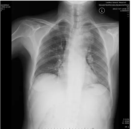
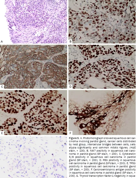
[image:2.629.100.530.455.670.2]a similarly round shadow was uneven density, within low-density areas, rough border, and showed filopodia-like changes among the sur-rounding lung tissue in the left upper lobe. It was next to mediastinum and unclear boundar-ies aortic arch, which size was approxi- mately 3.3 × 3.4 cm, with multiple lymph nodes in the mediastinum appearing enlarged. After consulting with the patient, we performed a core needle biopsy guided by ultrasound on the right parotid swelling. The biopsy pathology reported a squamous-cell lung cancer meta- stases to parotid (Figure 5A) and immunohi- stochemistry (IHC) showed Ki67≈20% (Figure
[image:3.629.97.535.79.510.2]5B), cytokeratin 5/6 (CK5/6, ++) (Figure 5C), P63 (++) (Figure 5D), TP53 (++) (Figure 5E), carcinoembryonic antigen (CEA, +) (Figure 5F) and thyroid transcription factor-1 (TTF-1, -) (Figure 5G). Therefore, we recommended that the patient receive palliative chemotherapy with nedaplatin (80 mg/m2 IV Day 1), nimus- tine (100 mg/m2 IV Day 1) and supportive ther-apy was given. After the first course of che- motherapy, the patient was showed II de- gree myelosuppression and the swelling on the right parotid basically subsided and local pain relief. Currently she remains under follow-up.
Parotid gland metastasis of lung cancer
Discussion
In Western countries, approximately one-third of all cancer-related mortalities are caused by lung cancer. Lung squamous cell carcinoma (SCC) is a common type of non-small-cell lung cancer (NSCLC) and the second leading cause of death related to lung cancer, presenting as locally advanced disease in 25-30% [9-11]. Pulmonary SCC is generally a centrally located lung carcinoma that has been classified histo-logically by World Health Organization (WHO) into four broad categories: Clear cell, small cell, papillary and basaloid. Metastasis is defined as the transfer of disease cells from one organ or part to another site not directly connected with it [12]. Metastasis of lung cancer to the cranio-facial region is not a common characteristic of this tumor. Despite many case reports on me- tastasis of lung cancer to the parotid gland, a
invade blood vessels into the blood circulation resulting in distant metastasis. Despite Nuyens et al. [13] found that the predominant amount of metastatic diseases to the parotid gland were squamous cell carcinomas and malignant melanomas, the metastasis of non small cell lung cancer to the parotid gland is rare in the literature. The most frequent metastasis sites are the liver, the bone, the brain, and adrenal glands.
[image:4.629.97.408.79.389.2]Diagnosis of parotid tumors is made via need- le aspiration or excisional biopsy or ultrasound-guided core needle biopsy which is as a very safe and effective tool in cases of parotid swell-ing in which fine needle aspiration cytology has failed to give a definitive diagnosis [14, 15]. However, complications after these operations can not be ignored, such as facial nerve injury, fistula of parotid duct, hematoma, local swell-ing, pain, infection and other [16].
Figure 4. A lung computational tomography (CT) scan revealed a similarly round shadow was uneven density, within low-density areas, rough border, and showed filopodia-like changes among the surrounding lung tissue in the left upper lobe. It was next to mediastinum and unclear boundaries aortic arch, which size was approximately 3.3 × 3.4 cm, with multiple lymph nodes in the mediastinum ap-pearing enlarged.
In the past few decades, despite numerous advances in cancer treatment, the efficacy of chemotherapy for stage IV NSCLC is only sli- ght improvement. For patients with an adequ- ate performance status, modern
chemothera-py regimens can realistically achieve a tumor response rate of 20 to 30%, median overall sur-vival of 8 to 13months, and a 1-year sursur-vival rate of 30 to 50% [17]. The general principle for the treatment of stage IV NSCLC patients is
[image:5.629.98.535.81.648.2]Parotid gland metastasis of lung cancer
liative care. It goal is to improve the quality of life for patients, and prolong tono obvious symptoms of survival as long as possible. The main method of stage IV is systemic chemo-therapy [18]. However, chemochemo-therapy will bring myelosuppression gastrointestinal symptoms and liver and kidney dysfunction. Compared with best supportive care, the platinum-based chemotherapy can improve the quality of life and prolong the median survival [19-21]. The patient was treated combination chemotherapy with nedaplatin (80 mg/m2 IV Day 1) and Nimustine (100 mg/m2 IV Day 1), because of metastasis to cerebellum from lung cancer. Although the parotid swelling gradually disap-pears and the treatment effect is acceptable, but she still needs to continue post-treatment and long-term follow-up.
Although metastasis to the parotid gland from any distant primary site is quite unusual and extremely rare, a potential metastasis of lung cancer should not be ignored in the diagnosis of parotid tumor. For this reason, in addition to the histopathologic findings, it is important to scan the other systems too. Core needle biopsy guided by ultrasound is a very safe and effec-tive tool in cases of parotid swelling. Although the management of the metastatic tumor to the parotid gland was controversial, the combina-tion chemotherapy with platinum-based mod-els has a certain effect.
Disclosure of conflict of interest
None.
Address correspondence to: Dr. Long Chen, De- partment of Oncology, General Hospital of Lanzhou Military Command, 333 Binhe South Road, Qilihe District, Lanzhou 730050, China. Tel: +86-931-899- 4237; E-mail: wy163_lanzhou@163.com
References
[1] Nonaka D. A study of ΔNp63 expression in lung non-small cell carcinomas. Am J Surg Pathol 2012; 36: 895-899.
[2] Yasar F, Oz G, Dolanmaz D, Akgünlü F. Man-dibular metastasis in a patient with pulmo- nary adenocarcinoma. Dentomaxillofac Radiol 2006; 35: 383-385.
[3] Claramunt R, Englebert A, Laka A, Delos M, Remacle M. Small-cell carcinoma of the parot-id gland. Apropos of a case. Ann Otolaryngol Chir Cervicofac 1994; 111: 223-227.
[4] Takatsugi K, Komuta K, Hosen N, Kitada S, Iida S, Nishihara K, Kimura R, Maeda K, Igarashi T.
Metastasis of small cell lung cancer to the pa-rotid gland as the initial clinical manifestation, followed by metastases to the pituitary gland and lumber spinal cord. Nihon Kokyuki Gakkai Zasshi 1998; 36: 246-250.
[5] Boeger D, Hocke T, Esser D. The interesting case--case no. 68. Metastasis of a small-cell bronchial carcinoma to the parotid gland. La-ryngorhinootologie 2005; 84: 117-20.
[6] Ulubas B, Ozcan C, Polat A. Small cell lung cancer diagnosed with metastasis in parotid gland. J Craniofac Surg 2010; 21: 781-783. [7] Shi S, Fang QG, Liu FY, Sun CF. Parotid gland
metastasis of lung cancer: a case report. World J Surg Oncol 2014; 12: 119.
[8] Debnath CR, Shahjahan SM, Debnath MR, Alam MM, Moshwan MM, Khan MF, Rana MS, Himel RR, Ahmed S. Parotid gland metastasis-an unusual presentation of adenocarcinoma of lung. Mymensingh Med J 2015; 24: 175-177.
[9] Siegel RL, Miller KD, Jemal A. Cancerstatistics, 2015. CA Cancer J Clin 2015; 65: 5-29. [10] Ginsberg MS, Grewal RK and Heelan RT. Lung
cancer. Radiol Clin North Am 2007; 45: 21-43. [11] Cancer Genome Atlas Research Network. Com-prehensive genomic characterization of squa-mous cell lung cancers. Nature 2012; 489: 519-25.
[12] Rossi G, Marchioni A, Sartori G, Longo L, Picci-ni S, Cavazza A. Histotype in Non-small lung cancer therapy and staging: The emerging role of an old and underrated factor. Curr Respir Med Rev 2007; 3: 69-77.
[13] Nuyens M, Schüpbach J, Stauffer E, Zbären P. Metastatic disease to the parotid gland. Oto-laryngol Head Neck Surg 2006; 135: 844-848.
[14] Henke AC, Cooley ML, Hughes JH, Timmerman TG. Fine-needle aspiration cytology of small cell carcinoma of the parotid. Diagn Cytopathol 2001; 25: 126Y129.
[15] Piccioni LO, Fabiano B, Gemma M, Sarandria D, Bussi M. Fine-needle aspiration cytology in the diagnosis of parotid lesions. Acta Otorhino-laryngol Ital 2011; 31: 1-4.
[16] Qayyum A, Ahmed N, Jani P, Berman LH. Ultra-sound-guided core needle biopsy of parotid land swellings. J Laryngol Otol 2009; 123: 449-52.
[17] Scheff RJ, Schneider BJ. Non-small-cell lung cancer: treatment of late stage disease: che-motherapeutics and new frontiers. Semin In-tervent Radiol 2013; 30: 191-198.
clinical oncology clinical practice guideline up-date. J Clin Oncol 2015; 33: 3488-515. [19] Shan J, Xiong Y, Wang D, Xu M, Yang YI, Gong
K, Yang Z, Wang GE, Yang X. Nedaplatin-versus cisplatin-based chemotherapy in the survival time of patients with non-small cell lung can-cer. Mol Clin Oncol 2015; 3: 543-549.
[20] Maneechawakajorn J, Suksuperm J. Quality of life in advanced non-small cell lung cancer re-ceiving chemotherapy of platinum combina-tion in oldversus new standard chemotherapy regimen. J Med Assoc Thai 2014; 97 Suppl 11: S69-75.
[21] Soria JC, Mauguen A, Reck M, Sandler AB, Saijo N, Johnson DH, Burcoveanu D, Fukuoka M, Besse B, Pignon JP; meta-analysis of beva-cizumab in advanced NSCLC collaborative group. Systematic review and meta-analysis of randomised, phase II/III trials adding
bevaci-zumab to platinum-based chemotherapy as