Original Article
Function and mechanism of combined PARP-1 and
BRCA genes in regulating the radiosensitivity
of breast cancer cells
Wei Zhao1,2*, Hongbo Hu1,3*, Qiyan Mo1, Ying Guan1, Ye Li1, Youqing Du1, Ling Li1
1Department of Radiation Oncology, Affiliated Tumor Hospital of Guangxi Medical University, Nanning, Guangxi Autonomous Region, China; 2Guangxi Colleges and Universities Key Laboratory of Biological Molecular Medicine Research, Nanning, Guangxi Autonomous Region, China; 3Department of Oncology, The People’s Hospital of Chongzuo City, Longxiashan Road, Chongzuo, Guangxi Autonomous Region, China. *Co-first authors.
Received June 11, 2019; Accepted August 29, 2019; Epub October 1, 2019; Published October 15, 2019
Abstract: Objective: To study the function and mechanism of combined PARP-1 and BRCA genes in regulating the radiosensitivity of breast cancer cells by poly ADP-ribose polymerase-1 (PARP-1) inhibitor 3-amion benzamide (3-AB) onBRCA mutant and non-mutant breast cancer cells. Methods: Four groups of BRCA mutant cells MDA-MB-436 and BRCA non-mutant cells MDA-MB-231 were divided respectively into control (CTRL), ionizing radiation alone (IR),
3-AB alone (3-AB), and ionizing radiation combined with 3-AB (IR+3-AB) groups. The γ-H2AX foci were detected by immunofluorescence assay to show the DNA double-strand damage. The clonogenic cell survival assay was applied to evaluate the radiosensitivity of breast cancer cells, and flow cytometry was used to assess the percentage of apoptosis cells. Results: The apoptosis rate of MB-436 cells was significantly increased compared with MDA-MB-231 cells treated with irradiation, and 3-AB could further enhance the effect. Similarly, the result of γ-H2AX foci detection showed that DNA double-stranded damage of the MDA-MB-436 cells was significantly greater than that of
MDA-MB-231 cells (t = 4.57, P < 0.05), and the DNA damage of MDA-MB-436 cells in IR+3-AB group was the most
remarkable. The difference was significant (t = 3.26, P < 0.05). In the same group, compared with MDA-MB-231
cells, MDA-MB-436 cells had the significantly greater apoptosis rate (t = 2.96, P < 0.05). The apoptosis rate of
MDA-MB-436 cells in the IR+3-AB group showed by flow cytometry was highest (t = 3.81, P < 0.05). Conclusions: Compared with non-BRCA mutant MDA-MB-231 cells, the BRCA mutant breast cancer MDA-MB-436 cells could
in-cur significantly greater DNA damage, and therefore the radiosensetivity of MDA-MB-436 cells is higher than that of
MDA-MB-231 cells. The inhibitor of PARP-1, which can block the repair of single-strand damage caused by radiation, can further enhance the level of apoptosis and radiosensitivity of BRCA-mutant cells.
Keywords: PARP-1 inhibitor, BRCA gene, radiosensitivity, breast cancer
Introduction
Breast cancer is a common malignant tumor in women, whose annual incidence in the world is almost 2%, and the incidence and mortality in China are also rising. Radiotherapy is an impor-tant component of treatment for breast cancer, and is also the main means of palliative treat-ment for advanced breast cancer. Studies have shown that radiotherapy works as adjuvant treatment after surgery and could reduce the local recurrence rate and death risk of breast cancer [1]. However, in the course of radiother-apy, cancer cells will produce resistance to
phe-nomenon of synthetic lethality was used to study the role of PARP-1 inhibitor 3-AB in the radiosensitivity of breast cancer cells with BRCA gene mutation to explore the mechanism of action of PARP-1 and BRCA genes in DNA damage repair induced by irradiation in order to effectively enhance the radiosensitivity of BRCA mutant breast cancer.
Materials and methods
Reagents and cell culture
The BRCA mutant cells MDA-MB-436 and BRCA non-mutant cells MDA-MB-231 were purcha- sed from the Typical Cell Culture Preservation Committee of Chinese Academy of Sciences (Shanghai, China). The cells were maintained in RPMI 1640 medium supplemented with 10% fetal bovine serum (Hyclone, Logan, UT) and a combination of penicillin (50 U/ml) and strepto-mycin (50 μg/ml), and maintained at 37°C in a 5% CO2 atmosphere humidified to 95-100%. High-energy linear accelerator (Precise 1120, Elekta Instrument AB, Stockholm, Sweden) was applied to provide 6 MV X-ray exposure. The source-tumor distance was SSD = 100 cm, and the dose rate was 200 cGy/min.
The PARP-1 inhibitor 3-amino benzamide (3- AB) was purchased from Sigma (St. Louis, USA), Annexin V-FITC was purchased from Bibo Company (Nanjing, China). The γ-H2AX mon-oclonal antibody and the secondary antibodies (goat anti-mouse IgG) were purchased from Cell Signaling Technology (Danvers, MA).
Flow cytometry assay
The cells were divided into four groups: Control group (CTRL group), 3-AB group (3-AB, 10 mmol/L), Irradiation group (IR group, 8 Gy) and irradiation combined 3-AB group (IR+3-AB group). Cells were grown in 6 well plates and treated with drug or irradiation. 48 hours after treatment, the cells were trypsinized to give a single-cell suspension, and were pelleted by centrifugation. Then the cells were washed twice in cold PBS buffer, fixed in ice-cold 70% ethanol for at least 24 h at -20°C. The ethanol was removed by centrifugation, and the cells were resuspended in 0.5 ml propidium iodide staining solution (0.1% sodium citrate, 0.3% Triton X-100, 100 mg/ml RNase A, 100 mg/ml propidium iodide) and incubated at 37°C for 30 min at a concentration of 5 * 105 cells/mL.
Cellular fluorescence was measured by FASort flow cytometry (Becton Dickinson, USA). The data were analyzed by CELL Quest software. Experiments were repeated three times.
Clonogenic assay
The cells were trypsinized and counted using a hemacytometer. In a typical experiment, 50 to 5,000 cells were plated per well of a six-well plate for irradiation with doses ranging from 0, 2, 4, 6, 8, and 10 Gy, respectively. The plates were then incubated at 37°C for 10 to 14 days; the cells were stained with Giemsa and colo-nies with more than 50 cells per colony were scored under a light microscope. Survival frac-tion under different doses was calculated based on the colonies according to dose. Based on a simple multi-target model, survival curves were constructed by plotting colony-forming ability on a logarithmic scale as a function of the radiation dose administered on a linear scale. Then the radiation biology parameter D0 value (mean lethal dose), Dq value (quasi-threshold dose), N value (target number) and SF2 (cell survival fraction on 2 Gy) were calcu-lated. All experiments were repeated three times.
siRNA
The scrambled RNAi oligonucleotides and siR-NAs targeting mouse Atg5, Atg7 (ONTARGETplus SMARTpoolTM RNAi reagents) were obtained from Dharmacon (Layfayette, CO). All siRNAs were transfected into cells using the Dhar- maFECT 4 Transfection Reagent (Dharmacon, Layfayette, CO) according to the manufactur-er’s protocol siRNA effectiveness was validated by wsestern blotting.
Western blot
with the Kodak Image Station 440CF (Kodak). The band density was quantified using the ImageJ image processing program developed by NIH and normalized to that of the control group.
γ-H2AX detection
After 1 hour of treatment, cells were fixed in ice cold 50% CH3OH and 50% (CH3)2CO for 20 minutes at room temperature. After fixation cells were permeabilized with 0.5% Triton X100: PBS and then blocked with 0.2% skimmed milk, 0.1% Triton-X 100, 5% FBS in Phosphate Saline Buffer (PBS). Cells were then stained with anti-γ-H2AX antibody (Up-state) and anti-mouse AlexaFluor-488 second-ary antibody (Molecular Probes) for the kinetics experiments. Coverslips were mounted with VECTASHIELD® Mounting Medium containing DAPI, to counterstain cellular nuclei. γ-H2AX foci were scored manually by the same opera-tor throughout the cell nuclei using a Zeiss Apotome fluorescence microscope with 63X objective and the average number of foci per cell was calculated from a minimum of 250 cells per dose/time point. Experimental data represent the average of 3 independent experiments.
Statistical analysis
All experiments were done in triplicate, and all data are presented as mean ± standard devia-tion (SD). Significance was determined by Student t-test.
Result
The effects of PARP-1 inhibitor 3-AB on the apoptosis of MDA-MB-436 and MDA-MB-231 cells
The result showed that the 3-AB alone had no significant effect on the apoptosis of these two
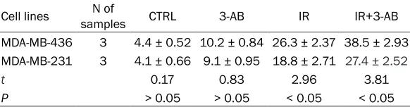
same cells, the apoptosis rate of the IR+3-AB group was significantly increased compared with the IR group,. The difference was signifi -cant (t = 3.81, P < 0.05); especially obvious was the increased apoptosis rate of MDA-MB-436 cells (Table 1).
Effects of 3-AB on the radiosensitivity of MDA-MB-436 and MDA-MB-231 cells
After treatment, survival curves were obtained and radiosensitivity parameters were calculat-ed using the multi-target single-hit model. The experimental results showed that the values of D0, Dq, N, SER, and SF2 of MDA-MB-436 cells were less than that of MDA-MB-231 cells. The shoulder region of the cell survival curve of MDA-MB-436 cells was more narrow, suggest-ing that the radiosensitivity of BRCA mutant breast cancer is higher than that of BRCA non-mutant cells. After treatment, cells were irradi-ated with different doses. The results show that compared with the irradiation group, the shoul-der region of survival curve, and D0, Dq, N val-ues of IR+3-AB group all decreased, sugges- ting that PARP-1 inhibitor 3-AB can increase the radiosensitivity of BRCA mutated (MDA-MB-231) and BRCA non-mutated breast can- cer cells (MDA-MB-436), with marked increase of radiation sensitivity in MDA-MB-231 (Figure 1).
Difference of DNA double strand breaks (DNA-DSBs) between MB-436 and MDA-MB-231 cells
Two groups of cells were given or not given 2 Gy irradiation, and 1 h after irradiation, the γH2AX foci formation was detected by immunofluores -cence. The results showed that in the two groups, the number of γH2AX foci was not significantly affected by the PARP inhibitor 3-AB alone. Compared with the non-irradiation group, irradiation can cause cell DNA
double-Table 1. Apoptosis of MDA-MB-436 and MDA-MB-231 cells treated with radiation and 3-AB (%, _x ± S)
Cell lines samplesN of CTRL 3-AB IR IR+3-AB MDA-MB-436 3 4.4 ± 0.52 10.2 ± 0.84 26.3 ± 2.37 38.5 ± 2.93 MDA-MB-231 3 4.1 ± 0.66 9.1 ± 0.95 18.8 ± 2.71 27.4 ± 2.52
t 0.17 0.83 2.96 3.81
P > 0.05 > 0.05 < 0.05 < 0.05
CTRL, control group; 3-AB, 3-AB treatment group; IR, irradiation group; IR+3-AB, irradiation+3-AB group.
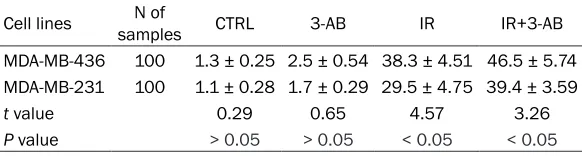
[image:3.612.92.383.98.174.2]strand breaks, and at the same dose, the γH2AX foci of MDA-MB-436 cells was higher than that of MDA-MB-231 cells (t = 4.57, P < 0.05). For the same cells, the γH2AX foci of the cells in IR+3-AB group is much higher than that of irradiation alone group (t = 3.26, P < 0.05); especially in MDA-MB-436 cells, the DNA-DSBs increased significantly (Figure 2; Table 2).
PARP-1 and BRCA control the repair of single and double DNA strand pathways, which play a key role in the repair of DNA damage induced by irradiation, directly affecting the sensitivity of tumor cells [5, 6].
[image:4.612.91.526.74.260.2]Because of the important role of PARP-1 in DNA repair, use of PARP-1 inhibitors occurs two Figure 2. The γH2AX foci of MDA-MB-436 and MDA-MB-231 cells were detected 1 hour after irradiation with 2 Gy X rays.
Table 2. The DNA-DSB of MDA-MB-436 and MDA-MB-231 cells treated with radiation and 3-AB (%, _x ± S)
Cell lines samplesN of CTRL 3-AB IR IR+3-AB MDA-MB-436 100 1.3 ± 0.25 2.5 ± 0.54 38.3 ± 4.51 46.5 ± 5.74 MDA-MB-231 100 1.1 ± 0.28 1.7 ± 0.29 29.5 ± 4.75 39.4 ± 3.59
t value 0.29 0.65 4.57 3.26
P value > 0.05 > 0.05 < 0.05 < 0.05
CTRL, control group; 3-AB, 3-AB treatment group; IR, irradiation group; IR+3-AB, irradiation+3-AB group.
Discussion
[image:4.612.93.525.298.446.2] [image:4.612.91.382.522.600.2]ways. The first is the PARP-1 for DNA inhibitors as a radiotherapy or chemotherapy sensitizing agent [7, 8]; the second is the specific genetic characteristics of some tumors by chemical synthetic lethality to cause DNA damage, but the overall treatment effect is not ideal [9]. Therefore, this study introduced the synthetic lethal phenomenon: namely, when inhibited PARP-1 coexists with a BRCA defect, a large number of DNA strand breaks induced by radia-tion will lead to synthetic lethality. The specific mechanism is: when PARP-1 protein of breast cancer cells is inhibited, frequent DNA-SSBs in the cells fail to be repaired in a timely way and would accumulate increasingly, which causes the disintegration of the replication fork, and finally produces large amounts of DNA-DSBs. DNA-DSBs with strong cytotoxicity could be repaired in normal cells by the homologous recombination (HR) repair pathway mediated by BRCA gene. However, in the cells with BRCA mutation, DNA-DSBs could not be repaired, or just be repaired by non homologous end joining pathway (NHEJ) alternatively due to lack of HR, thereby greatly increasing the probability of cell death [10, 11].
This study explores these two key DNA repair genes of PARP-1 and BRCA by natural BRCA mutant breast cancer cells to study the role of two genes in DNA damage repair, apoptosis, and radiosensitivity after irradiation. We found that the DNA damage, cell apoptosis, and radio-sensitivity of BRCA mutant cells MDA-MB-436 after irradiation increased significantly com -pared with BRCA non-mutant cells MDA-MB-231, which verified the important role of BRCA gene in DNA damage repair. However, the PARP-1 inhibitor 3-AB combined with irradiation could further cause DNA damage, cell apopto-sis, and increasing radiosensitivity of the two kinds of cells, especially of BRCA mutant cells MDA-MB-436. This phenomenon suggests that PARP-1 inhibitors and ionizing radiation have a synergistic effect. When the DNA-SSBs and DNA-DSBs repair pathways are blocked simul-taneously, both of SSB and DSB cannot be repaired timely, leading to increased DNA dam-age significantly, cell apoptosis and final increased radiosensitivity.
Therefore, PARP-1 inhibitors can not only be used as a general radiotherapy sensitizing agent, but also can be used as specific drugs
for certain tumors with suppressor gene muta-tions, such as BRCA mutation breast cancer, which will greatly increase the treatment effects [6, 12]. The increase of DNA damage, cell apop-tosis and radiosensitivity of BRCA mutation breast cancer cells in our study provide the theoretical guidance for improving the radio-therapy efficacy of BRCA mutant breast cancer by using PARP-1 inhibitors. However, there are still some problems, such as how to select bet-ter concentration of PARP-1 inhibitor to avoid adverse drug reactions. To solve these prob-lems, we need further in-depth study of specific regulatory mechanisms of PARP1 inhibitors in BRCA mutated breast cancer cells. The results of numerous in vitro studies also need thror-ough animal experiments and clinical trials to further verify.
Acknowledgements
This work was supported by a grant from Guangxi Colleges and Universities Key La- boratory of Biological Molecular Medicine Research (Grant Number: GXBMR201604), grant from Natural Science Foundation of Guangxi Province (Grant Number: 2017GXN-SFAA198056), grant from Guangxi Key La- boratory of Bio-targeting Theranostics (Grant Number: GXSWBX201808), and a grant from the Medical and Health Appropriate Technology Development and Promotion Project of Guangxi Zhuang Autonomous Region (Grant Number: S2018008).
Disclosure of conflict of interest
None.
Address correspondence to: Ling Li, Department
of Radiation Oncology, Affiliated Tumor Hospital of
Guangxi Medical University, Nanning 530021, Guangxi Autonomous Region, China. E-mail: [email protected]
References
for 10,801 women in 17 randomised trials. Lancet 2011; 378: 1707-1716.
[2] Matsuda Y and Tobari I. Radiosensitivity and
effects of repair inhibitors for X-ray-induced
chromosomal damage in mouse zygotes in S and G2 phases. Int J Radiat Biol 1995; 68: 615-623.
[3] Pristauz G, Petru E, Stacher E, Geigl JB, Schwarzbraun T, Tsybrovskyy O, Winter R and Moinfar F. Androgen receptor expression in breast cancer patients tested for BRCA1 and BRCA2 mutations. Histopathology 2010; 57: 877-884.
[4] Vuong B, Hogan-Cann AD, Alano CC, Stevenson M, Chan WY, Anderson CM, Swanson RA and
Kaup-pinen TM. NF-κB transcriptional activa
-tion by TNFα requires phospholipase C, extra -cellular signal-regulatedkinase 2 and poly
(ADP-ribose) polymerase-1. J Neuroinflamma -tion 2015; 12: 229.
[5] Stilmann M, Hinz M, Arslan SC, Zimmer A, Sch-reiber V and Scheidereit C. A nuclear poly (ADP-ribose)-dependent signalosome confers
DNA damage-induced IκB kinase activation.
Mol Cell 2009; 36: 365-378.
[6] Bryant HE, Schultz N, Thomas HD, Parker KM, Flower D, Lopez E, Kyle S, Meuth M, Curtin NJ
and Helleday T. Specific killing of BRCA2-defi -cient tumours with inhibitors of poly(ADP-ri-bose) polymerase. Nature 2005; 434: 913-7.
[7] Schlicker A, Peschke P, Bürkle A, Hahn EW and Kim JH. 4-Amino-1, 8-naphthalimide: a novel inhibitor of poly(ADP-ribose) polymerase and radiation sensitizer. Int J Radiat Biol 1999; 75: 91-100.
[8] Khan K, Araki K, Wang D, Li G, Li X, Zhang J, Xu
W, Hoover RK, Lauter S, O’Malley B Jr, Lapidus RG and Li D. Head and neck cancer radiosen-sitization by the novel poly(ADP-ribose) poly-merase inhibitor GPI-15427. Head Neck 2010; 32: 381-391.
[9] Simons A, Dafni N, Dotan I, Oron Y and Ca-naani D. Establishment of a chemical synthet-ic lethality screen in cultured human cells. Ge-nome Res 2001; 11: 266-273.
[10] Iglehart JD, Silver DP. Synthetic lethality--a new direction in cancer-drug development. N Engl J Med 2009; 361: 189-91.
[11] Guha M. PARP inhibitors stumble in breast cancer. Nat Biotechnol 2011; 29: 373-4. [12] Farmer H, McCabe N, Lord CJ, Tutt AN,