Int J Clin Exp Pathol 2017;10(9):9262-9272 www.ijcep.com /ISSN:1936-2625/IJCEP0058864
Original Article
MiR-100-5p, miR-199a-3p and miR-199b-5p induce
autophagic death of endometrial carcinoma
cell through targeting mTOR
Junhong Cai1, Ying Zhang2, Sizhe Huang2, Mengdan Yan3, Jingjie Li3, Tianbo Jin3,4, Shan Bao2
1Central Laboratory, 2Department of Gynecology and Obstetrics, Hainan General Hospital, Haikou, Hainan, China; 3Key Laboratory of Resource Biology and Biotechnology in Western China (Northwest University), Ministry of
Edu-cation, 4School of Life Sciences, Northwest University, Xi’an, Shaanxi, China
Received June 6, 2017; Accepted June 22, 2017; Epub September 1, 2017; Published September 15, 2017
Abstract: Objective: The study aimed to explore the association between three miRNAs (miR-100-5p, miR-199a-3p and miR-199b-5p) and mTOR induced autophagy in EEC cells. The expression of three miRNAs and autophagy-related genes Beclin1 and LC3 in Ishikawa and KLE cells were detected. Methods: The effects of three miRNAs on proliferation and apoptosis in EEC cells were analyzed in KLE and ISK cells transfected with miR-100-5p, miR-199a-3p and miR-199b-5p mimics and inhibitors. Quantitative real-time polymerase chain reaction (qRT-PCR), CCK-8 method, flow cytometry and luciferase reporter assay were used to assess the effects of three miRNAs on cell viability, proliferation, apoptosis, and autophagy. Results: We found an increased expression of Beclin1 and LC3
in Ishikawa and KLE cells transfected by miR-100-5p, miR-199a-3p and miR-199b-5p mimics compared with NC (P<0.05). Additionally, Ishikawa and KLE cells transfected with miR-100-5p, miR-199a-3p and miR-199b-5p mim-ics grew more slowly than mock and mimmim-ics control (P<0.05); we also found an increased apoptosis incidence of Ishikawa cell transfected with miR-100-5p, miR-199a-3p and miR-199b-5p mimics (P<0.05). Finally, luciferase reporter results showed miR-100-5p, miR-199a-3p and miR-199b-5p were all down-regulated the luciferase activity in cells transfected with miRNA mimics compared with mock. All results suggested that miR-100-5p, miR-199a-3p and miR-199b-5p may induce the autophagic death of EEC cell through targeting mTOR. Conclusions: Our results suggested that miR-100-5p, miR-199a-3p and miR-199b-5p may induce the autophagic death of EEC cell through targeting mTOR.
Keywords: mTOR, autophagy, microRNA (miRNA), Beclin1, LC3B, endometrial endometrioid adenocarcinoma (EEC)
Introduction
Endometrial carcinoma (EC) is the sixth most common cancer in women worldwide, and its incidence is rapidly increasing in China [1]. In some developed areas of China, the incident of EC has exceeded cervical cancer and is being the leading form of female reproductive system cancer [2]. Endometrial endometrioid adeno-carcinoma (EEC) is the main form of EC, which accounts for more than 80% of all EC patients [3]. Alteration of some genes has been con-nected with etiology of EEC, and may also serve as targets for drug delivery and therapy [4, 5]. Further knowledge of the molecular signaling pathways actively involved in the occurrence and development of EEC may provide new
tion of EEC by targeting mTOR [10]. Based on these results, we intend to identify the associa-tion between three miRNAs (100-5p, miR-199a-3p, miR-199b-5p) and mTOR induced autophagy in EEC cells.
Autophagy is a very complicated metabolism process regulated by several autophagy-relat-ed genes. Beclin1 is the first identified mam-malian autophagy mediated gene involved in autophagic initiation [11]. LC3 is ubiquitously distributed and essential to the formation of autophagosomes in mammalian cells [12]. In this study, we examined the expression of three miRNAs (100-5p, 199a-3p and miR-199b-5p) and autophagy-related genes Beclin1
and LC3 in Ishikawa and KLE cells, through real-time quantitative PCR, CCK-8 assay, flow cytometer and luciferase reporter assay. To our knowledge, this study is the first to investigate the expression of miR-100-5p, miR-199a-3p, miR-199b-5p and their relationship with mTOR induced autophagy in EEC cells.
Materials and methods
Cell culture
Human EEC Ishikawa (well differentiated) and KLE (poorly differentiated) cell lines were pur-chased from Biofavor Biotech Co. Ltd, Wuhan, China. Both of cell lines were grown in Dulbecco modified Eagle medium (HyClone, Uath, USA) containing 10% fetal bovine serum (HyClone) and the penicillin-steptomycin solution (100 U/ ml penicillin; 100 μg/ml streptomycin; HyClone) in a humidified incubator with 5% CO2 at 37°C.
Transfection
Before transfection, Ishikawa and KLE cells were seeded in 24-well plates and grown to 80% confluence. Using Lipofectamine 2000 Tr- ansfection Reagent (Thermo Fisher, Waltham, USA), the cells were transfected with miR-100-5p, miR-199a-3p, miR-199b-5p miRNA mimics, inhibitor and NC mimics/inhibitors (Shanghai GenePharma Co. Ltd, Shanghai, China), as well as normal control, in accordance with the instructions provided by Thermo Fisher. The Ishikawa cells were treated with 10-ng/mL ra- pamycin (Sigma-Aldrich, St Louis, MO). Cells were collected at different time points for anal-ysis after transfection.
RNA isolation and reverse transcription of total RNA
Isolation of total RNA was used the TRIzol Plus RNA Purification Kit (Invitrogen) according to manufacturer protocol. Total RNA (500 ng) was reverse transcribed using Precision nanoScript Reverse Transcription kit (Primer Design) acc- ording to manufacturer protocol.
Real-time qPCR for Beclin1e and LC3 expres-sion
Amplification of Beclin1e, LC3 and reference gene GADPH was performed in 20 μL reac-tions. Each reaction consisted of: 1 μL te- mplate, 10 μL SuperReal PreMix Plus (SYBR Green), 0.6 μL Forward primer, 0.6 μL Reverse primer and 7.8 μL ddH2O. Reactions were car-ried out in Rotor Gene 6000 2-plex HRM ther-mo cycler (Corbett Research) using the fol- lowing protocol: 95°C for 15 min and 45 quan-tification cycles of 95°C for 10 sec, 60°C for 20 sec, and 72°C for 30 sec.
miRNA reverse transcription and real-time qPCR
miRNA was reversed using miRcute Plus miRNA First-Strand cDNA Synthesis Kit (TIANGEN, Beijing, China) according to manufacturer pro-tocol. qPCR reactions for miRNA expression analysis were prepared as follows: 1 μL miRNA first strand cDNA, 5 μL 2× miRute miRNA pre-mix, 0.2 μL Forward primer, 0.2 μL Reverse primer and 3.6 μL Rnase free ddH2O. All qPCR reactions were performed in duplicates in ViiA7 Real-Time PCR System (Applied Biosystems) using the following PCR protocol: 94°C for 2 min, 5 cycles of 94°C for 20 sec, 65°C for 30 sec, and 72°C for 34 sec and 40 cycles of 94°C for 20 sec and 60°C for 34 sec.
Cell viability assay
MiRNAs induce autophagic death of EEC cells
MiRNAs induce autophagic death of EEC cells
Analysis of cell apoptosis
Annexin V-FITC apoptosis Detection kit (KeyGEN BioTECH, Jiangsu, China) was used to deter-mine the cellular apoptosis. Ishikawa cells (4×10⁶ per well) were cultured in 6-well plate for overnight, and treated respectively. Acc- ording to manufacturer’s instruction, cells were collected by trypsinization, then washed twice with phosphate-buffered saline (PBS) and centrifuged at 1000 rpm 5 min. Finally, cells were suspended in 500 μl of 1× binding buffer, and incubated in the dark with 5 μl Annexin V-fluorescein isothiocyanate (FITC) and 5 μl propidium iodide (PI) for 10 min at room
tem-perature. Flow cytometer (FACSCalibur Becton Dickinson, Franklin Lakes, NJ, USA) was used to analyze cellular apoptosis.
Luciferase reporter assay
The influence of miR-100-5p, miR-199a-3p miR-199b-5p miRNA mimics to mTOR gene was performed used Dual-Glo Luciferase Assay System (Promega, Madison, USA).
Statistical analysis
[image:5.612.94.524.66.492.2]Nonparametric one-way analysis of variance and Student t test were performed, with P<0.05 considered statistically significant.
MiRNAs induce autophagic death of EEC cells
Results
Overexpression of miR-100-5p, miR-199a-3p and miR-199b-5p in Ishikawa and KLE cells
To analyze the potential role of miR-100-5p, miR-199a-3p and miR-199b-5p in EEC cells, we used miR-100-5p, miR-199a-3p, miR-199b-5p miRNA mimics, inhibitor and NC mimics/inhibi-tors, to transfect the Ishikawa and KLE cells, respectively. The expression levels of miR-100-5p, miR-199a-3p and miR-199b-5p in Ishikawa and KLE cells after transfection were deter-mined by SYBR-Green stem-loop real-time PCR analysis. The results showed that miR-100-5p, miR-199a-3p and miR-199b-5p expression lev-els were all increased in the Ishikawa and KLE cells compared with NC (P<0.01). The expres-sion levels of three miRNAs in miRNA inhibitor and NC, which were not significantly different (Figure 1).
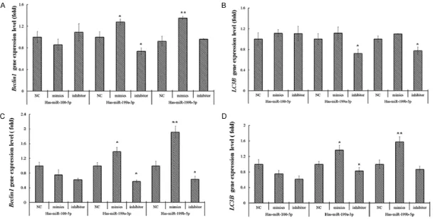
Beclin1 and LC3 mRNA expression in Ishikawa and KLE cells
Beclin1 and LC3 were autophagy related genes. To investigate the association between miR-100-5p, miR-199a-3p and miR-199b-5p and cell autophagy, we also detected the expres-sion level of Beclin1 and LC3 mRNAs in Is- hikawa and KLE cells after transfection. We found an increased expression of Beclin1 and
LC3 mRNA in Ishikawa and KLE cells transfe- cted by 100-5p, 199a-3p and miR-199b-5p mimics compared with NC (P<0.05), and the expression levels of Beclin1 and LC3
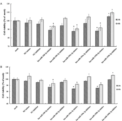
and miR-199b-5p mimics grew more slowly (P<0.05). Reduction of the intracellular miR-100-5p, miR-199a-3p and miR-199b-5p level by transfection with the miRNA inhibitor inc- reased their growth rate (Figure 3), suggesting that overexpression of miR-100-5p, miR-199a-3p and miR-199b-5p could suppress the prolif-eration of Ishikawa and KLE cells.
Apoptosis of Ishikawa cell was induced by miR-100-5p, miR-199a-3p and miR-199b-5p
To clarify the association between three miR-NAs and apoptosis of Ishikawa cell, we carried out flow cytometry. Compared with the mock and the miRNA mimics control, we found an increased apoptosis incidence of Ishikawa cell transfected with miR-100-5p, miR-199a-3p and miR-199b-5p mimics (P<0.05). In contrast, the apoptosis incidence of Ishikawa cell trans-fected with miR-199a-3p inhibitor was lower than NC inhibitor control (P<0.05) (Figure 4).
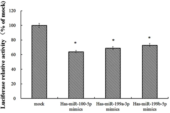
mTOR is a target of miR-100-5p, miR-199a-3p and miR-199b-5p
[image:8.612.92.375.72.260.2]To directly investigate the relationship between three miRNAs and its targets, we constructed the luciferase reporter vectors in HEK 293 cell, miR-100-5p, miR-199a-3p and miR-199b-5p were all down-regulated the luciferase activity in cells transfected with miRNA mimics com-pared with mock (Figure 5). These results indi-cating that the mTOR 3’UTR was the target directly regulated by miR-100-5p, miR-199a-3p and miR-199b-5p.
Figure 5. mTOR is a Target of miR-100-5p, miR-199a-3p and miR-199b-5p.
mRNA were lower in Ishika- wa and KLE cells transfected by 199a-3p and miR-199b-5p miRNA inhibitors compared with NC (P<0.05) (Figure 2).
Proliferation of Ishikawa and KLE cells were inhibited by miR-100-5p, miR-199a-3p and miR-199b-5p
MiRNAs induce autophagic death of EEC cells
Discussion
In the present study, we investigated the expr- ession levels of three miRNAs (miR-100-5p, miR-199a-3p and miR-199b-5p) and autopha-gy-related genes Beclin1 and LC3 in Ishikawa and KLE cells. We found an increased expres-sion of Beclin1 and LC3 in Ishikawa and KLE cells transfected by miR-100-5p, miR-199a-3p and miR-199b-5p mimics compared with NC. Additionally, Ishikawa and KLE cells transfect-ed with 100-5p, 199a-3p and miR-199b-5p mimics grew more slowly; we also found an increased apoptosis incidence of Ishikawa cell transfected with miR-100-5p, miR-199a-3p and miR-199b-5p mimics. Finally, luciferase reporter results showed miR-100-5p, miR-199a-3p and miR-199b-5p were all down-regulated the luciferase activity in cells trans-fected with miRNA mimics compared with mock. All results suggested that miR-100-5p, miR-199a-3p and miR-199b-5p may induce the autophagic death of EEC cell through targeting mTOR.
MicroRNAs usually bind to the 3’UTR of target mRNAs, which inhibit mRNA translation or induce mRNA degradation [8]. Several studies have found abnormal expression of miRNAs in tumors cells, suggesting that miRNAs may have similar functions like proto-oncogenes or tumor suppressor genes [13-15]. MiR-100-5p exhibit-ed a low expression level in head and neck squamous cancer cell [16]. abnormal expres-sion of miR-199a-3p has been found in several kinds of cancer [17]. MiRNA-199b-5p is involved in the Notch signaling pathway, and it has been found associated with risk of breast [18], ovar-ian [19], lung [20] and renal cancers [21]. In our study, we identified that overexpression of miR-100-5p, miR-199a-3p and miR-199b-5p in transfected cells significantly reduced EEC cell proliferation and accelerated cell apoptosis. Combined with the previous results, we sug-gested that 100-5p, 199a-3p, miR-199b-5p may play important roles in the dev- elopment of human cancer, and the results identified here need to be confirmed in further studies.
Autophagy is a conserved, programmed res- ponse to metabolic and environmental stress, which could regulate the survival and death of cancer cells [22]. There are several important
genes in different stages of autophagy, such as Beclin1 and LC3B. Beclin1 is involved in autophagosome formation at an early stage, rather than the expansion step [23]. Previous studies have found a lower expression level of Beclin1 in breast, ovarian and colorectal can-cer cells [24, 25]. LC3B is closely associated with the number of autophagosomes, which pl- ay an indicator role in autophagosome forma-tion [26]. In our present study, the expression level of Beclin1 and LC3 were both increased in Ishikawa and KLE cells transfected by miR-100-5p, miR-199a-3p and miR-199b-5p mim-ics. We speculated that miR-100-5p, miR-199a-3p and miR-199b-5p may have influence on autophagy by targeting mTOR, so we further investigated the association between three miRNAs and mTOR using luciferase reporter assay.
mTOR kinase is a major target in the autophagy signaling pathway, which regulates cell tran-scription, translation, and cytoskeletal organi-zation [27]. An increased expression level of mTOR increases has been found in several types of cancers [28]. In our study, miR-100-5p, miR-199a-3p and miR-199b-5p were all down-regulated the luciferase activity in cells trans-fected with miRNA mimics, which indicating that mTOR 3’UTR was the target directly regu-lated by miR-100-5p, miR-199a-3p and miR- 199b-5p.
In conclusion, after Ishikawa and KLE cells transfected by mTOR targets miRNA (miR-100-5p, miR-199a-3p, miR-199b-5p), autophagy-related genes Beclin1 and LC3 were overex-pressed, cell grow more slowly, and an incr- eased apoptosis incidence were detected. Our results suggested that miR-100-5p, miR-199a-3p and miR-199b-5p may induce the autopha-gic death of EEC cell through targeting mTOR. These findings help us better understand the molecular mechanisms underlying the occur-rence and development of EEC, and also impli-cate that 100-5p, 199a-3p and miR-199b-5p could be considered as promising biomarkers for early detection and prognosis of EEC.
Acknowledgements
Disclosure of conflict of interest
None.
Address correspondence to: Dr. Shan Bao, Depart- ment of Gynecology and Obstetrics, Hainan General Hospital, 19 Xiuhua Road, Haikou 570311, Hainan, China. Tel: 68642629; Fax: +86-898-68642629; E-mail: baoshan3@126.com
References
[1] Ervik M, Soerjomataram I and Ferlay J. GLO- BOCAN 2012: estimated cancer incidence, mortality and prevalence worldwide, in 2012. 2015.
[2] Wei KR, Chen WQ and Zhang SW. An analysis of incidence and mortality of corpus uteri can-cer in China, 2009. China Cancan-cer 2013. [3] Gao J, Yang G, Wen W, Cai QY, Zheng W, Shu XO
and Xiang YB. Impact of known risk factors on endometrial cancer burden in Chinese women. Eur J Cancer Prev 2016; 25: 329-34.
[4] Moreno-Bueno G, Hardisson D, Sarrió D, Sán-chez C, Cassia R, Prat J, Herman JG, Esteller M, Matías-Guiu X and Palacios J. Abnormalities of E- and P-cadherin and catenin (β-, γ-catenin, and p120ctn) expression in endometrial can-cer and endometrial atypical hyperplasia. J Pathol 2003; 199: 471.
[5] Mcconechy MK, Ding J, Senz J, Yang W, Melnyk N, Tone AA, Prentice LM, Wiegand KC, Mcalp-ine JN and Shah SP. Ovarian and endometrial endometrioid carcinomas have distinct CTN-NB1 and PTEN mutation profiles. Mod Pathol 2014; 27: 128-34.
[6] Saxton RA and Sabatini DM. mTOR signaling in growth, metabolism, and disease. Cell 2017; 168: 960-976.
[7] Darb-Esfahani S, Faggad A, Noske A, Weichert W, Buckendahl AC, Müller B, Budczies J, Röske A, Dietel M and Denkert C. Phospho-mTOR and phospho-4EBP1 in endometrial adenocarcino-ma: association with stage and grade in vivo and link with response to rapamycin treatment in vitro. J Cancer Res Clin Oncol 2009; 135: 933-941.
[8] Spitzer IB, Tuschl T, Spitzer WJ, Farazi TA and Morozov P. miRNAs in Human Cancer 2010. [9] Torres A, Torres K, Pesci A, Ceccaroni M,
Pasz-kowski T, Cassandrini P, Zamboni G, Maciejew-ski R. Deregulation of miR-100, miR-99a and miR-199b in tissues and plasma coexists with increased expression of mTOR kinase in endo-metrioid endometrial carcinoma. BMC Cancer 2012; 12: 369.
[10] Wu D, Huang HJ, He CN and Wang KY. MicroR-NA-199a-3p regulates endometrial cancer cell
proliferation by targeting mammalian target of rapamycin (mTOR). Int J Gynecol Cancer 2013; 23: 1191-1197.
[11] Jiang ZF, Shao LJ, Wang WM, Yan XB and Liu RY. Decreased expression of Beclin-1 and LC3 in human lung cancer. Mol Biol Rep 2012; 39: 259-67.
[12] Sou YS, Tanida I, Komatsu M, Ueno T and Ko- minami E. Phosphatidylserine in addition to phosphatidylethanolamine is an in vitro target of the mammalian Atg8 modifiers, LC3, GAB-ARAP, and GATE-16. J Biol Chem 2006; 281: 3017-24.
[13] Kasinski AL and Slack FJ. Epigenetics and ge-netics. MicroRNAs en route to the clinic: prog-ress in validating and targeting microRNAs for cancer therapy. Nat Rev Cancer 2011; 11: 849-64.
[14] Rawlings-Goss RA, Campbell MC and Tishkoff SA. Global population-specific variation in miR-NA associated with cancer risk and clinical bio-markers. BMC Med Genomics 2014; 7: 53. [15] Kara M, Yumrutas O, Ozcan O, Celik OI,
Bozgeyik E, Bozgeyik I and Tasdemir S. Differ-ential expressions of cancer-associated genes and their regulatory miRNAs in colorectal carci-noma. Gene 2015; 567: 81-86.
[16] Cancer Genome Atlas Network. Comprehen-sive genomic characterization of head and neck squamous cell carcinomas. Nature 2015; 517: 576-82.
[17] Fornari F, Milazzo M, Chieco P, Negrini M, Calin GA, Grazi GL, Pollutri D, Croce CM, Bolondi L and Gramantieri L. MiR-199a-3p regulates mTOR and c-Met to influence the doxorubicin sensitivity of human hepatocarcinoma cells. Cancer Res 2010; 70: 5184-93.
[18] Fang C, Zhao Y and Guo B. MiR-199b-5p tar-gets HER2 in breast cancer cells. J Cell Bio-chem 2013; 114: 1457.
[19] Liu MX, Siu MK, Liu SS, Yam JW, Ngan HY and Chan DW. Epigenetic silencing of microR-NA-199b-5p is associated with acquired che-moresistance via activation of JAG1-Notch1 signaling in ovarian cancer. Oncotarget 2014; 5: 944-958.
[20] Shen Q, Cicinnati VR, Zhang X, Iacob S, Weber F, Sotiropoulos GC, Radtke A, Lu M, Paul A and Gerken G. Role of microRNA-199a-5p and dis-coidin domain receptor 1 in human hepatocel-lular carcinoma invasion. Mol Cancer 2010; 9: 227.
MiRNAs induce autophagic death of EEC cells
[22] Yu L, Strandberg L and Lenardo MJ. The selec-tivity of autophagy and its role in cell death and survival. Autophagy 2008; 4: 567-73.
[23] Pattingre S, Espert L, Biardpiechaczyk M and Codogno P. Regulation of macroautophagy by mTOR and Beclin 1 complexes. Biochimie 2008; 90: 313-323.
[24] Wu S, Sun C, Tian D, Li Y, Gao X, He S and Li T. Expression and clinical significances of Be-clin1, LC3 and mTOR in colorectal cancer. Int J Clin Exp Pathol 2015; 8: 3882-91.
[25] Miracco C, Cevenini G, Franchi A, Luzi P, Cosci E, Mourmouras V, Monciatti I, Mannucci S, Bi-agioli M and Toscano M. Beclin 1 and LC3 autophagic gene expression in cutaneous me-lanocytic lesions. Hum Pathol 2010; 41: 503-512.
[26] Kabeya Y, Mizushima N, Ueno T, Yamamoto A, Kirisako T, Noda T, Kominami E, Ohsumi Y and Yoshimori T. LC3, a mammalian homologue of yeast Apg8p, is localized in autophagosome membranes after processing. EMBO J 2014; 22: 4577-4577.
[27] Gao W, Li JZ, Chan JY, Ho WK and Wong TS. mTOR pathway and mTOR inhibitors in head and neck cancer. ISRN Otolaryngol 2012; 2012: 953089.