Original Article
Diagnostic value of long non-coding RNA H19, UCA1,
and HOTAIR as promising biomarkers in
human bladder cancer
Wei Wang1, Zhihua Yin2
1Department of Drug Control, Criminal Investigation Police University of China, Shenyang, P. R. China; 2 Depart-ment of Public Health, China Medical University, Shenyang, P. R. China
Received July 26, 2017; Accepted October 17, 2017;Epub December 1, 2017; Published December 15, 2017
Abstract: Aim: Currently, there are no satisfactory markers for bladder cancer available in clinics. In this study, we selected 3 long noncoding RNAs, H19, UCA1, and HOTAIR, to assess their diagnostic value in bladder cancer screen-ing. Method: In this research, a total of 96 cancer tissues and paired non-cancer tissues from patients with bladder cancer (BC) were collected. Total RNA was isolated from tissues using TRIzol reagent and performance of three selected lncRNAs as cancer markers were analyzed by reverse transcription-PCR. A new lncRNA-based score was conducted by Logistic regression model. Receiver operating characteristic (ROC) curves and area under the ROC curve (AUC) was generated to assess the diagnostic values of the lncRNAs. Result: Expression levels of lncRNAs H19, UCA1 and HOTAIR were remarkably increased in bladder cancer tissues compared with those in normal tissues (all P<0.001). The AUC (95% CI) of H19, UCA1, and HOTAIR were 0.717 (0.647-0.779), 0.787 (0.722-0.843), 0.713 (0.643-0.776), respectively. We conducted a new score named lncRNA-score, based on three selected LncRNAs: Lnc-Score =0.48*H19+0.49*UCA1+1.2*HOTAIR, which had the best diagnosability performance with AUC of 0.870
(0.814-0.914), sensitivity of 70.8%, specificity of 88.5%, +LR of 3.67 and -LR of 0.22. Conclusion: The 3 selected
lncRNAs were remarkably increased in bladder cancer tissues and have great potential to be new sensitive, reliable biomarkers for diagnosis of bladder cancer.
Keywords: lncRNA, bladder cancer, diagnose, ROC analysis
Introduction
As one of the most common types of genitouri-nary tumors, human bladder cancer accounts for one half of all tumors of the urinary system [1, 2]. Despite increased early detection of bladder cancer and more frequent surgery, the incidence and mortality of bladder cancer has not changed significantly in the past several decades [3]. Therefore, an urgent need of searching new sensitive, reliable biomarkers is emphasized for bladder cancer.
Deep sequencing recently facilitated the dis-covery of thousands of novel transcripts, now classified as long noncoding RNAs (lncRNAs), in many vertebrate and invertebrate species [4]. Like microRNAs, lncRNA are also proving to be the key mediators of cellular differentiation, cell lineage choice, organogenesis and tissue
homeostasis [4, 5]. Dysregulation of lncRNA expression has been shown to be important in carcinogenesis and cancer metastasis [6-8]. A study that considered samples from 5,037 human tumor specimens from the cancer genome atlas project, demonstrated that, lncRNAs are highly cancer type specific com-pared with protein-coding genes, which pro-vides a resource for investigating lncRNA in cancer and lays the groundwork for the devel-opment of new sensitive, reliable biomarkers for diagnosis and treatment.
expres-sion level in cancer tissues and pair-matched adjacent normal tissues.
Materials and methods
Subjects
Total of 96 cancer tissues and paired non-can-cer tissues (defined as >2 cm distance from tumor edge) from patients with urothelial carci-noma were collected. Then tissues were ali-quoted into microcentrifuge tubes, marked and stored at -80°C within 2 hours of collection. Clinical data were collected, including gender, age, smoking, drinking and TNM stage. This research was conducted in strict accordance with the protocol approved by the Ethics Committee of Peking Union Medical College Hospital, and a written informed consent was obtained from each subject before their partici-pation in the study.
Diagnosis of primary BC was clinically con-firmed by histopathology or biopsy and patients who meet the following criteria were excluded 1) who has severe infection, active clinical comorbidities, or a history of any other malig-nancy; 2) who has received any chemotherapy, radiotherapy, or operation; 3) older than 75 years or younger than 40 years.
RNA isolation
Total RNA was isolated from tissues using TRIzol reagent according to the manufacturer’s
protocol (Invitrogen). Briefly, 1 ml TRIzol was added to 100 mg of sample tissues, and then homogenated completely by a power homoge-nizer. 0.2 mL isopropanol was added to the mix-ture and incubate at room temperamix-ture for 10 minutes. Then the sample was centrifuged at 12,000×g for 10 minutes at 4°C. Wash the pel-let, with 1 mL of 75% ethanol and centrifuge the tube at 7500×g for 5 minutes at 4°C. Finally, the RNA pellet was resuspended with RNase-free water. The concentration and purity of the RNA solution was measured by detecting its absorbance at 260/280 and 260/230 nm with NanoDrop 1000A spectrophotometer. (NanoDrop Technologies, Wilmington, DE). All the purified RNA samples were stored at -80°C for further processing.
Reverse transcription and quantitative real-time PCR (q-RTPCR)
To synthesize single-stranded cDNA from total RNA, we used the High Capacity cDNA Re- verse Transcription Kits (Thermo). Prepare the 2×RT master mix using the kit com- ponents before preparing the reaction pl- ate. 2 μg of total RNA was added to 20 μL reac-tion. The reverse transcription was performed on a MJ Research PTC-200 Peltier Thermal Cycler (Global Medical Instrumentation) at 25°C for 10 min, 37°C for 120 min, and 85°C for 5 min.
[image:2.612.91.290.96.295.2]Then qRT-PCR was performed to quantify the expression level of Long non-coding RNA H19, UCA1, and HOTAIR with SYBR Green PCR Master Mix (Thermo) following the manufactur-er’s instructions. The amplifications reaction contained: master mix, forward primer, reverse primer, and nuclease-free water. GAPDH was used as an intrinsic control. H19 for- ward, 5’-ATCGGTGCCTCAGCGTTCGG-3’ and re- verse, 5’-CTGTCCTCGCCGTCACACCG-3’; UCA1 forward, 5’-ACGCTAA CTGGCACCTTGTT-3’ and reverse, 5’-TGGGGATTACTGGGGTAGGG-3’; HO- TAIR forward, 5’-GCTGCTCCGGAATTTGAGAG-3’ and reverse, 5’-TGCTGC CAGTTAGAAAAGCG-3’; GAPDH forward, 5’-AGCCA CATCGCTCAGACAC-3’ and reverse, 5’-GCCCAATACGACCAAATCC-3’. The RT-PCR reaction was performed at 95°C for 2 min and in 40 cycles at 95°C for 15 s and 60°C for 1 min on an ABI 7500 thermocycler (Applied Biosystems). Relative gene expression level of each LncRNA was analyzed using the 2-∆∆Ct method.
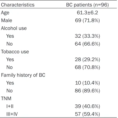
Table 1. Characteristics of bladder cancer patients
Characteristics BC patients (n=96)
Age 61.3±6.2
Male 69 (71.8%)
Alcohol use
Yes 32 (33.3%)
No 64 (66.6%)
Tobacco use
Yes 28 (29.2%)
No 68 (70.8%)
Family history of BC
Yes 10 (10.4%)
No 86 (89.6%)
TNM
I+II 39 (40.6%)
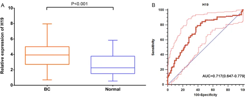
Figure 1. Diagnostic performance of lncRNA H19 in human bladder cancer (BC) tissue. A: Relative expression levels of lncRNA H19 in BC tissue and normal tissues. B: ROC curve analysis of lncRNA H19 in BC tissue and normal tis-sues.
Figure 2. Diagnostic performance of lncRNA UCA1 in human bladder cancer tissue. A: Relative expression levels of lncRNA UCA1 in BC tissue and normal tissues. B: ROC curve analysis of lncRNA UCA1 in BC tissue and normal tissues.
Statistical analysis
Continuous variables are expressed as mean ± standard deviation and categorical variables are expressed as absolute relative frequencies. We compared two groups using the t test for continuous variables and χ2 test for categorical
variables. Receiver operating characteristic (ROC) curves were constructed and area under the ROC curve (AUC) was generated to assess the diagnostic values of the candidate RNAs. We used the Logistic regression model to do the multivariable analysis, and calculate a new LncRNA-based score using the three selected LncRNAs. P value <0.05 was considered as sta-tistically significant. All statistical analysis were
performed by R software version 3.0.1 (MathSoft Inc., USA), and the graphs were obtained from GraphPad Prism 5.0 (GraphPad Software Inc., CA).
Results
Clinical characteristics of study population
LncRNAs level in bladder cancer tissues
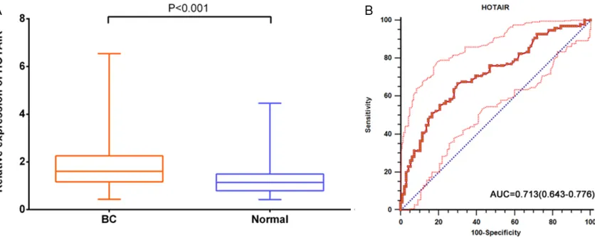
The expression level of three selected lncRNAs was detected by quantitative real-time PCR in BC tissues and pair-matched adjacent normal tissues form 96 BC patients. As Figures 1A, 2A, 3A shows, the relative expression of H19, UCA1, and HOTAIR in BC tissues were 3.93 (2.75, 5.00), 6.83 (5.25, 8.94), 1.61 (1.17, 2.22), respectively. While the relative ex- pression in the normal tissues were lo- wer, with the media (quartile) of 2.26 (1.53, 3.75), 3.99 (2.46, 5.66), 1.14 (0.80, 1.49), respectively. Statistically significant differen- ce can be observed between BC tissues and pair-matched adjacent normal tissues (all P<0.001). Then we performed the Logistic regression model to calculate a new LncRNA-based score using the three selected LncRNAs: Lnc-Score =0.48*H19+0.49*UCA1+ 1.2*HOTAIR.
Diagnostic performance of LncRNAs in blad-der cancer detection
Receiver operating characteristic (ROC) curves were constructed and area under the ROC curve (AUC) was generated to assess the diag-nostic values of the three selected LncRNAs and the Lnc-Score. As Figures 1B, 2B, 3B shows, the AUC of H19, UCA1, and HOTA- IR were 0.717 (0.647-0.779), 0.787 (0.722-0.843), 0.713 (0.643-0.776), respectively. Table 2 shows the detail information about the ability of the three LncRNAs and Lnc-Score to diagnose the BC in patients. Among 3 selected
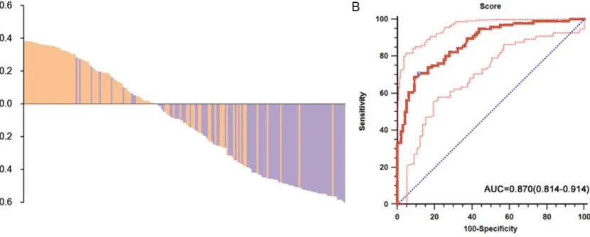
LncRNAs, UCA1 had the highest sensitivity of 80.2% while HOTAIR had the highest specificity of 69.9%. We also can observe that UCA1 had the highest +LR of 2.41 and the lowest -LR of 0.30. Figure 4A shows the distribution of risk for BC, which suggested that BC tissues had higher Lnc-Score than that of adjacent normal tissues. Compared with these 3 separate LncRNAs, Lnc-Score showed the highest AUC, with value of 0.870 (0.814-0.914). Also, the best diagnosability parameters were observed in Lnc-Score, with relative higher sensitivity of 70.8%, the highest specificity of 88.5%, highest +LR of 3.67 and lowest -LR of 0.22.
Discussion
[image:4.612.100.518.72.239.2]Early diagnosis and surveillance for metastasis of bladder cancer are critical issues. Dere- gulation of long non-coding RNAs (lncRNAs) has been implicated in urologic malignancies and represents potential markers. In this study, we detected the relative expression levels of lncRNAs H19, UCA1 and HOTAIR in tissues from patients with bladder cancer. Higher expres-sion of these three lncRNAs were observed in the BC tissues than those in normal tissues. Then we performed receiver operating charac-teristic (ROC) curves and area under the ROC curve (AUC) to assess the diagnostic values of the three lncRNAs. We can observe ideal diag-nosability of the three selected lncRNAs, with AUCs of 0.717, 0.787 and 0.713, respectively. Furthermore, we constructed a new score based on these lncRNAs, named lncRNA-score, which show greater diagnosability than 3
selected miRNAs alone. Collectively, our study provides evidences that serum level of lncRNAs H19, UCA1 and HOTAIR have great clinical value as promising biomarkers in BC.
[image:5.612.92.524.85.151.2] [image:5.612.102.517.197.364.2]Overexpression of non-coding RNAs are impli-cated in metastasis of human tumors, but most are shorter non-coding like microRNAs (miR-NAs) [9]. Recently, an accumulating body of evi-dence has have linked specific lncRNA gene mutations with cancer, either by overlap of sequencing libraries with previously an- notated GENCODE lncRNAs, or by de novo assembly of all available public datasets, rais-ing the possibility of lncRNA-based cancer diag-nostics and therapy [6, 10-13]. A study in- cluding more than 7,000 RNA-Seq libraries from 25 independent studies found that am- ong 91,013 expressed genes, over 68% (58,648) of genes were classified as lncRNAs, of which 79% (48,952) were previously unan-notated [12]. Sequencely, summary of lncRNAs with experimental data supporting the expres-sion and functions of promoting tumor inva- sion and metastasis was made in some studies [6, 14].
Recently, a number of lncRNAs proved to be able to modulate Urologic neoplasms. The lncRNA RCCRT1 is upregulated remarkably in renal cell carcinoma (RCC) compared with adja-cent noncancerous tissues, particularly in hi- gh-grade RCC tissues. Furthermore, siRNA-induced depletion of RCCRT1 expression suppressed migration and invasion in RCC cell lines [15]. Another study, which identified a novel lncRNA named SChLAP1, demonstrated that SChLAP1 expression increases with pros-tate cancer progression, and high SChLAP1 expression is associated with poor outcome after radical prostatectomy by both univariate and multivariate analysis [16].
In present study, we investigated three select-ed lncRNAs H19, UCA1 and HOTAIR in the blad-der cancer tissues and pair-matched adjacent normal tissues from 96 patients. H19, the old-est known lncRNA, was widely studied in can-cer biology even before lncRNAs had gained the attention of cancer researchers [6]. The oncogenic properties of H19 were strongly associated with antagonism of the tumor sup-pressor miRNA let-7 and forced EMT mediated Table 2. The receiver operating characteristic (ROC) analysis of lncRNA in human bladder cancer miRNAs AUC 95% CI P value Youden Cut-off Sensitivity Specificity +LR -LR H19 0.717 0.647-0.779 <0.001 0.395 2.65 77.8% 62.5% 2.06 0.37 UCA1 0.787 0.722-0.843 <0.001 0.469 4.94 80.2% 66.7% 2.41 0.30 HOTAIR 0.713 0.643-0.776 <0.001 0.365 1.34 66.7% 69.9% 2.21 0.48 Lnc-Score 0.870 0.814-0.914 <0.001 0.594 6.53 70.8% 88.5% 3.67 0.22 Note. AUC, area under the receiver operating characteristic curve; CI, confidence interval; +LR, positive likelihood ratio; -LR,
negative likelihood ratio; P-value, compared with AUC of 0.5. LncRNA-Score =0.48*H19+0.49*UCA1+1.2*HOTAIR.
by the non-histone chromosomal transcription-al regulator HMGA2 [17, 18]. Also, H19 expres-sion is directly induced by the v-myc avian myelocytomatosis viral oncogene homolog (c-MYC) and loss of the p53 tumor suppressor further supporting the importance of H19 as a potent oncogene [19, 20]. Urothelial carcino-ma-associated 1 (UCA1), another upregulated lncRNA in bladder cancer, is significantly corre-lated with greater tumor depth and apoptosis escape [21]. Overexpression of UCA1 enhances ERK1/2 MAPK and PI3-K/AKT kinase activity, causing increased expression of the coactiva-tor p300 and its coactivacoactiva-tor cAMP response element-binding protein (CREB), which pro-motes cell cycle progression, carcinogenesis, and cancer invasion [22, 23]. HOTAIR is a non-coding 2.2-kb RNA gene located downstream, acts as a scaffold for histone modification com-plexes [6]. In cancer cells, HOTAIR partners with Polycomb Repressive Complex 2 (PRC2, a his-tone methyltransferase) to induce genome-wide gene silencing and increased cancer inva-siveness and metastasis in a manner dependent on PRC2 [24, 25]. Additionally, HOTAIR has been shown to regulate several genes involved in epithelial-to-mesenchyme transition (EMT) including Snail family zinc fin-ger 1 (SNAI1), Poly r(C)-Binding Protein 1 (PCBP 1), Junctional adhesion molecule 2 (JAM2) and ABL proto-oncogene 2 (ABL2) [25-27]. These findings indicate that lincRNAs have active roles in modulating the bladder cancer and may be important targets for cancer diagnosis and therapy.
Conclusion
In conclusion, above all, our results extend the findings of previous studies about lncRNAs H19, UCA1 and HOTAIR in bladder cancer patients. Our data provide the 3 selected lncRNAs were significantly higher in bladder cancer tissues compared to normal tissues, and demonstrated the ideal diagnostic value to distinguish cancer tissues from normal tissues. However, whether this correlation is exactly pro-portional requires carefully scrutiny and study on a larger sample is needed to confirm this results.
Acknowledgements
This study was supported by the National Natural Science Foundation of China (8720321).
Disclosure of conflict of interest
None.
Address correspondence to: Dr. Wei Wang, Department of Drug Control, Criminal Investigation Police University of China, No. 83, The Tawan Street, Huanggu District, Shenyang 110035, P. R. China. Fax: +86-024-86982468; E-mail: zhigangji352@ sina.com
References
[1] Witjes JA, Comperat E, Cowan NC, De Santis M, Gakis G, Lebret T, Ribal MJ, Van der Heijden AG, Sherif A; European Association of Urology. EAU guidelines on muscle-invasive and meta-static bladder cancer: summary of the 2013 guidelines. Eur Urol 2014; 65: 778-792. [2] Yang L, Parkin DM, Li LD, Chen YD and Bray F.
Estimation and projection of the national
pro-file of cancer mortality in China: 1991-2005.
Br J Cancer 2004; 90: 2157-2166.
[3] Kim WJ and Bae SC. Molecular biomarkers in urothelial bladder cancer. Cancer Sci 2008; 99: 646-652.
[4] Schmitz SU, Grote P and Herrmann BG. Mech-anisms of long noncoding RNA function in de-velopment and disease. Cell Mol Life Sci 2016; 73: 2491-2509.
[5] Lorenzen JM and Thum T. Long noncoding RNAs in kidney and cardiovascular diseases. Nat Rev Nephrol 2016; 12: 360-373.
[6] Jiang C, Li X, Zhao H and Liu H. Long non-cod-ing RNAs: potential new biomarkers for predict-ing tumor invasion and metastasis. Mol Can-cer 2016; 15: 62.
[7] Evans JR, Feng FY and Chinnaiyan AM. The bright side of dark matter: lncRNAs in cancer. J Clin Invest 2016; 126: 2775-2782.
[8] Schmitt AM and Chang HY. Long noncoding RNAs in cancer pathways. Cancer Cell 2016; 29: 452-463.
[9] Bullock MD, Sayan AE, Packham GK and Mirnezami AH. MicroRNAs: critical regulators of epithelial to mesenchymal (EMT) and mes-enchymal to epithelial transition (MET) in can-cer progression. Biol Cell 2012; 104: 3-12. [10] Beermann J, Piccoli MT, Viereck J and Thum T.
Non-coding RNAs in development and disease: background, mechanisms, and therapeutic ap-proaches. Physiol Rev 2016; 96: 1297-1325. [11] Silva A, Bullock M and Calin G. The clinical
rel-evance of long non-coding RNAs in cancer. Cancers (Basel) 2015; 7: 2169-2182.
Iyer HK and Chinnaiyan AM. The landscape of long noncoding RNAs in the human transcrip-tome. Nat Genet 2015; 47: 199-208.
[13] Yan X, Hu Z, Feng Y, Hu X, Yuan J, Zhao SD, Zhang Y, Yang L, Shan W, He Q, Fan L, Kan-dalaft LE, Tanyi JL, Li C, Yuan CX, Zhang D, Yuan H, Hua K, Lu Y, Katsaros D, Huang Q, Montone K, Fan Y, Coukos G, Boyd J, Sood AK, Rebbeck T, Mills GB, Dang CV and Zhang L. Comprehensive genomic characterization of long non-coding RNAs across human cancers. Cancer Cell 2015; 28: 529-540.
[14] Bartonicek N, Maag JL and Dinger ME. Long noncoding RNAs in cancer: mechanisms of ac-tion and technological advancements. Mol Cancer 2016; 15: 43.
[15] Song S, Wu Z, Wang C, Liu B, Ye X, Chen J, Yang Q, Ye H, Xu B and Wang L. RCCRT1 is correlat-ed with prognosis and promotes cell migration and invasion in renal cell carcinoma. Urology 2014; 84: 730, e731-737.
[16] Mehra R, Shi Y, Udager AM, Prensner JR, Sahu A, Iyer MK, Siddiqui J, Cao X, Wei J, Jiang H, Feng FY and Chinnaiyan AM. A novel RNA in situ hybridization assay for the long noncoding RNA SChLAP1 predicts poor clinical outcome after radical prostatectomy in clinically local-ized prostate cancer. Neoplasia 2014; 16: 1121-1127.
[17] Ma C, Nong K, Zhu H, Wang W, Huang X, Yuan Z and Ai K. H19 promotes pancreatic cancer metastasis by derepressing let-7’s suppres-sion on its target HMGA2-mediated EMT. Tu-mour Biol 2014; 35: 9163-9169.
[18] Yan L, Zhou J, Gao Y, Ghazal S, Lu L, Bellone S, Yang Y, Liu N, Zhao X, Santin AD, Taylor H and Huang Y. Regulation of tumor cell migration and invasion by the H19/let-7 axis is antago-nized by metformin-induced DNA methylation. Oncogene 2015; 34: 3076-3084.
[19] Barsyte-Lovejoy D, Lau SK, Boutros PC, Khos-ravi F, Jurisica I, Andrulis IL, Tsao MS and Penn LZ. The c-Myc oncogene directly induces the
H19 noncoding RNA by allele-specific binding
to potentiate tumorigenesis. Cancer Res 2006; 66: 5330-5337.
[20] Dugimont T, Montpellier C, Adriaenssens E, Lottin S, Dumont L, Iotsova V, Lagrou C, Stehe-lin D, Coll J and Curgy JJ. The H19 TATA-less
promoter is efficiently repressed by wild-type
tumor suppressor gene product p53. Onco-gene 1998; 16: 2395-2401.
[21] Xue M, Li X, Li Z and Chen W. Urothelial carci-noma associated 1 is a hypoxia-inducible fac-tor-1alpha-targeted long noncoding RNA that enhances hypoxic bladder cancer cell prolifer-ation, migrprolifer-ation, and invasion. Tumour Biol 2014; 35: 6901-6912.
[22] Wang Y, Chen W, Yang C, Wu W, Wu S, Qin X and Li X. Long non-coding RNA UCA1a(CUDR) promotes proliferation and tumorigenesis of bladder cancer. Int J Oncol 2012; 41: 276-284. [23] Yang C, Li X, Wang Y, Zhao L and Chen W. Long
non-coding RNA UCA1 regulated cell cycle dis-tribution via CREB through PI3-K dependent pathway in bladder carcinoma cells. Gene 2012; 496: 8-16.
[24] Kogo R, Shimamura T, Mimori K, Kawahara K, Imoto S, Sudo T, Tanaka F, Shibata K, Suzuki A, Komune S, Miyano S and Mori M. Long non-coding RNA HOTAIR regulates
polycomb-de-pendent chromatin modification and is associ -ated with poor prognosis in colorectal cancers. Cancer Res 2011; 71: 6320-6326.
[25] Gupta RA, Shah N, Wang KC, Kim J, Horlings HM, Wong DJ, Tsai MC, Hung T, Argani P, Rinn JL, Wang Y, Brzoska P, Kong B, Li R, West RB, van de Vijver MJ, Sukumar S and Chang HY. Long non-coding RNA HOTAIR reprograms chromatin state to promote cancer metastasis. Nature 2010; 464: 1071-1076.
[26] Wang J, Zhou Y, Lu J, Sun Y, Xiao H, Liu M and Tian L. Combined detection of serum exosomal miR-21 and HOTAIR as diagnostic and prog-nostic biomarkers for laryngeal squamous cell carcinoma. Med Oncol 2014; 31: 148. [27] Zhang ZZ, Shen ZY, Shen YY, Zhao EH, Wang M,