NAR Breakthrough Article
Prespacer processing and specific integration in a
Type I-A CRISPR system
Clare Rollie, Shirley Graham, Christophe Rouillon and Malcolm F. White
*Biomedical Sciences Research Complex, School of Biology, University of St Andrews, North Haugh, St Andrews, Fife KY16 9ST, UK
Received October 31, 2017; Revised November 22, 2017; Editorial Decision November 27, 2017; Accepted November 29, 2017
ABSTRACT
The CRISPR–Cas system for prokaryotic adaptive immunity provides RNA-mediated protection from viruses and mobile genetic elements. Adaptation is dependent on the Cas1 and Cas2 proteins along with varying accessory proteins. Here we analyse the pro-cess in Sulfolobus solfataricus, showing that while Cas1 and Cas2 catalyze spacer integration in vitro, host factors are required for specificity. Specific in-tegration also requires at least 400 bp of the leader sequence, and is dependent on the presence of hy-drolysable ATP, suggestive of an active process that may involve DNA remodelling. Specific spacer inte-gration is associated with processing of prespacer 3 ends in a PAM-dependent manner. This is reflected in PAM-dependent processing of prespacer 3 ends in vitroin the presence of cell lysate or the Cas4 nucle-ase, in a reaction consistent with PAM-directed bind-ing and protection of prespacer DNA. These results highlight the diverse interplay between CRISPR–Cas elements and host proteins across CRISPR types.
INTRODUCTION
CRISPR–Cas systems are present in around half of bacte-rial and 90% of archaeal genomes sequenced to date and form an adaptive immune system important in defence against invasion by foreign nucleic acids. Key to CRISPR– Cas immunity is the ability to adapt to new threats by in-corporating short segments of foreign DNA, called spacers, into the CRISPR array of the host. These spacers constitute immunological memories that are then used by CRISPR-associated (Cas) proteins to mount sequence-specific de-fence on subsequent infection. The process of acquiring new spacers is termed Adaptation and can be divided into two
main stages: firstly, the generation and capture of a pres-pacer by Cas1, Cas2 (and potentially other) proteins and secondly, the docking of this nucleoprotein complex at the leader:repeat site, leading to integration of the new spacer by transesterification. The integration process is completed by DNA polymerase and DNA ligase. The overall process has been reviewed recently (1–3) and a schematic represen-tation of the steps involved in adaprepresen-tation inSulfolobus sol-fataricusis shown in Figure1A.
The first stage in adaptation is the capture of a prespacer from foreign DNA. Prespacers have no identified conserved sequences, but are found next to a short PAM (protospacer adjacent motif), which is required to guide the Cas adapta-tion and later interference machinery. The addiadapta-tion of a new spacer requires the staggered nicking of the CRISPR locus at the 5ends of the first repeat and the co-ordinated joining of a prespacer to the repeat ends. The joining of both ends of the spacer to the host genome occurs by two ‘half-site’ reactions, one 3end of the incoming DNA will be joined to the 5end of the first repeat, proximal to the leader se-quence (site 1), and the second 3end of the prespacer will be joined to the leader-distal 5 end of the first repeat on the complementary strand (site 2). Both nicking and joining occur through a one-step transesterification reaction medi-ated by Cas1, in which the 3hydroxyl residues (3OH) of the incoming prespacer are used to attack the host locus (4). PAM sequences are crucial for prespacer selection and in-tegration in the correct orientation to license interference, with the prespacer end that was previously adjacent to the PAM always being inserted proximal to the leader sequence (5).
Structural studies of theEscherichia coliCas1–Cas2 com-plex in the presence and absence of bound DNA revealed that two Cas1 dimers are joined by a central Cas2 dimer (6–8). In the DNA:Cas1–Cas2 ternary structure, tyrosine residues from two Cas1 subunits were found to bracket a 23 bp duplex and act as wedges to splay the remaining 5 bp of
*To whom correspondence should be addressed. Tel: +44 1334 463432; Fax: +44 1334 462595; Email: [email protected]
Present address: Clare Rollie, Environment and Sustainability Institute, University of Exeter, Penryn Campus, Penryn, Cornwall TR10 9FE, UK.
C
The Author(s) 2017. Published by Oxford University Press on behalf of Nucleic Acids Research.
A
[image:2.612.133.460.70.441.2]B
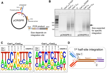
Figure 1. Adaptation inS. solfataricus. (A) Model for the integration of a new spacer by Cas1–Cas2 during adaptation (based on (1)). 1. A short segment of DNA containing a PAM sequence is captured and bound by a complex of Cas1 and Cas2 proteins. The ends of the captured prespacer may be splayed and trimmed by nucleases. 2. The prespacer-bound adaptation complex docks sequence-specifically at the leader-repeat junction of the host CRISPR array. The PAM may provide a polarity to the complex, as the PAM-proximal end of the prespacer must be integrated at the leader-distal end of repeat 1 (site 2) to allow transcription of crRNA in the correct orientation for interference. 3. During integration of the prespacer the 3ends may be trimmed. 4. A transesterification reaction mediated by Cas1 joins one 3hydroxyl of the incoming spacer to the leader-proximal 5end of 1st repeat (site 1). 5. A second transesterification joins the other end of the prespacer to the 5repeat end at site 2. 6. Gap filling and ligation. (B) Representation of the CRISPR–Cas system ofS. solfataricus. The Cas1ABand Cas2ABproteins are encoded by thesso1405andsso1404genes associated with CRISPR loci A and B. The
sso1450andsso1450agenes encoding Cas1CDand Cas2CDare associated with CRISPR loci C and D. The number of spacers contained in the CRISPR
arrays is indicated in subscript after the array name. Leader regions are shown in red and indicated by the letter ‘L’. There are three type I-A, one type III-B and one type III-D effector modules.
duplex DNA at either end into single strands. The single-stranded 3ends are bound tightly in an arginine-rich cleft of Cas1 and there is some evidence that they are cut five nucleotides from the end of the 23 bp duplex, at PAM se-quences (5-CTT-3) (8). This proposed cleavage would re-sult in the 3hydroxyl residues being positioned exactly in the metal-binding active site, poised to perform nucleophilic attack at the leader:repeat junction (7,8).
The insertion of new spacers is polarized and almost always occurs between leader and repeat 1 (9,10), which suggests that these elements contain important motifs that guide docking of the Cas1 and Cas2 proteins. In support of this hypothesis, the last 60 bp of the leader and the first re-peat inE. coliwere shown to be essential and sufficient for integration of new spacers (11).Escherichia coli Cas1 has
coliadaptation complexes have revealed the molecular de-tails of the integration event, highlighting the requirement for structural distortion of the target DNA, and explaining the importance of an upstream recognition motif, brought into contact with Cas1 due to IHF-mediated DNA binding, for the integration process (17).
Information on the mechanism of adaptation in organ-isms other thanE. coliis more patchy, but nucleic acid se-quences around the leader:repeat junction appear generally important (18–20). Recent structural studies of the Entero-coccus faecalistype II-A adaptation process have provided a molecular framework for each stage of the integration path-way, including ternary complex formation, integration at site 1 and subsequent DNA distortion leading to full inte-gration (21).
The work presented here focuses on the CRISPR–Cas system of S. solfataricus, which includes three different CRISPR–Cas types (type I-A, III-D and III-B), two dif-ferent repeat families (AB and CD) and adaptation cas-settes made up of genes coding for Cas1, Cas2, Csa1 and Cas4 proteins (Figure1B). Previous studies inS. solfatari-cushave suggested that the AB and CD loci may be active for adaptation under different conditions (22). Here, we re-constitute integrationin vitroand demonstrate that the in-trinsic specificity of Cas1 is augmented by host factors in an ATP-dependent reaction. Cas1 is shown to protect pres-pacer DNA ends from degradation by cellular nucleases or Cas4 in a manner influenced by PAM sequences.
MATERIALS AND METHODS
Cloning, expression and purification
The following proteins were expressed and purified as described previously: Cas1CD and Cas2CD (12); Cas2AB
(23); Sso7 (24); Alba1 (25); SSB (26). The CRISPR DNA repeat binding protein (Cbp1) was a kind gift from Dr Xu Peng, and was expressed and purified as described (27). The Cas1AB gene Sso1405 was
am-plified from S. solfataricus genome DNA by PCR using the following primer pair: (forward primer: 5 -GGCGCCATGGATAAGAAAATAGCGTTCG; reverse primer: 5-GGTTGGATCCTCACTTCGCTAGGTATGG) and cloned into expression plasmid pEHisTev using the introduced NcoI and BamHI sites, allowing expression with a cleavable N-terminal polyhistidine tag in E. coli (28). The Cas1AB protein was expressed and purified as
described previously for Cas1CD (12), with the addition
of a heparin-sepharose chromatography step following removal of the polyhistidine tag. Site directed mutagenesis to generate variants of Cas1AB (D234A variant), Cas1CD
(E142A variant) and Cas2AB (D10A variant) was carried
out using standard methodology and the sequences of the oligonucleotides used are available from the corresponding author on request. Sulfolobus solfataricusCas4 (Sso1391) was purified as described previously (29).
DNA substrate preparation
DNA oligonucleotides and double-stranded gBlocks were ordered from Integrated DNA Technologies (Coralville,
IA, USA). If required, oligonucleotides were 5-32
P-radiolabelled and gel purified as described previously (16). Double-stranded prespacer substrates were formed by heat-ing equimolar concentrations (20M) of complementary strands at 95◦C for 5 min and then slow cooling to room temperature overnight in a heating block. The assembled substrates were purified by native polyacrylamide (12%) gel electrophoresis with 1×Tris–borate–EDTA (TBE) buffer, followed by band excision, gel extraction, ethanol precipita-tion, as described previously (12). gBlocks were cloned into a pUC19 backbone according to manufacturer’s instruc-tions using EcoRI and BamHI restriction sites. All plas-mid constructs were verified by sequencing (GATC Biotech, Konstanz, Germany) and gBlock sequences are available from the corresponding author on request (pCRISPR A and derivatives, pCRISPR C, pLeadArepC)
Integration assay with radiolabelled prespacer
Cas1 and Cas2, both at 20 M, were incubated together at 55◦C for 30 min. 1l of this solution was then added to a reaction containing 1l 532P-radiolabelled DNA
sub-strates (2M final) for integration (∼1% is labelled), 1l (100 ng/l) plasmid DNA, 1l 10×integration buffer (200 mM Tris (pH 7.5), 100 mM NaCl), 1l MnCl2 (50 mM)
and 5l water making the total reaction volume up to 10
l. This reaction was then incubated at 55◦C for 30 min. Following the incubation, 1l of proteinase K (20 mg/ml) (ThermoFisher Scientific) was added and the digest was in-cubated at 37◦C for 1 h, before phenol extraction of the DNA. 10l of the aqueous phase containing the DNA was removed, mixed with 2l of 6×DNA loading dye and run on a 1% agarose gel, pre-stained with ethidium bromide, at 100 V for 1 h in 1×TBE buffer and photographed under UV light. The gel was dried for 4 h on a slab gel drier (Savant) and phosphorimaged. Plasmids were nicked with nickase Nt.BspQI (New England BioLabs) according to manufac-turer instructions and run on agarose gels alongside integra-tion assay products to act as a marker for the nicked form of the pCRISPR or pUC19 plasmids.
PCR amplification of integration sites
A 9 l reaction was prepared containing 200 ng of the pCRISPR A/pCRISPR C plasmids, 5 mM MnCl2, 1X
98◦C for 30 s, 55◦C for 30 s and 72◦C for 30 s, with a final extension for 2 min at 72◦C and an infinite hold step at 4◦C. The products of the PCR reaction were separated on a 1.5% agarose gel, which allowed rough localisation of the in-tegration sites. PCR products selected for sequencing were cleaned up using the Wizard SV Gel and PCR Clean-Up System (Promega). Products were then digested with 1 l NcoI and 1l XhoI FastDigest enzymes in a 20l reac-tion containing 1X FastDigest buffer at 37◦C for 1 h. 1
g of the pEHISTEV vector was also restricted using the same method with NcoI and XhoI to produce compati-ble ends for ligation of the insert. The digested inserts and plasmid were ligated and the ligation products were trans-formed into DH5␣E. colicells. Transformants were selected by overnight growth at 37◦C on LB agar plates containing 35g/ml kanamycin. Plasmids were extracted from posi-tive clones by Miniprep and sent for sequencing using the T7 primer (GATC Biotech). The sequences around the in-sertion site were used to make a sequence logo on the We-bLogo server (30).
Integration assays withS. solfataricuslysate
Integration assays coupled to PCR were modified by the addition of S. solfataricuslysate, Sso7, Alba or Cbp1 be-fore Cas1 and Cas2 proteins. The reaction mix was set up as above without the addition of Cas proteins or RNase-free water. 1l of purified host proteins (from stock concentra-tions of 12.5–100M) or increasing volumes ofS. solfatar-icus cell lysate (1–5l) (prepared as described previously (31)) were added to the reaction mix and the total volume was made up to 9l with RNase-free H2O before the
addi-tion of 2M Cas1 and Cas2. The reaction was completed and the products resolved as described above.
Preparation and size exclusion chromatography of cell lysate
3.5 g of S. solfataricus cell pellet was resuspended in 10 ml of lysis buffer (20 mM Tris (pH 7.5), 150 mM KCl, 1 EDTA-free mini protease inhibitor tablet) and sonicated for 6 × 30 s bursts at 10 m. The lysed cells were cen-trifuged at 35 000 rpm, 4◦C for 30 min using the Optima L-90 K Ultracentrifuge and 70Ti rotor (Beckman Coulter). The lysate was then decanted and filtered before being used in assays. Lysate was fractionated by size exclusion chro-matography and eluted in 2 ml fractions from a Superdex 200 prep grade column (GE Healthcare). Fractions were concentrated from 1.5 ml to 75 l and 3 l added to in-tegration assays. Inin-tegration assays with fractionated lysate were supplemented with ATP or an ATP analogue (see fig-ure legends for species and concentration) to retain specific integration.
Processing of prespacer substrates
Cas1 (and where indicated Cas2) proteins (final concentra-tion of 2M) were added to 20 nM prespacer substrate in a buffer containing 20 mM Tris (pH 7.5), 10 mM NaCl, 5 mM MnCl2(50 mM) and 5 mM ATP. 3lS. solfataricuscell
lysate or Cas4 (Sso1391) (1.5M) was then added and the
reaction incubated at 60◦C for 30 min before phenol extrac-tion of the products and separaextrac-tion on a 15% denaturing polyacrylamide gel and phosphorimaging.
RESULTS
Reconstitution of prespacer integration by S. solfataricus Cas1 and Cas2
To characterise the process of adaptation in theS. solfatar-icustype I-A system, an integration assay was developed with Cas1, Cas2 and prespacer DNA with a 5-32P radioac-tive label. These were incubated with two supercoiled plas-mid DNA species, pUC19 and pCRISPR, which is derived from pUC19 with an insert containing the CRISPR ar-ray leader, repeat and first spacer. The experiment was car-ried out separately with both sets of Cas1–Cas2 proteins (Cas1ABand Cas2ABor Cas1CDand Cas2CD) together with
the corresponding pCRISPR A or C plasmids. Wild-type Cas1 caused an increase in conversion of supercoiled (SC) to nicked (N) plasmid (Figure2, top panel). The position of the nicked form of the plasmid corresponded with the migration of the radiolabelled prespacer, suggestive of in-tegration (Figure2, bottom panel). Integration was clearly enhanced by the addition of the Cas2 protein. No integra-tion was mediated by active site variants of either Cas1CDor
Cas1AB. Both the pUC19 and the pCRISPR plasmids were
good substrates for integration, suggesting that the reaction was not specific for the leader:repeat junction.
Intrinsic specificity of Cas1 influences integration site choice
To assess where prespacers were being integrated into the plasmid DNA, a spacer integration (SPIN) assay was de-veloped by coupling a standard integration reaction to PCR amplification of the integration site (Figure3A). A forward primer complementary to one strand of the inserted pres-pacer with an internal NcoI site, and a reverse primer com-plementary to the pCRISPR plasmid with an internal XhoI site were used to amplify through the prespacer insertion sites in plasmid DNA. Integration at site 1 (the leader:repeat junction) produces a product of 323 bp for pCRISPR A and 341 bp for pCRISPR C. In the presence of active Cas1, a smear of PCR products was obtained (Figure3B). This is consistent with integration taking place at hundreds of sites at different distances from the reverse primer, leading to the amplification of a range of products of varying sizes.
Table 1. DNA substrates used in this study
Double-stranded prespacer Oligo components Sequence (5to 3)
3overhang 3-f TCGCCATGGTGAGCACAGAGGATAATGTAACACT
3-r TACATTATCCTCTGTGCTCACCATGGCGACGAGC
5overhang 5-f ACACTTCGCCATGGTGAGCACAGAGGATAATGTA
5-r CGAGCTACATTATCCTCTGTGCTCACCATGGCGA
Duplex Dup-f CGAGCTCGCCATGGTGAGCACAGAGGATAATGTAACACT
Dup-r AGTGTTACATTATCCTCTGTGCTCACCATGGCGAGCTCG
triplePAM prespacer triplePAM TCGCCATGGTGAGCACAGAGGATAATGTACGACGACGA
polyT TACATTATCCTCTGTGCTCACCATGGCGATTTTTTTTT
Primers used in SPIN assays Sequence
IntFor TCGCCATGGTGAGCACAGAGGATA
pUC19Rev1 AATTCTCGAGTTGGCCGATTCATTAATGC
pUC19Rev2 AATTCTCGAGGGATAACCGTATTACCGCC
IntRev TATCCTCTGTGCTCACCATGGCGA
Leader269 AATTCTCGAGGAGATAAAGAGAAAACCGG
specificity of Cas1CDdetermined using a disintegration
as-say (12) and with the nucleotide sequences present at site 1 and site 2 of thebona fideintegration site (Figure3D). The limited intrinsic specificity of Cas1, in the presence or ab-sence of Cas2, is clearly not sufficient to direct integration to the cognate leader:repeat site on its own, suggesting that other factors are requiredin vivo.
Archaeal chromatin proteins do not confer specific integra-tion
Integration host factor (IHF) was shown to be important in guiding specificity of theE. coliCas1–Cas2 complex to the leader-repeat junction by binding a consensus site in the leader and causing a sharp bend in this region (14,15). Given the low sequence specificity observedin vitrofor in-tegration by theS. solfataricusCas1–Cas2 proteins, we in-vestigated whether a similar host protein factor might be required for specific integration in this system. There is no IHF-type protein coded byS. solfataricus; however, the abundant DNA-binding proteins Alba1 (32) and Sso7 (33) are involved in DNA bending and compaction (34,35), and the archaeal SSB binds single-stranded DNA (36). Addi-tionally, the protein Cbp1 (CRISPR DNA repeat-binding protein) binds specifically to the CRISPR repeats inS. solfa-taricus,opening the DNA duplex around these sites (27,37). We hypothesized that one or more of these proteins could play a role analogous to IHF in S. solfataricus adapta-tion. Accordingly, we carried out SPIN assays in the pres-ence of increasing concentrations (0–10M) of these DNA-binding proteins (Figure4A). However, no specific integra-tion was observed in the presence of these proteins. In high concentrations of Alba1 a reduction in the smear caused by non-specific integration was observed, which may be due to this protein coating the plasmid DNA and blocking non-specific integration.
Host factors facilitate site-specific integration of prespacers
To test the possibility that unknown host factors are re-quired for specific integration by Cas1, SPIN assays were carried out with the addition of clearedS. solfataricuscell lysate to the integration reaction. As increasing volumes of lysate were added to the Cas1–2ABreactions, the smear of
non-specific products obtained following PCR amplifica-tion was reduced and a specific (323 bp) band appeared, consistent with integration specifically at the CRISPR A leader–repeat1 junction (Figure4B), an observation subse-quently confirmed by DNA sequencing. The absence of a specific integration product in the lysate-only condition is consistent with low expression levels of Cas1–2 in the ab-sence of infection (38). These results confirmed that a cel-lular factor, or factors, guides specific integration during adaptation by Cas1–2ABproteins inS. solfataricus. The
ad-dition ofS. solfataricuslysate did not confer the same speci-ficity to the integration reaction performed by Cas1–2CD
into the CRISPR C array, although a reduction in non-specific integration was observed (Figure4C). Furthermore, Cas1–2AB was specific for the AB locus and did not
inte-grate prespacers specifically at the CD locus (Figure4D). To probe this further, we designed a chimeric integration sub-strate by fusing the CRISPR A leader to the CRISPR C re-peat (pLeadArepC). Cas1–2ABintegrated prespacers
specif-ically into this chimera (Figure4E), suggesting that the dif-ferences in the leader regions, rather than repeats, are crucial for this specificity.
Prespacer structure influences integration
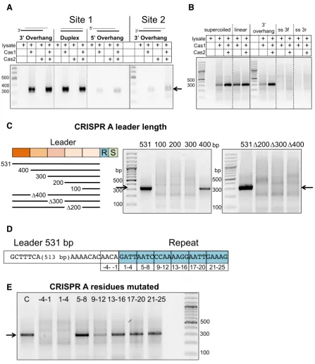
Prespacer end structures were varied from 5 nt single-stranded 3or 5overhangs to complete duplex ends. SPIN assays were carried out with Cas1–2AB in the presence of
cell lysate and primers used to amplify integrations at either site 1 (leader proximal), or site 2 (leader distal). Prespac-ers with 3single-stranded ends or blunt duplex ends were integrated efficiently at site 1 (Figure5A). However, those with 5single-stranded ends resulted in very low levels of integration. The right hand panel of the image shows the products at site 2 of CRISPR A following a SPIN assay in the presence of cell lysate. A weak amplification product is present at the correct size (338 bp) in the presence of Cas1– 2AB. We conclude that integrationin vitrois much less
Figure 2. Integration of spacers into supercoiled DNAin vitro.Cas1–2CDor Cas1–2AB(2M) were incubated with supercoiled plasmid (100 ng) and 32P-labelled prespacer (3 overhang (Table1), 2M) at 55◦C for 30 min in the presence of 5 mM MnCl
2. The products of the assay were separated on a
1% agarose gel containing ethidium bromide and visualised under UV light (top). The agarose gel was then dried and phosphorimaged (middle). The scan of the pre-stained EtBr agarose and the phosphorimage of the dried gel were combined to create a composite image (bottom), with the ethidium bromide signal shown in sepia. The first lane for each substrate is a control without protein and the last lane for the pCRISPR substrates (N) is the result of nicking the plasmid with the nickase Nt.BspQI. Reactions were also set up with the Cas1 proteins alone or Cas1 active site variants, E142A Cas1CDor D234A
Cas1AB.
of supercoiling is not a major factor for the type I-A sys-tem. Integration was very weak with single-stranded DNA prespacers (Figure5B), as observed recently for the type I-F system (16), consistent with the expected requirement for partially duplex DNA with two 3ends for full integration.
A long leader is required for specific integration
In the well characterised type I-E system, leaders are gener-ally less than 100 bp in length, and only 60 bp of the leader proximal to the first repeat is required for adaptation (11).
In contrastS. solfataricus, in common with many other ar-chaeal types, has much longer leader sequences (39). To as-sess the importance of the long (531 bp) CRISPR A leader sequence to integrationin vitro, truncated versions of the leader were designed (Figure5C) and used in SPIN assays with Cas1–2AB and cell lysate. Assays with the truncated
com-A
B
[image:7.612.89.538.70.376.2]C
D
Figure 3. Identifying sequence motifs at Cas1–Cas2 integration sites. (A) Schematic of the SPIN (spacer integration) assay used to amplify integration sites. An integration reaction into the pCRISPR C or A plasmids was performed with Cas1–2CDor Cas1–2ABand a prespacer with 5 nt 3single-stranded
ends (3overhang (Table1)) followed by spacer-specific PCR amplification (primers IntFor and pUC19Rev1). (B) Analysis of PCR amplification products by agarose gel electrophoresis. The control lanes (C) show amplification from integration reactions without Cas1–2, followed by reactions with Cas1 active site mutants, Cas1 alone and Cas1–2. (C) A sequence logo was generated on the WebLogo server following PCR amplification, cloning and sequencing (n=45) of the integration sites selected by Cas1–2CDor Cas1–2AB. The residues are numbered as in D. The sequence of thein vivointegration site 1
of CRISPR A or CRISPR C is shown above the WebLogo. (D) Schematic of the two half-site integrations carried out by Cas1–2 during adaptation at CRISPR loci. The first half-site reaction takes place at the leader-proximal 5end of repeat 1 (site 1) and the second at the leader-distal 5end of repeat 1 (site 2). Incoming prespacer ends are shown in blue.
pared to the full length 531 bp substrate. Deletion of 100 bp sections internal to the leader also abolished integration. These results indicate that the full length of the long leader sequences found in systems such as S. solfataricusare im-portant for specific integration, in marked contrast to the situation in types I-E, I-F and II-A.
An intact leader-repeat junction is required for integration
To assess the importance of the repeat sequence for pres-pacer integration, we generated variants of the repeat-proximal leader sequence and repeat1 sequence with blocks of four nucleotides mutated (A’s were changed to C’s, T’s to G’s, andvice versa) (Figure5D) for SPIN assays. When the last four nucleotides of the leader (–4 to –1) or first four nucleotides of the first repeat (1–4) were altered, integra-tion was abolished (Figure5E). This is perhaps unsurprising given the strong sequence selection already identified for the residues at positions –2 of the leader and +1 of the repeat imposed by Cas1 during both the disintegration and inte-gration reaction (12). Changing the sequence of the repeat between position 9 and 12 also reduced integration at site 1. This suggests that internal motifs in the repeat are
im-portant docking sites for the adaptation complex, similar to the repeat motifs suggested to be important for adapta-tion complex binding and accurate repeat duplicaadapta-tion inH. hispanica(19). Mutations at positions 5–8 and position 13 onwards had little effect on integration.
Specific integration requires ATP hydrolysis
A
B
D
E
[image:8.612.48.550.64.598.2]C
Figure 4. A host factor inS. solfataricuslysate leads to specific integration by Cas1–2AB. (A) Abundant DNA binding proteins ofS. solfataricuswere
added to SPIN assays to assess whether these factors influenced integration specificity. The leftmost lane contains a 100 bp DNA ladder, followed by lanes with the amplification products from integration assays containing: cell lysate only (c); Cas1–2AB(–); positive control for specific integration (c2);
Cas1–2ABand 1.25, 2.5, 5 or 10M of either Alba1 (sso0962), Sso7d (sso10610), SSB (sso2364) or Cbp1 (sso0454). (B) SPIN assay products following
integration into the pCRISPR A plasmid by Cas1–2ABwith or without the addition ofS. solfataricuscell lysate. Lanes are: DNA ladder, lysate-only control
(c), Cas1–2AB, and Cas1–2ABwith 1 to 5l ofS. solfataricuscell lysate added. A 323 bp product indicates correct integration at the leader-proximal 5
end of repeat 1 (site 1). (C) As in B, but showing assays with Cas1–2CDand pCRISPR C,where no specific products were observed. (D) SPIN assay with
Cas1–2ABand lysate, as indicated, integrating into pCRISPR A or pCRISPR C. (E) SPIN assay with Cas1–2ABorCDand lysate, as indicated, integrating
A
C
D
E
[image:9.612.89.537.66.583.2]B
Figure 5. DNA requirements for specific integration. (A) SPIN assays carried out with prespacers with a 29 bp duplex region and either blunt ends or 3 or 5single-stranded ends (5 nt) (Duplex, 3overhang, 5 overhang (Table1)), Cas1–2ABand lysate, as indicated, and pCRISPR A. The first three
panels show integration at site 1, the fourth shows integration at site 2 (PCR primers for site 2 amplification were IntFor and Leader269). (B) SPIN assays following integration of double (3 overhang) or single-stranded prespacers (3-f or 3-r) by Cas1–2ABinto pCRISPR A in supercoiled on linearised forms,
as indicated (see Table1for sequences). (C) SPIN assays following integration by Cas1–2ABof prespacer DNA into the pCRISPR A leader variants in
A
[image:10.612.124.479.87.517.2]B
C
Figure 6. ATP is needed for host-factor mediated specific integration. (A) Products of a SPIN assay carried out with either rawS. solfataricuslysate or fractions of lysate separated by size exclusion chromatography. A trace of the peak fractionsabsorption at 280 nm is shown in the top panel. The bottom panel shows the effect addition of column fractions to an integration reaction with Cas1–2AB. From left to right, the lanes contain a DNA ladder, PCR
from assay with raw lysate, followed by PCR from assay containing a concentrated sample of every third fraction across the elution peak. Assays with fractionated lysate were carried out in the presence of 5 mM ATP. The fractions that led to an integration product of the correct size are boxed. (B) Active fractions from A only facilitated specific integration by Cas1AB–Cas2ABin the presence of hydrolysable ATP. From left to right the lanes contain a 100
bp ladder, PCR amplifications from integration assays in the presence of raw lysate, a gel filtration elution pool in presence of increasing concentrations of ATP (0, 1.25, 2.5, 5 mM). The last lane is the result of PCR amplification from the products of an assay with GF elution and the non-hydrolysable ATP analogue ATP␥S (5 mM). (C) Comparison of integration sites chosen by Cas1–Cas2 in the presence of raw lysate or an ‘active’ pool of fractionation lysate supplemented with ATP (5 mM) and/or two non-hydrolysable ATP analogues (ATP␥S and AMP-PNP) (both at 15 mM). Expected product for integration at site 1 is 449 bp here as primers IntFor and pUC19Rev2 (see Table1) were used.
mechanism for specific integration inS. solfataricus. Unfor-tunately, the host factor could not be purified further as the activity was lost on subsequent chromatography steps.
Prespacers are frequently processed in a PAM-specific man-ner during integration
DNA sequencing revealed that new spacers were almost in-variably inserted correctly at site 1 during integration
whilst the complementary strand has a 9T overhang (Figure
7A). Before integrationin vivothese PAM sequences must be removed from the prespacer end inserted at site 2 in or-der to license effective interference (see Figure 1A). InE. colithe removal of the PAM is thought to be carried out by Cas1 after a prespacer substrate is bound (8).
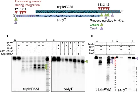
The sequencing analysis showed that the 3end contain-ing the triplePAM was trimmed to remove at least one and frequently two or more of the three PAMs before integra-tion (Figure7A, red triangles). In contrast, when integra-tions of the complementary poly-T 3end were sequenced, much less processing was observed. In 17 of the 25 integra-tions sequenced no processing of the poly-T 3end had oc-curred and in the remaining sequences only 2-4 nts had been trimmed from the end before integration (Figure7A).
To investigate this further, the same prespacer substrates were run on denaturing polyacrylamide gels following incu-bation with Cas1, Cas2 andS. solfataricuslysate in the same conditions used in the integration assays. Prespacers con-taining the triplePAM were almost completely degraded in the presence of cell lysate (Figure7B, middle panel). How-ever, when both Cas1 and Cas2 were present in the reac-tion, two products several nucleotides shorter than the full-length prespacer predominated. Both Cas1ABand Cas2AB
together were required for the generation of these products, but their production did not require the active site of either protein, as the inactive mutants D234A Cas1AB or D10A
Cas2AB still led to the appearance of the same processed
products. In contrast, the complementary strand contain-ing a poly-T 3end was not processed in the same way in the presence of Cas1–2AB(Figure7B).In the presence of lysate
this strand was completely degraded, and addition of Cas1– 2ABresulted in protection of the full-length strand, with no
partly truncated products observed. These data are consis-tent with Cas1–2AB mediated, PAM-specific processing of
prespacers by cellular nucleases. This pattern of prespacer processing would result in integrated spacers with a mean size around 39 bp, in good agreement with that observed in practice (10,41)
Type I-A systems typically include a Cas4 gene as part of the adaptation module (42). The Cas4 enzyme associated with Cas1–2AB, encoded bysso1391, is a nuclease with both
bi-directional exonuclease and Mn-dependent endonucle-olyic activities (43) and is therefore a candidate for the nu-clease activity detected in the cell lysate in these experi-ments. We therefore tested the effect of Cas4 (Sso1391) in our prespacer processing assays (Figure7C). Just as for cell lysate, we observed a PAM-dependent processing of pres-pacers, with Cas4-mediated DNA cleavage at the PAM site (purple arrows). In marked contrast, no corresponding pro-cessing was observed in the polyT strand of the prespacer.
Together, these results indicate that the shortening of the prespacer 3end results from processing by a nuclease and is halted when a bound Cas1–2ABcomplex is encountered.
The presence of PAM sequences seems to direct the posi-tioning of the Cas1–2 proteins, leading to the removal of at least one, and frequently two of the PAM sequences, while poly-T ends were fully protected by Cas1–2AB. The data
ob-tained from sequencing integration sites agrees well with the processing we observed from denaturing gel electrophoresis, as the triplePAM was processed to remove PAM residues
before integration, while the poly-T strand was often in-serted without processing (Figure7A).
DISCUSSION
The CRISPR–Cas system of S. solfataricusis one of the most complex studied to date, with multiple CRISPR re-peats and loci, adaptation modules and effector complexes (44). Here, we have focussed on the biochemistry of Adap-tation, and specifically the prespacer processing and in-tegration processes. This work has revealed a number of commonalities with other adaptation types: in particu-lar, the requirement for key sequence motifs in the re-peat and leader:rere-peat junction, the importance of PAMs and the preference for partially duplex prespacers with 3 -overhangs. However, in several respects adaptation in the type I-A systems appears quite fundamentally different from the well-studied I-E, I-F and II-A systems.
Firstly, there is a clear requirement for the full 531 bp length of the leader sequence for specific integrationin vitro. Previously, a naturally occurring deletion of about 20 bp around position -50 in a CD-family leader (locus E) asso-ciated with defective adaptation inS. solfataricus was de-scribed (39). This CRISPR locus is very short, and new spacers have not been added since the divergence of theS. solfataricusP1 and P2 strains (45): observations consistent with the loss of leader sequence essential for adaptation. Extensive deletion analysis has revealed that each 100-bp section of the AB-family leader is important, with only the region beyond 400 bp non-essential. This is markedly dif-ferent from the situation in I-E and II-A types, where short leaders are the norm. In type II-A systems, integration is ob-served with only∼10 bp of leader sequence (21). InE. coli adaptation, extreme bending of the leader over a very short DNA length (∼60 bp) is accomplished by IHF binding, al-lowing distal regions of the leader to contact Cas1 bound at the leader:repeat junction to ensure a productive integration event (17). The abundant chromatin proteins inS. solfatari-cus, Alba1 and Sso7, do not fulfil the same function as IHF in vitro, as they are capable of only limited amounts of DNA bending.
A
[image:12.612.62.536.83.400.2]B
C
Figure 7. Prespacer processing is influenced by PAM sequences. (A) A prespacer with 3 PAM complementary sequences (5-CGA-3) in the 3 single-stranded region of one strand (triplePAM) and a poly-T at the other 3end (polyT) (triplePAM prespacer (Table1) was integrated into pCRISPR A in a SPIN assay containingS. solfataricuscell lysate. The products of integration of either strand at site 1 were cloned and sequenced to identify processing events at the 3end of the prespacers. All processing events occurred in the 3overhangs and are indicated by red arrows, the number above indicates how many integration events were processed at each site. Green arrows indicate processing sites following incubation with Cas1–2ABand cell lysate (see B), and
purple arrows indicate processing in the presence of Cas1–2ABand Cas4 (Sso1391) (see C). (B) The prespacer used in A was also incubated with Cas1–2AB,
5 mM MnCl2andS. solfataricuslysate at 60◦C and products run on a 15% denaturing urea–TBE polyacrylamide gel. The left hand panel shows the result
of labeling the triplePAM strand and the right the result of labeling the polyT strand. The first lane is a control with only the labeled substrate loaded, followed by the products of incubation with Cas1–2ABor Cas proteins in combination withS. solfataricus(Sso) lysate. Inactive mutant D234A Cas1 or
D10A Cas2 were also included in incubations as indicated. An A+G ladder (L) was also loaded to map the products of triplePAM processing. Green arrows indicate the major processing products. (C) The same prespacer was assayed with Cas1–2ABand cell lysate or Cas4 (Sso1391) to compare processing. The
first lane is a control without protein (C) and an A+G ladder (L) was also loaded for each labelled species. Purple arrows indicate the major processing products.
The pathway of prespacer processing and capture is much less well defined than the integration process that follows. The limited evidence available on the final stages of pacer processing points to a role for Cas1 in trimming pres-pacers to generate short 3-overhangs suitable for integra-tion. This presumed nuclease activity of Cas1 is consistent with the activity of other integrases (50)––it is essentially the same chemistry as the transesterification reaction catal-ysed during integration and can take place in the same ac-tive site. It also provides a neat explanation for the detection and removal of PAM sequences, but direct observation of this activity is difficult in studies linking processing to inte-gration, as the Cas1 active site performs both roles. There is one report of PAM-directed nuclease activity byE. coli Cas1in vitro(8), and recent studies of the type I-F system are consistent with a role for Cas1 (and not Cas3) in PAM-dependent processing of prespacer 3ends (16). In the type
sequences are largely untrimmed. Together, these observa-tions are consistent with PAM-directed DNA binding of prespacers by Cas1–2 leading to protection of a spacer-sized DNA fragment adjacent to a PAM by a combination of se-quence specific and ruler mediated DNA binding. The com-plexes may be trimmed by Cas4, potentially in combination with other host nucleases.In vivo, final prespacer processing could take place once the Cas1–2-prespacer complex has docked to a target DNA site, ensuring correct orientation with respect to the PAM site.
In conclusion, our study highlights the diversity in CRISPR Adaptation mechanisms across the prokaryotic domains of life. Specific integration in a type I-A system is shown to be an ATP-dependent process requiring long leaders, pointing to a possible role for active DNA remod-elling. The capture and processing of prespacers, leading to integration, is one of the least understood elements of the CRISPR–Cas system. Here, we have demonstrated that the presence of a PAM sequence is a key determinant in pres-pacer processing, observed both fromin vitronuclease as-says and sequencing of integration products. The nuclease activity of Cas1 is not required for this processing, but the Cas4 nuclease has been shown to possess the relevant activ-ity, pointing to a mechanism involving PAM-directed pres-pacer footprinting by Cas1–2 coupled with Cas4 dependent DNA cleavage.
ACKNOWLEDGEMENTS
Thanks to Sabine Gr ¨uschow for critical reading and helpful discussions.
FUNDING
Biotechnology and Biological Sciences Research Coun-cil [BB/M021017/1 to M.F.W.]. Funding for open access charge: Research Councils UK.
Conflict of interest statement.None declared.
REFERENCES
1. Jackson,S.A., McKenzie,R.E., Fagerlund,R.D., Kieper,S.N., Fineran,P.C. and Brouns,S.J. (2017) CRISPR–Cas: adapting to change.Science,356, eaal5056.
2. Sternberg,S.H., Richter,H., Charpentier,E. and Qimron,U. (2016) Adaptation in CRISPR–Cas systems.Mol. Cell,61, 797–808. 3. Amitai,G. and Sorek,R. (2016) CRISPR–Cas adaptation: insights
into the mechanism of action.Nat. Rev.,14, 67–76.
4. Arslan,Z., Hermanns,V., Wurm,R., Wagner,R. and Pul,U. (2014) Detection and characterization of spacer integration intermediates in type I-E CRISPR–Cas system.Nucleic Acids Res.42, 7884–7893. 5. Mojica,F.J., Diez-Villasenor,C., Garcia-Martinez,J. and
Almendros,C. (2009) Short motif sequences determine the targets of the prokaryotic CRISPR defence system.Microbiology,155, 733–740. 6. Nunez,J.K., Kranzusch,P.J., Noeske,J., Wright,A.V., Davies,C.W. and
Doudna,J.A. (2014) Cas1–Cas2 complex formation mediates spacer acquisition during CRISPR–Cas adaptive immunity.Nat. Struct. Mol. Biol.,21, 528–534.
7. Nunez,J.K., Harrington,L.B., Kranzusch,P.J., Engelman,A.N. and Doudna,J.A. (2015) Foreign DNA capture during CRISPR–Cas adaptive immunity.Nature,527, 535–538.
8. Wang,J., Li,J., Zhao,H., Sheng,G., Wang,M., Yin,M. and Wang,Y. (2015) Structural and Mechanistic Basis of PAM-Dependent Spacer Acquisition in CRISPR–Cas Systems.Cell,163, 840–853.
9. Pourcel,C., Salvignol,G. and Vergnaud,G. (2005) CRISPR elements in Yersinia pestis acquire new repeats by preferential uptake of
bacteriophage DNA, and provide additional tools for evolutionary studies.Microbiology,151, 653–663.
10. Lillestol,R.K., Redder,P., Garrett,R.A. and Brugger,K. (2006) A putative viral defence mechanism in archaeal cells.Archaea (Vancouver, B.C.),2, 59–72.
11. Yosef,I., Goren,M.G. and Qimron,U. (2012) Proteins and DNA elements essential for the CRISPR adaptation process in Escherichia coli.Nucleic Acids Res.,40, 5569–5576.
12. Rollie,C., Schneider,S., Brinkmann,A.S., Bolt,E.L. and White,M.F. (2015) Intrinsic sequence specificity of the Cas1 integrase directs new spacer acquisition.eLife,4. e08716.
13. Nunez,J.K., Lee,A.S., Engelman,A. and Doudna,J.A. (2015) Integrase-mediated spacer acquisition during CRISPR–Cas adaptive immunity.Nature,519, 193–198.
14. Nunez,J.K., Bai,L., Harrington,L.B., Hinder,T.L. and Doudna,J.A. (2016) CRISPR immunological memory requires a host factor for specificity.Mol. Cell,62, 824–833.
15. Yoganand,K.N., Sivathanu,R., Nimkar,S. and Anand,B. (2017) Asymmetric positioning of Cas1–2 complex and Integration Host Factor induced DNA bending guide the unidirectional homing of protospacer in CRISPR–Cas type I-E system.Nucleic Acids Res.,45, 367–381.
16. Fagerlund,R.D., Wilkinson,M.E., Klykov,O., Barendregt,A., Pearce,F.G., Kieper,S.N., Maxwell,H.W.R., Capolupo,A., Heck,A.J.R., Krause,K.L.et al.(2017) Spacer capture and
integration by a type I-F Cas1–Cas2-3 CRISPR adaptation complex.
Proc. Natl. Acad. Sci. U.S.A.,114, E5122–E5128.
17. Wright,A.V., Liu,J.J., Knott,G.J., Doxzen,K.W., Nogales,E. and Doudna,J.A. (2017) Structures of the CRISPR genome integration complex.Science357, 1113–1118.
18. Wei,Y., Terns,R.M. and Terns,M.P. (2015) Cas9 function and host genome sampling in Type II-A CRISPR–Cas adaptation.Genes Dev.,
29, 356–361.
19. Wang,R., Li,M., Gong,L., Hu,S. and Xiang,H. (2016) DNA motifs determining the accuracy of repeat duplication during CRISPR adaptation in Haloarcula hispanica.Nucleic Acids Res.,44, 4266–4277.
20. Wright,A.V. and Doudna,J.A. (2016) Protecting genome integrity during CRISPR immune adaptation.Nat. Struct. Mol. Biol.,23, 876–883.
21. Xiao,Y., Ng,S., Nam,K.H. and Ke,A. (2017) How type II CRISPR–Cas establish immunity through Cas1–Cas2-mediated spacer integration.Nature550, 137–141.
22. Erdmann,S. and Garrett,R.A. (2012) Selective and hyperactive uptake of foreign DNA by adaptive immune systems of an archaeon via two distinct mechanisms.Mol. Microbiol.,85, 1044–1056. 23. Oke,M., Carter,L.G., Johnson,K.A., Liu,H., McMahon,S.A.,
Yan,X., Kerou,M., Weikart,N.D., Kadi,N., Sheikh,M.A.et al.(2010) The Scottish Structural Proteomics Facility: targets, methods and outputs.J. Struct. Funct. Genomics,11, 167–180.
24. Kvaratskhelia,M., Wardleworth,B.N., Bond,C.S., Fogg,J.M., Lilley,D.M. and White,M.F. (2002) Holliday junction resolution is modulated by archaeal chromatin componentsin vitro.J. Biol. Chem.,
277, 2992–2996.
25. Wardleworth,B.N., Russell,R.J., Bell,S.D., Taylor,G.L. and White,M.F. (2002) Structure of Alba: an archaeal chromatin protein modulated by acetylation.EMBO J.,21, 4654–4662.
26. Wadsworth,R.I. and White,M.F. (2001) Identification and properties of the crenarchaeal single-stranded DNA binding protein from Sulfolobus solfataricus.Nucleic Acids Res.,29, 914–920.
27. Deng,L., Kenchappa,C.S., Peng,X., She,Q. and Garrett,R.A. (2012) Modulation of CRISPR locus transcription by the repeat-binding protein Cbp1 in Sulfolobus.Nucl Acids Res.,40, 2470–2480. 28. Liu,H. and Naismith,J.H. (2009) A simple and efficient expression
and purification system using two newly constructed vectors.Protein Express. Purif.,63, 102–111.
29. Zhang,J., Kasciukovic,T. and White,M.F. (2012) The CRISPR associated protein Cas4 Is a 5’ to 3’ DNA exonuclease with an iron-sulfur cluster.PLoS One,7, e47232.
30. Crooks,G.E., Hon,G., Chandonia,J.M. and Brenner,S.E. (2004) WebLogo: a sequence logo generator.Genome Res.,14, 1188–1190. 31. G ¨otz,D., Paytubi,S., Munro,S., Lundgren,M., Bernander,R. and
32. Bell,S.D., Botting,C.H., Wardleworth,B.N., Jackson,S.P. and White,M.F. (2002) The interaction of Alba, a conserved archaeal chromatin protein, with Sir2 and its regulation by acetylation.
Science,296, 148–151.
33. Baumann,H., Knapp,S., Lundback,T., Ladenstein,R. and Hard,T. (1994) Solution structure and DNA-binding properties of a thermostable protein from the archaeon Sulfolobus solfataricus.Nat. Struct. Biol.,1, 808–819.
34. Driessen,R.P., Meng,H., Suresh,G., Shahapure,R., Lanzani,G., Priyakumar,U.D., White,M.F., Schiessel,H., van Noort,J. and Dame,R.T. (2013) Crenarchaeal chromatin proteins Cren7 and Sul7 compact DNA by inducing rigid bends.Nucleic Acids Res.,41, 196–205.
35. Laurens,N., Driessen,R.P., Heller,I., Vorselen,D., Noom,M.C., Hol,F.J., White,M.F., Dame,R.T. and Wuite,G.J. (2012) Alba shapes the archaeal genome using a delicate balance of bridging and stiffening the DNA.Nat. Commun.,3, 1328.
36. Morten,M.J., Peregrina,J.R., Figueira-Gonzalez,M., Ackermann,K., Bode,B.E., White,M.F. and Penedo,J.C. (2015) Binding dynamics of a monomeric SSB protein to DNA: a single-molecule multi-process approach.Nucleic Acids Res.,43, 10907–10924.
37. Peng,X., Brugger,K., Shen,B., Chen,L., She,Q. and Garrett,R.A. (2003) Genus-specific protein binding to the large clusters of DNA repeats (short regularly spaced repeats) present in Sulfolobus genomes.J. Bacteriol.,185, 2410–2417.
38. Quax,T.E., Voet,M., Sismeiro,O., Dillies,M.A., Jagla,B., Coppee,J.Y., Sezonov,G., Forterre,P., van der Oost,J., Lavigne,R.et al.(2013) Massive activation of archaeal defense genes during viral infection.J. Virol.,87, 8419–8428.
39. Alkhnbashi,O.S., Shah,S.A., Garrett,R.A., Saunders,S.J., Costa,F. and Backofen,R. (2016) Characterizing leader sequences of CRISPR loci.Bioinformatics,32, i576–i585.
40. Erdmann,S., Shah,S.A. and Garrett,R.A. (2013) SMV1 virus-induced CRISPR spacer acquisition from the conjugative plasmid pMGB1 in Sulfolobus solfataricus P2.Biochem. Soc. Trans.,41, 1449–1458. 41. Lintner,N.G., Kerou,M., Brumfield,S.K., Graham,S., Liu,H.,
Naismith,J.H., Sdano,M., Peng,N., She,Q., Copie,V.et al.(2011) Structural and functional characterization of an archaeal clustered regularly interspaced short palindromic repeat (CRISPR)-associated complex for antiviral defense (CASCADE).J. Biol. Chem.,286, 21643–21656.
42. Makarova,K.S., Wolf,Y.I., Alkhnbashi,O.S., Costa,F., Shah,S.A., Saunders,S.J., Barrangou,R., Brouns,S.J., Charpentier,E., Haft,D.H.
et al.(2015) An updated evolutionary classification of CRISPR–Cas systems.Nat. Rev. Microbiol.,13, 722–736.
43. Lemak,S., Beloglazova,N., Nocek,B., Skarina,T., Flick,R., Brown,G., Popovic,A., Joachimiak,A., Savchenko,A. and Yakunin,A.F. (2013) Toroidal structure and DNA cleavage by the CRISPR-associated [4Fe-4S] cluster containing Cas4 nuclease SSO0001 from Sulfolobus solfataricus.J. Am. Chem. Soc.,135, 17476–17487.
44. Sokolowski,R.D., Graham,S. and White,M.F. (2014) Cas6 specificity and CRISPR RNA loading in a complex CRISPR–Cas system.
Nucleic Acids Res.,42, 6532–6541.
45. Lillestol,R.K., Shah,S.A., Brugger,K., Redder,P., Phan,H., Christiansen,J. and Garrett,R.A. (2009) CRISPR families of the crenarchaeal genus Sulfolobus: bidirectional transcription and dynamic properties.Mol. Microbiol.,72, 259–272.
46. Shah,S.A. and Garrett,R.A. (2011) CRISPR/Cas and Cmr modules, mobility and evolution of adaptive immune systems.Res. Microbiol.,
162, 27–38.
47. Brinkers,S., Dietrich,H.R., de Groote,F.H., Young,I.T. and Rieger,B. (2009) The persistence length of double stranded DNA determined using dark field tethered particle motion.J. Chem. Phys.,130, 215105. 48. Durr,H., Korner,C., Muller,M., Hickmann,V. and Hopfner,K.P.
(2005) X-ray structures of the Sulfolobus solfataricus SWI2/SNF2 ATPase core and its complex with DNA.Cell,121, 363–373. 49. Gruber,S. (2014) Multilayer chromosome organization through DNA
bending, bridging and extrusion.Curr. Opin. Microbiol.,22, 102–110. 50. Delelis,O., Carayon,K., Saib,A., Deprez,E. and Mouscadet,J.F.
(2008) Integrase and integration: biochemical activities of HIV-1 integrase.Retrovirology,5, 114.
51. Liu,T., Liu,Z., Ye,Q., Pan,S., Wang,X., Li,Y., Peng,W., Liang,Y., She,Q. and Peng,N. (2017) Coupling transcriptional activation of CRISPR–Cas system and DNA repair genes by Csa3a in Sulfolobus islandicus.Nucleic Acids Res.,45, 8978–8992.
52. Li,M., Wang,R., Zhao,D. and Xiang,H. (2014) Adaptation of the Haloarcula hispanica CRISPR–Cas system to a purified virus strictly requires a priming process.Nucleic Acids Res.,42, 2483–2492. 53. Levy,A., Goren,M.G., Yosef,I., Auster,O., Manor,M., Amitai,G.,
Edgar,R., Qimron,U. and Sorek,R. (2015) CRISPR adaptation biases explain preference for acquisition of foreign DNA.Nature,