FACTORS RELATED TO SURGICAL SITE INFECTION FOLLOWING
CESAREAN SECTION IN A BAGHDAD’S WOMEN
1
Dr. Bushra A. Najm* and 2Dr. Khawla Ali Majeed
1
Ministry of Health - Baghdad Medical office - Al-Karkh, - Karkh Maternity Hospital,
Baghdad, Iraq. 2
(M.B.Ch. - D.O.G.), Ministry of Health - Baghdad Medical office - Al-Karkh, Al - Dorah
District - Al-Seha Primary Health Center, Baghdad, IRAQ.
ABSTRACT
Background: Cesarean Section (CS) is one of the most commonly
performed surgical procedures in an obstetrical and gynecological
department. Surgical site infection (SSI) after a cesarean section
increases maternal morbidity with prolongs hospital stay and increased
medical costs. Objective: The aim of this study was to identify the
associated factors with surgical site infection among cesarean section
cases. Methods and patients: A prospective, descriptive study was
conducted at Al-Krkh Hospital, Department of Obstetrics and
Gynecology from first of January to 30th of Jun 2017, we selected
convenient sample of 200 women who underwent a surgical procedure
for delivery during the study period were included in the study. Data
were collected from the patient by direct interview with the participants using a structured
questionnaire and examination of wound till discharge was done. Data Statistical analyses
were done using SPSS. Result: Out of the 200 women involved, 23(11.5%) of them had SSI,
which represents the incidence rate of SSI post-CS in our setting and 177 (88.5%) of them
were non-infected wound. The mean age was 29 years and the most prevalent age was from
29-40 years (74%), the mean BMI of women was 27.20 and without any significantly
associated between age women, BMI and SSI after cesarean section (p=0.066), (P=0.509).
SSI was found to be common in women who had an emergency cesarean section (p=0.005),
rupture of membrane before surgery (p=0.020), the women who had vertical skin incision
(p=0.001) and subcuticular skin suturing (p=0.001) during surgery. Conclusion: The most
Volume 8, Issue 2, 101-124. Research Article ISSN 2277– 7105
Article Received on 05 Dec. 2018,
Revised on 27 Dec. 2018, Accepted on 18 Jan. 2019
DOI: 10.20959/wjpr20192-14162
*Corresponding Author
Dr. Bushra A. Najm
Ministry of Health -
Baghdad Medical office -
Al-Karkh, - Karkh
Maternity Hospital,
important factors that related to SSI after CS, were modifiable factors, would lead
obstetricians to pay more attention during daily practice.
KEYWORDS: Cesarean Section (CS), surgical site infection (SSI).
1. INTRODUCTION
Caesarean section (CS): is a surgical procedure in which one or more incisions are made
through anterior abdominal wall (laparotomy) and uterus (hysterotomy) to deliver one or
more babies or rarely to deliver a dead fetus[1] Cesarean section (CS) is one of the most commonly undertaken operations worldwide and accounts for up to 60% of deliveries in
some countries, Million women who undergo this operation per year face substantially
increased risks of maternal morbidity and mortality compared with women who deliver
vaginally.[2]
The global rate of caesarean section is not known, but, if it is 10%, 13 million cesarean
sections are performed each year, equivalent to 24 each minute.[3] so the incidence of re-laparotomy after caesarean section is 0.12–1.04% and the most common indications being
intra-abdominal bleeding, intra-abdominal abscess or bladder and bowel complications.[4]
Also caesarean section is the most important risk factor for postpartum sepsis, which may
arise from a number of sources. Wound infection and endometritis are the commonest sites of
postoperative infection and the risk of sepsis is, unsurprisingly, higher for emergency
compared with elective caesarean section.[5]
Skin incision type
General approaches to CD are (Pfannenstiel, Joel-Cohen)
Pfannenstiel skin incision is slightly curved, two to three cm or two fingers above the
symphysis pubis, with the midportion of the incision within the shaved area of the pubic
hair.[6]
Joel-Cohen incision is straight, three cm below the line that joins the anterior superior
Uterine incision type
Transverse incision
We recommend using a transverse incision along the lower uterine segment (Monroe-Kerr or
Kerr incision).
Advantages of the transverse incision include less blood loss, less need for bladder
dissection, easier reapproximation, and lower risk of rupture in subsequent pregnancies as
compared with vertical incisions.[2]
The major disadvantage of the transverse incision is that significant lateral extension is not
possible without risking laceration of major vessels. A ―J‖ or inverted ―T‖ extension is
often required if a larger incision is needed.[2]
Vertical incision
There are two types of vertical incision, the low vertical (Kronig, De Lee, or Cornell) and the
classical vertical.
The low vertical is performed in the lower uterine segment and appears to be as strong as
the low transverse incision.[8]
The major disadvantage of the low vertical incision is the possibility of extension cephalic
into the uterine fundus or caudally into the bladder, cervix or vagina. It’s also difficult to
determine that the low vertical incision is truly low, as the separation between lower and
upper uterine segment is easily identifiable clinically.[9]
The classical incision that extend into the upper uterine segment(fundus).This incision is
rarely performed at or near term because in subsequent pregnancies its associated with higher
frequency of uterine dehiscence (rupture).
Infections[10]
Once skin is injured, micro-organisms that are normally sequestered at the skin surface obtain
access to the underlying tissues. The state of infection and replication standing of the
microorganisms verify whether or not the wound is classed as having, colonization, local
infection, Contamination and or spreading invasive infection.
Colonization is the presence of replicating microorganisms on the wound without tissue
damage.
Local critical colonization is an intermediate stage, with microorganism replication and the
beginning of local tissue responses.[10]
There are different levels of wound infections
Superficial infection: the infection is in the skin area only.
Deep infection: the infection goes deeper than the skin into the muscle and tissue.
Organ/space: the infection is deep and involves the organ and space where you had surgery.[11]
Criteria for diagnosing a superficial incisional SSI[12]
• Superficial Incisional SSI
Infection occurs within 30 days after caesarean section and Infection involves only skin or
subcutaneous tissue of the incision and at least one of the following.
1. Septic voidance, with or without laboratory confirmation, from the superficial incision.
2. Organisms isolated from associate degree aseptically obtained culture of fluid or tissue
from the superficial incision.
3. A minimum of one in every of the subsequent signs or symptoms of infection: pain or
tenderness, localized swelling, redness, or heat and superficial incision is deliberately opened
by surgeon, unless incision is culture-negative.
4. Diagnosing of superficial incisional SSI by the physician or attending medical practitioner.
Criteria for diagnosing a deep incisional SSI[13]
• Deep Incisional SSI
Infection occurs within 30 days after caesarean section and Infection involves deep soft
tissues (e.g. Fascial and muscle layers) of the incision and a minimum of one in every of the
following.
1. Purulent drainage from the deep incision by not from the organ/space component of the
surgical site.
2. A deep incision spontaneously or is deliberately opened by a surgeon when the patient
has at least one of the following signs or symptoms: fever (>38°C), localized pain, or
3. Associate degree symptom or alternative proof of infection involving the deep incision is
found on direct examination, during reoperation, or by histopathologic or radiologic
examination.
4. Diagnosing of a deep incisional SSI by a physician or a attending medical practitioner.
Criteria for diagnosing an organ/space SSI[14]
Organ/Space Incisional SSI: Infection occurs within 30 days after caesarean section and
Infection involves any part of the anatomy (e.g. organs or spaces), other than the incision,
which was opened or manipulated during the caesarean section and at least one of the
following.
1. Septic voidance from a drain that's placed through a stab wound into the organ/space.
2. Organisms isolated from associate degree aseptically obtained culture of fluid or tissue
within the organ/space.
3. Associate degree symptom or alternative proof of infection involving the organ/space
that's found on direct examination, during reoperation, or by histopathologic or radiologic
examination.
4. Diagnosing of associate organ/space SSI by a physician or attending medical practitioner
Risk factors for C-section wound infection[15,16,17]
Some ladies are more possible than others to induce a post-cesarean wound infection with
more risk factors that can included
1. Obesity.
2. Diabetes mellitus.
3. Immunosuppressive disorder.
4. Chorioamnionitis (infection of the amniotic fluid and fetal membrane) during labor.
5. Taking long-term steroids (by mouth or intravenously).
6. Poor prenatal care (few visits to a doctor).
7. Previous cesarean deliveries.
8. Lack of cautionary antibiotics or pre-incision antimicrobial care.
9. Prolong labor or surgery.
10.Excessive blood loss during labor, delivery, or surgery.
According to a 2012 study revealed within the South African Medical Journal, women who
Staple sutures can also be problematic. Sutures made up of polyglycolide (PGA) are preferred
as a result of they're each absorbed and perishable.
Healing: Wound healing is a complex and dynamic process of replacing devitalized and
missing cellular structures and tissue layers, highly dependent on the coordinated functions of
soluble mediators, blood cells, extracellular matrix, and parenchymal cells.[18]
Process of wound healing
Wound healing proceeds through four, but overlapping, phases, such as hemostasis,
inflammation, proliferation (also known as replication and synthesis stage), and remodeling,
four stages were created because of practical reasons.[19]
Hemostasis phase: The first stage of wound healing starts immediately after an injury
appears. It begins with narrowing the damaged vessels, which is caused by the activity of
vasoconstriction factors, such as serotonin, thromboxane A2, or adrenaline being, on the
other hand, connected with adhesion, aggregation, and platelets‟ activation in the damaged
place.[19]
Inflammatory phase: Inflammatory part of the healing method develops throughout
twenty four hours from the instant once associate injury occurred and lasts for up to forty
eight hours on the average. This phase is accompanied by characteristic inflammatory
symptoms, such as redness, body heat, swelling, and pain around the wounded place.[20]
Proliferation phase: During this point, the number of cells in the wound bed increases,
which is connected with migration and proliferation of fibroblasts and endothelial cells as
well as keratinocytes. The proliferation phase is connected with the activity of fibroblasts
which, in the presence of newly formed blood vessels, proliferate and synthesize
Extracellular matrix (ECM) components. Endothelial cells proliferate and migrate above the
granulation tissue-closing‖ the wound surface.[21]
Remodeling phase: Remodeling is that the last part of the healing method. In its course,
the wound surface is contracted. The key phenomenon of wound contracture is phenotypic
Elements of Wound Healing: Platelets: Platelets play a critical role in wound healing,
actively promoting cell recruitment, tissue regeneration and matrix remodeling, angiogenesis
and blood vessel maturation.[23]
Neutrophils: Neutrophils are the first inflammatory cells which appear in the wound area.
These cells create the first line of defense against infections phagocytosing and killing the
bacteria by generating reactive oxygen and nitrogen species and digesting, by released
proteases (elastase, collagenase, and cathepsin G) the damaged, during the injury, connective
tissue components.[24]
Thrombin: Thrombin is the protease involved in blood coagulation. Its deregulation can lead to hemostatic abnormalities, which range from subtle subclinical to serious life-
threatening coagulopathies, i.e., during septicemia.[25]
Macrophages: Macrophages are cells of a great importance for the process of healing,
macrophages play a double role in the healing process. On one hand, they participate in
phagocytosis and process of killing bacteria or removing debris, by secreting matrix
metalloproteinase.[23]
Collagen: Collagen is a protein, accounting about 30% of the total protein in the human
body. In normal tissues, collagen provides strength, integrity and structure.[25]
Fibroblasts and keratinocytes: Fibroblasts and keratinocytes are two of the major cell
types that respond to the inflammatory phase in the cutaneous repair/regeneration process.[20]
Factors Affecting Wound Healing: Multiple factors can lead to impaired wound healing. In
general terms, the factors that influence repair can be categorized into local and systemic.
Local factors are those that directly influence the characteristics of the wound itself, while
systemic factors are the overall health or disease state of the individual that affect his or her
ability to heal. Many of these factors are related, and the systemic factors act through the
local effects affecting wound healing.
Prevent wound infection in C-section[26,27]
Most follow the instructions of wound care and postoperative medication directions given
Prophylactic antibiotics to prevent an infection, don’t skip doses or stop using them until
you have finished the entire course of treatment.
Wound cleaning and change the dressings regularly.
Chick temperature regularly and inspection for incision sites that contain pus, swell, or
tenderness, or show redness on the skin that spreads from the incision site.
Maintain a healthy weight and avoid pregnancy with an obese body mass index.
2. Aim of study: identified factors that associated with surgical site infection after cesarean
section.
3. METHODOLOGY
3. 1 Study design: A Prospective descriptive study.
3.2 Study setting: The study was conducted in Baghdad Governorate which is the capital of
the Republic of Iraq, with a total area of 204.2 square kilometers.
The city is located in the heart of Iraq on a vast plain bisected by the Tigris river into two
halves; with the eastern half being called 'Risafa' and the Western half known as 'Karkh'
(Appendix 1).
Al-Karkh health directorate in (Al-karkh side of Baghdad) had seven hospitals distributed
according to the geographical location and population divisions in the region. Each hospital
has department of obstetrics and gynecology. The study was conducted at one hospital were
selected by using a random sampling technique, which were in center of city; called alkarkh
obstetric hospital.
3.3. Time Data Collection: The study was carried out during the period from first of January
to 30th of Jun 2017.
3.4. Study population: pregnant women within selected criteria.
1. Inclusion criteria
1- Aged 18 -40 years as identified from card.
2- All pregnant women who had labor pain for delivery with induced CS.
3- All gestational ages were determined by first day of the last menstrual period and
4- Full-term pregnancy :( 37—42 wks.)
5- Primigravida and multigravida were included with natural pregnancy.
6- Pregnant women with indicated or elective cesarean section.
2.2 Exclusion Criteria
1- Pregnant women with a chromosomal or anatomic abnormality, luteal phase defect, known
cardiac disease, renal dysfunction, confirmed peptic ulcer, SLE, kidney stones, rheumatoid
arthritis diabetes mellitus, abnormal results of an oral glucose tolerance test, previous
thromboembolism, known malignancy, malabsorption syndrome, sensitivity to aspirin,
hypertension or current treatment with antihypertensive drugs, previous prednisone therapy,
an abnormal chest radiographic result, or a positive result of a tuberculin skin test.
2- Spontaneous vaginal delivery.
3- Premature rupture membrane: before the onset of labor.
4- Preterm labor: labor at<37 weeks of gestation.
5- Post term labor: labor at >42wks of gestation.
6- Still birth (intrauterine death at or after 28 weeks of gestation).
7- -teenage mother with twin baby.
3.5 Sample Size: 1. The minimum sample size for the study was calculated using the Epi-
Info statistical software. Sample size is based on the following assumptions: the overall
satisfaction is 50%, the confidence level at 95%, the power of the study to be taken at the
high level of 80% for more precision and the P value of equal or < 0.05 as a cut point for
significance. This gave an estimated sample size of 178 women, which was approximated to
200 women. An extra 10% of the estimated sample size was added for incomplete or
unreliable answers and to reduce sample bias, giving a final sample size of 200 pregnant
women, selected as convenience sample.
3.6 Sampling Method: A multistage sampling technique will be adopted starting with the 3
hospitals in alkarkh side of Baghdad city as a first stage, we will choose (2 in the city center
and 1 in the rural areas). In the second stage and by using a simple random sampling, we will
select 1 hospitals from each of the 3 hospitals to ensure a representative sample.
3.7 Tools of the study: Two modified forms of questionnaire that draw from WHO/MOH.
Exit interview questionnaire by using an Arabic language consist of two components, one of
Time Data Collection: The study was carried out during the period from first of March to
end of Jun, 2018.
Data Collection: This study will be carried out by direct interview with the participants using
a structured questionnaire. A structured questionnaire is developed to collect information
from the pregnant women or accompanier. The subtending was conducted by the researcher.
Some information of maternal factors and certain other information was obtained from the
records, while other information was obtained from the participant.
Data Collection Tool
1- Questionnaire: A structured questionnaire is the base for data collection developed by the
researcher and reviewed by the supervisor and it is consisting of; The demographic
information includes: code NO., gender, birthday, weight, height and BMI, age of women in
pregnancy and Parity was considered as number of gestations resulting in live birth or still
birth.
Maternal characteristics such as education was coded as not education and, or primary or
secondary education and high education, types of CS, mother occupation, maternal smoking
during pregnancy, weight pre- pregnancy, and family history of diabetes mellitus
(DM)/hypertension, history of abortion and presence of gestational diabetes mellitus were
obtained from the pregnancy chart.
Wound observation was done for the development of SSI (surgical site infection) on third,
fifth post-operative day and on the day of discharge. All the suspected surgical sites were
evaluated irrespective of the day of operation.
2. Categorization of surgical site infection for this study
Superficial surgical site infection - infection involves only skin or subcutaneous tissue
which is treated by dressing and antibiotics.
Deep incisional SSI - infection involves deep soft tissue (e.g. fascial and muscle layers) or
presence of wound dehiscence which requires secondary suturing.
Organ/space SSI - infection involves any part of the anatomy (e.g. organs or spaces), other
than the incision, which was opened or manipulated during an operation which required
2. Anthropometric measurements
1. Weight: taken by a sensitive digital UNICEF Scale, with the patients wearing the lightest
possible clothes results were taken to the nearest 0.1 kg.
2. Height: will be measured by using a measuring without shoes, results were taken to the
nearest 0.1 cm.
3. Body mass index (BMI) percentile, was determined using the 2000Centers for Disease
Control and Prevention (CDC). BMI is a ratio between height and weight and is calculated by
dividing weight in kg by height2 in m2 (BMI = kg/m2).
3.8 Operational definition
Age: The age of each pregnant is taken from the family card in the primary health care
centers or from parents, calculated in months.
Sex: The state of being male or female.
Parity: The number of births that a woman has had after 20 weeks’ gestation.
Primipara: A woman who has given birth to one child or who is giving birth for the first
time.
Multigravida: A woman who has been pregnant more than one time.116
Gravidity: Describes the total number of confirmed pregnancies that a woman has had,
regardless of the outcome.
3.9 STATISTICAL ANALYSIS
Data was translated into a computerized database structure. Statistical analyses was done
using SPSS (Statistical Package for Social Sciences). Version 20 computer software for
windows. Categorical variables were presented as frequency and percentage, Chi-square was
used to test the significance of the association between categorical variables. with considered
P. Value of ≤ 0.05 was statistically significant.
3.10 Ethical consideration
1. Agreement of MOH-Iraq, and Baghdad Al Karkh directorate of health.
2. The collection of data was kept confidential and not be divulged except for the purpose of
the study.
3. The Participant’s agreement will be considered and they will be informed that the
participation is voluntary and they can withdraw from the study after having agreed to
RESULT
The total sample of current study was 200 women underwent CS. The mean age was 29 years
and the most prevalent age was from 29-40 years (74%) as show in figure 1. Out of 200
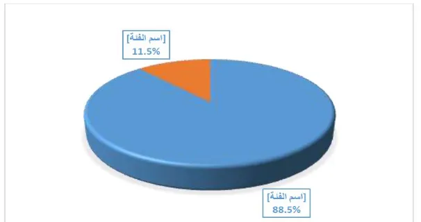
women involved, 23(11.5%) of them had SSI, which represents the incidence rate of SSI post
CS in our setting and 177 (88.5%) of them were non infected wound (figure 2, table 1). the
demographic characteristics of study sample were 24.5% Ladies whom within aged 18- 28
years old and 73.5% were in age 29-40 while they lowest proportion were at aged >40 years
old (2%). Regarding to education level the study founded highest prevalence were in
secondary level (27.5%) while 22.5% women were illiterate. Also the highest prevalence of
study sample were non employed women 109 (54.5%) but the employed women were 91
(45.5%) and finally very small percent of smoker women were found in our sample compares
[image:12.595.146.449.337.497.2]to nonsmoker women as (2% and 98% respectively ). Table 2.
[image:12.595.77.494.545.772.2]Figure 1: distribution of study sample according to age. N=200.
Table 1: Distribution of study sample according to Sociodemographic. N=200.
Age at delivery Frequency Percent
18-29 49 24.5
29-40 147 73.5
>40 4 2.0
Total 200 100.0
women Education Frequency Percent
0 45 22.5
primary 51 25.5
secondary 55 27.5
university 49 24.5
Total 200 100.0
Smoker women Frequency Percent
non smoke 196 98.00
Total 200 100.0
Employed women Frequency Percent
no 109 54.5
yes 91 45.5
Total 200 100.0
The result of current study showed in table (2) and table (3) the frequency of obstetric
characteristic and surgery site infection factors. In table (2) founded 117(58.5%) were one
parity women and 83(41.5%) were tow parity women, while the women that have antenatal
care in study sample were 178(89%) and 11% that don’t have any antenatal care. Regarding
to type of cesarean section, the result showed more prevalence among delivered women by
emergency cesarean section 164(78%) while the women have elective cesarean section were
30 (18%) as show in figure (3).also the women that have rapture membrane were 140(70%)
and 60(30%) were don’t have rupture membrane before caesarian section.
In table (3) show the factors related to surgery site of infection, regarding to types of skin
incision, (96%) were vertical incision and 8 (4%) were have horizontal incision. While the
types of skin suturing the result show 12 (6 %) were interrupted suture and 188(94%) were
[image:13.595.107.487.72.147.2]subcuticular suturing.
[image:13.595.147.450.439.589.2]Figure 3: distribution of study sample according to types of CS. N=200.
Table 1: Distribution of study sample according to obstetric factors. N=200.
Parity Frequency Percent
1 117 58.5
2 83 41.5
Total 200 100.0
ANC Frequency Percent
yes 175 89.0
no 25 11.0
[image:13.595.72.488.628.759.2]Type CS Frequency Percent
Emergency 164 78.0
elective 30 18.0
Total 200 100.0
Rupture of membranes Frequency Percent
yes 140 70.0
no 60 30.0
Total 200 100.0
Table 1: Distribution of study sample according to SSI factors. N=200.
Types of incision (skin) Frequency Percent
vertical 8 4.00
horizontal 192 96.0
Total 200 100.0
Types of suturing (skin) Frequency Percent
Interrupted 12 6.00
Subcuticular 188 94.0
Total 200 100.0
Concerning maternal BMI, it was found the mean BMI of women was 27.20 with SD= 5.089
without any significantly associated between overweight and obese women and increase the
site of infection after cesarean section (p=0.066) table 4.
Table 4: Relation between women BMI and SSI. N=200.
N Mean Std. Deviation
Women BMI
non infected 177 26.96319 5.06265
infected 23 29.03518 5.025713
T test =0.809 p= 0.066
Regarding to relation between sociodemographic factors and SSI. The study showed there are
not any significant association between education level of women and infection of site of
operation (p=0.107). also smoker women that have infected wound were one cases and
without significant association with surgery site infection in study sample (p=0.39). while the
employed women the study founded the infected wound in site of surgery double among
employed women than non-employed women with significant association between them
(p=0.036). But regarding to age of women the result don’t show any significant association
Table 5: relation between sociodemographic characteristic and SSI. N=200.
p-value
non infected infected
Mother Education
0 41 4
primary 45 6
secondary 52 3 0.107
university 39 10 df=3
Total 177 23
non infected infected p-value
Smoker mother non smoke 175 23 0.39
smoke 2 0 df=1
Total 177 23 F*
non infected infected
Employed mother No 101 8
Yes 76 15 0.036 df=1
Total 177 23
non infected infected
Age at delivery
<18 45 4
18-39 128 19 0.509
>40 4 0 df=2
Total 177 23
Figure 4: Distribution of study sample according to sociodemographic characteristic
and SSI. N=200.
In a comparison of obstetric characteristics between the study groups; we founded parity and
antenatal care were not significant associated with their site infection of surgery (p=0.532, p=
0.067 respectively). Also there weren’t significantly association founded between rapture
membrane and infection wound after operation (p=0.02). While there were significantly
association founded between wound infection and types of cesarean section (emergency or
Table 6: distribution of study sample according to obstetric characteristic and SSI.
N=200.
SSI
non infected infected
Parity 1 105 12
2 72 11
Total 177 23
Pearson Chi-Square 0.428, DF= 1, P= 0.532
SSI
non infected infected
ANC yes 162 13
no 15 10
Total 177 23
Pearson Chi-Square 10.243, DF= 1, P= 0.067
SSI
non infected infected
Type CS emergency 150 14
elective 27 9
Total 177 23
Pearson Chi-Square 7.862, DF= 1, P= 0.000
non infected infected
Rupture of yes 120 20
membranes no 57 3
Total 177 23
Pearson Chi-Square 1.160, DF= 1, P= 0.02
Figure 5: Distribution of study sample according to obstetrics characteristic and SSI.
N=200.
On other hand the current study showed the relationship between the wound characteristic
and surgical site infection, we founded the women who had vertical incision more infected
wound after surgery from the women who had horizontal incision with significant association
[image:16.595.85.503.102.626.2]Regarding to wound suturing, the results showed the women who had Subcuticular suturing
were more infected wound than women who electiveemergancy no yes 2 1 typeCS ANC
Parity suturing the wound by interrupted method with significant association between them
(p=0.001). as shown in in table (7). And figure (9).
Table 7: Distribution of study sample according to wound characteristic and SSI.
N=200.
Total non infected infected
Types of incision (skin) vertical 1 7 8
horizontal 171 11 192
Total 177 23 200
Pearson Chi-Square 77.297, DF= 1, P= 0.001
Total non infected infected
Types of suturing (skin) Interrupted 5 7 12
Subcuticular 172 11 111
Total 177 23 200
[image:17.595.65.530.200.541.2]Pearson Chi-Square 27.512, DF= 1, P= 0.001
Figure 8: Distribution of study sample according to types of incision and SSI. N=200.
Figure 9: Distribution of study sample according to types of wound suturing and SSI.
[image:17.595.150.448.587.722.2]DISCUSSION
Cesarean section (CS) is one of the commonest and most famous surgeries in obstetrics.
Surgical site infections (SSI) rise maternal morbidity, elongate hospital admission, and have a
direct effect on healthcare cost. Usually most data and articles come from modern world, with
high quality medical services and an effective health system.[28]
Surgical site infection following caesarean section found high rates in this study, which
comprises 11.5%. Comparing to other studies conducted in different parts of the world, this
result it is similar to study conducted in Iraq 2014.[29] but the surgical site infection following CS was found to be lower in other studies: Oman study 66%,[30] US 5%, 29 Norway 8.3%,[31] and UK 9.6%.32 also were Similar rates founded in other studies conducted in UK 11.2% and
Ethiopia 11.4%.[33] However, higher rate (16%) was found in studies conducted in US and India (24.2%) before intervention.[34]
In a retrospective study done in Patan hospital in Nepal, the surgical site infection rate was
found to be only 2.7% which is lower compared to this study. However, another study
conducted in Chitwan showed wound complications rate for the entire cohort was found
15.2%.[35,36] The possible explanation of these differences might be related to variations in study setting and sample constitution. In addition to wide different in health system
developing between the region, in Iraq the infection prevention and control program very
weak and increase nosocomial infection. Also the war on Iraq in the previous time, added
regarding patients whom internally moved due to terrorism after caused by appearance of
what was called [Islamic State in Iraq and Syria (ISIS)].
Developing SSI after cesarean-section have multiple risk factors and has been influence to
result of study. Sociodemographic characteristic of study sample in current study were
revealing the age of women, smoking women and education level, all these factors don’t have
any correlation to surgery site infection after cesarean section. this result similar to case
control study conducted in Baghdad 2015.[37] and another descriptive study in Kurdistan al-Iraq 2014.[29]
Regarding to obstetric factors that related to cesarean section outcome and Variable risk
factors of post cesarean section surgical site infection were investigated within this study,
In the present study parity was a obstetric factor that statically not significant with increase
the risk of wound infection after cesarean section (p=0.532), this result similar to study
conducted in Iraq37 (p=0.874), in Brussels, Belgium 2014.38 and another study conducted in Nigeria 12 (p=0.450, p=0.642 respectively).
Regarding to antenatal care, the result of study founded no any significant association
between women who had never attended ANC clinic throughout pregnancy and infection site
of surgery, this result similar to study conducted in Iraq.[29]
While the types of cesarean section were statically significant with SSI, The ratio of elective
and emergency surgery rate in this study observed was 1:5. So, more incidence of SSI was
observed in those who had undergone emergency CS (61%) compared to elective CS (39%)
and was statistically significant (p=0.005). this result was similar to study conducted in India
also revealed that Emergency caesarean section predisposes more to SSI as compared to
elective (80.16%).[29] Similar findings were identified in a study conducted in Ethiopia where emergency surgery had two times increased risk of surgical site infection (11.9% vs 5.4%)
than elective cases.[33] This finding could be attributable to the fact that in emergency cases membrane rupture and multiple vaginal examinations are frequent.
There is also increased risk of bacterial contamination or breaks in sterile technique or lack of
timely antibiotic prophylaxis. But another studies disagreement with our result, it found Type
of CS (either emergency or elective) was not a significant risk factor of SSI.[39,40] The possible explanation of these differences might be related to variations in study setting and
sample constitution.
The relation between rupture of the membranes and infection wound after cesarean section
also showed statistically significant risk for surgical site infection (P=0.02). This result
agreement with study conducted in Oman that revealed four-fold increased risk in the rate of
wound infection among patient after operation.2 also another case control study conducted in
Baghdad founded increase risk of SSI with rupture membrane.[37]
Rapture membrane lead to increase chance of the amniotic fluid infected. It is thought that the
non-sterile amniotic fluid may act as a transport medium by which bacteria come into contact
Overweight and obesity is consider risk factors of multiple disease like heart disease and
diabetic mellitus, also increase body weight causes more fat accumulation in skin and vessels,
in our study showed the women who had overweight and obesity more among the patients
that have infected wound in site of surgery from the women that non-infected but without any
significant association (p=0.066).this result similar to study conducted in USA[41], but in another study postulated BMI increase the risk of wound complications after Cesarean
delivery due to physical characteristics (increased tissue trauma, increased wound length,
increased tension on the wound) as well as biological characteristics (decreased vascularity
and oxygenation of adipose tissue, decreased penetration of prophylactic antibiotics). this
result disagreement to another study conducted in Iraq[37], the possible causes of this different might be attributed to different of study sample and study design.
In current study increased rate of surgical site infection was observed in women with vertical
incision than those with horizontal incision which was statistically significant (p=0.001). This
result similar to study conducted in Baghdad (p=0.001).[37] and another a study conducted in India, vertical incision of skin had been found to be a risk factor for developing SSI.[34] but this result disagreement with case control study conducted in brazil.[42] The possible explanation of Infection is transverse incisions are made in the lower part of the abdomen,
near the pubis, which naturally forms a skin fold. This, in turn, can accumulate moisture,
secretions and dirt, which could increase the chances of an SSI.
A vertical incision of CS was mentioned by many papers to increase the risk of SSI and may
lead to formation of a hematoma due to less vascular tissues, while a transverse incision was
associated with less wound dehiscence.[43]
Based hospital Study conducted in USA found a greater incidence of wound complications in
women with vertical skin incisions those with transverse incision.[44]
Suturing techniques played an important role in SSI development after CS, subcuticular
suturing was a good predictor of SSI, when compared to an interrupted technique which had
lower infection events. In current study, the SSI was also found to be significantly higher (P=
<0.001) in those where subcuticular suturing than women who had intracutaneous suture.
6.7% in 2010 and 10.7% in 2011.[45] but another study conducted in Iraq[29] disagreement with our result, interrupted technique more surgical site infection than women with
subcuticular suturing. The possible causes of this different might be attributed to different of
study sample and study design.
REFERENCES
1. Herald: Fear a factor in surgical births. Am JOG, 2007; 195: 514-20.
2. Dahlke JD, Mendez-FH, Rouse DJ, Berghella V, Baxter JK and Chauhan SP:
Evidence-based surgery for cesarean delivery: an updated systematic review, Am J Obstet Gynecol,
2013; 3: 294-306.
3. Brocklehurst P, Quigley M, Ayers S, Juszczak E, Anderson E and Bowler U: Caesarean
section surgical techniques: a randomisedfactorial trial BJOG, 2010; 117(11): 136-145.
4. Ragab A, Mousbah Y, Barakat R, Zayed A, Badawy A. Relaparotomy after caesarean
deliveries: risk factors and how to avoid? J Obstet Gynaecol, 2015; 35: 1–3.
5. Fawcus S, Moodley J. Postpartum haemorhage associated with caesarean section and
caesarean hysterectomy. Best Pract Res ClinObstet Gynaeco, 2013; 27: 233–49.
6. Naki MM, Api O, Celik H, Kars B, Yasar E and Unal O: Comparative study of
Misgav-Ladach and Pfannenstiel-Kerr cesarean techniques: a randomized controlled trial, J
Matern Fetal Neonatal Med., 2011; 24: 239-44.
7. Hoffman M, Harger A, Lenkowski A, Hedner U, Roberts HR, and Monroe DM:
-Cutaneous wound healing is impaired in hemophilia B, Blood, 2009; 108(9): 3053-3060.
8. Patterson LS, O’Connell CM and Baskett TF: maternal and perinatal morbidity associated
with classic and inverted T Cesarean incisions. Obstet Gynecol, 2002; 100: 633.
9. Cunningham FG, Larry CG, Norman FG and Kenneth JL: Cesarean delivery and
postpartum hysterectomy. In Williams Obstetrics: 23th.ed.appleton and Lange,
California, 2011; (2): 422-450.
10.Edwards R and Harding KG: Bacteria and wound healing. Curr Opin Infect Dis., 2004;
17: 91-96.
11.Kulaylat MN, Dayton MT. Surgical complications In: Townsend CM Jr, Beauchamp RD,
Evers BM, Mattox KL, eds. Sabiston Textbook of Surgery. 20th ed. Philadelphia, PA: Elsevier, 2017: 12.
12.Mangram AJ, Horan TC, Pearson ML, Silver LC, Jarvis WR. Guideline for prevention of
13.Berríos-Torres SI, Umscheid CA, Bratzler DW, et al. Centers for Disease Control and
Prevention Guideline for the Prevention of Surgical Site Infection. JAMA Surgery, 2017;
152(8): 784-91.
14.Couto RC, Pedrosa TM, Nogueira JM, Gomes DL, Neto MF, Rezende NA.
Post-discharge surveillance and infection rates in obstetric patients. Int J Gynaecol Obstet,
1998; 61(3): 227-31.
15.Anderson V, Chaboyer W, Gillespie B. The relationship between obesity and surgical site
infections in women undergoing caesarean sections: An integrative review, Midwifery,
2013; 29(12): 1331-8.
16.Lakhan P, Doherty J, Jones M, Clements A. A systematic review of maternal intrinsic risk
factors associated with surgical site infection following caesarean sections. Healthc
Infect, 2010; 15(2): 35-41.
17.Avila C, Bhangoo R, Figueroa R, Santorelli J, Ogburn P, Desan PH. Association of
smoking with wound complications after cesarean delivery. J Matern Fetal Neonatal
Med., 2012; 25(8): 1250-3.
18.Pesce M, Patruno A, Speranza L and Reale M: Extremely low frequency electromagnetic
field and wound Healing: implication of cytokinesas biologicalmediators, Eur. Cytokine
Netw 2013; 24: 1010.1684.
19.Wu Y and Chen S: ―Apoptotic cell: linkage of inflammation and wound healing, ‖
Frontiers in Pharmacology, 2014; 5(1).
20.Busti AJ, Hooper JS, AmayaCJ, and Kazi S: ―Effects of perioperative antiinflammatory
and immunomodulating therapy on surgical wound healing, Pharmacotherapy, 2005;
25(11 I): 1566–1591.
21.Guo and DiPietro: ―Critical review in oral biology & medicine: factors affecting wound
healing, Journal of Dental Research, 2010; 89(3): 219–229.
22.Sarrazy, Billet F, Micallef L, Coulomb B and Desmouli`ere A ―Mechanisms of
pathological scarring: role of myofibroblasts and current developments, ‖ Wound Repair
and Regeneration, 2011; 19(1): S10–S15.
23.Christmann RB, SampaioBarros P, Stifano G, Borges CL, DeCarvalho CR and Kairalla
R: Keyroles for interferon-and TGF-beta-regulated genes, and macrophage activation in
progressive lung fibrosis associated with Systemic Sclerosis, Arthritis Rheum, 2013; 2(4):
310-313.
24.Brancato SK and Albina J E: ―Wound macrophages as key regulators of repair: origin,
25.Danckwardt S, Hentze MW and Kulozik AE: Pathologies at the nexus of blood
coagulation and inflammation: thrombin in hemostasis, cancer and beyond, J. Mol. Med.,
2013; 91: 1257–1271.
26.Chunder A, Devjee J, Khedun SM, Moodley J, Esterhuizen T. A randomised controlled
trial of suture materials used for caesarean section skin closure: Do wound infection rates
differ? SAMJ: South African Medical Journal, 2012 Jun; 102(6): 374-83.
27.Gilbert SA, Grobman WA, Landon MB, Spong CY, Rouse DJ, Leveno KJ, Varner MW,
Caritis SN, Meis PJ, Sorokin Y, Carpenter M. Elective repeat cesarean delivery compared
with spontaneous trial of labor after a prior cesarean delivery: a propensity score analysis.
American journal of obstetrics and gynecology, 2012 Apr 1; 206(4): 311-e1.
28.Olsen MA, Butler AM, Willers DM, Devkota P, Gross GA and Fraser VJ. Risk factors for
surgical site infection after low transverse cesarean section. Infect Control Hosp
Epidemiol, 2008; 9: 477-84.
29.Shrestha S, Shrestha R, Shrestha B, Dongol A. Incidence and Risk Factors of Surgical
Site Infection Following Cesarean Section at Dhulikhel Hospital. Kathmandu Univ Med
J., 2014; 46(2): 113-6.
30.Dhar H, Busaidi AI, Rathi B, Nimre A E, Sachdeva V and Hamdi I. A Study of
Post-Caesarean Section Wound Infections in a Regional Referral Hospital, Oman. Sultan
Qaboos University Med J., 2014 May; (14)2: e211-21.
31.Eriksen HM, Sæther AR, Løwer HL, Vangen S, Hjetland R, Lundmark H, Aavitsland P.
Infections after caesarean sections. Journal of Norwegian Medical Association. Tidsskr
Nor Legeforen 2009; 129:618–22.
32.Wloch C, Wilson J, Lamagni T, Harrington P, Chalett A, Sheridan E. Risk factors for
surgical site infection following caesarean section in England: result from a multicentre
cohort study; BJOG an international Journal of Obstetrics and gynaecology, 2012; 119:
1324-1333.
33.Amenu D, Belachew T and Araya F. Surgical site infection rate and risk factors among
obstetric cases of Jimma University specialized hospital, Southwest Ethiopia. Ethiop
Journal of Health Science 2011 July; 21(2).
34.Cocoran S, Jackson V, Smith C S, Loughrey J. Kenna MC, Cafferkey M. Surgical site
infection after cesarean section: Implementing 3 changes to improve the quality of patient
35.De D, saxena S, Mehata G, Yadav R, and Dutta R. Risk factor analysis and microbial
etiology of surgical site infections following lower segment caesarean section.
International Journal of Antibiotics 2013; Volume 2013, Article ID 283025, 6 pages.
36.Shrestha A, Napit J, Neupane B and Sedhain LB. A randomized trial comparing skin
closure in cesarean section: Interrupted suture with nylon vs subcutucular suture with No
1 polyfilament. JNHRC, 2013 Sept; 11(3).
37.Shaymaa K J. Post Cesarean Section Surgical Site Infection; Incidence and Risk Factors
International Journal of Science and Research (IJSR) ISSN, 2015; 2319-7064.
38.Me´decins sans Frontie`res, rue Dupre´ 94, 1090 Brussels, Belgium (2014).
39.Jido TA, Garba ID. Surgical-site infection following cesarean section in Kano, Nigeria.
Annals of medical and health sciences research, 2012; 2(1): 33-6.
40.Mitt P, Lang K, Peri A, Maimets M. Surgical-site infections following cesarean section in
an Estonian university hospital: postdischarge surveillance and analysis of risk factors.
Infection Control & Hospital Epidemiology, 2005 May; 26(5): 449-54.
41.Diebold, Kasey Elaine. "Risk factors for wound complications following cesarean
delivery." MS (Master of Science) thesis, University of Iowa, 2014.
https://ir.uiowa.edu/etd/1311.
42.Rongetti RL, e Castro PD, da Costa Vieira RA, Serrano SV, Mengatto MF, Fregnani JH.
Surgical site infection: an observer-blind, randomized trial comparing electrocautery and
conventional scalpel. International Journal of Surgery, 2014 Jul 1; 12(7): 681-7.
43.Thawal A Y, and Waghmare M. A Comparative Study Between Midline Vertical and
Pfannestiel incision in lower Segment Caesarean Sec on With Reference to Wound
Complications. Indian Journal of applied research, 2014; 4(10).
44.Wall DP, Deucy E E, Glantz and Pressman E K. Vertical Skin Incisions and Wound
Complications in the Obese Parturient, The American College of Obstetricians and
Gynecologists Nov. 2003; 102(5).
45.Johnson A, Young D, Reilly JV. Caesarean section surgical site infection surveillance.