STUDY OF THYROID FUNCTION TESTS IN AGING MEN OF RURAL
AND URBAN AREAS OF JAMMU REGION
Ravinder Kumar Malhotra*, Anjali Nadir Bhat and Anil Sharma
Postgraduate Department of Physiology, Government Medical College, Jammu.
ABSTRACT
Aging is the progressive universal decline, first in functional reserve
and then in function that occurs in organisms over time. It is
heterogeneous and varies widely in different individuals and in
different organs within a particular individual. The two most important
clinical changes in endocrine activity during aging involve the
pancreas and the thyroid gland. There may be impaired glucose
tolerance or diabetes mellitus. Several changes in thyroid functions
occur during aging. Normal aging is associated with changes in thyroid
hormone production and metabolism. This results in an increased
prevalence of subclinical thyroid disease in the elderly. In general subclinical thyroid disease
is associated with an increased risk of overt thyroid dysfunction and different negative
clinical parameters. The present study was undertaken to study the rural-urban difference in
the thyroid function tests in healthy aging men of Jammu. The present prospective, one-year
study was conducted on 100 healthy male subjects aged 50 years and above each from urban
and rural areas of Jammu region. Subjects with any thyroid disorder or intake of drugs known
to affect thyroid function, persons with chronic illness such as renal failure, malignant
neoplasm, hepatic cirrhosis and diabetes mellitus were excluded from the study. In the
present study, the mean T3 value was found to increase slightly in age group D both in rural
and urban subjects but the difference in the mean values was statistically insignificant. the
mean T4 values were within normal range for laboratory, in all the age groups both in rural as
well as urban subjects. The difference in the mean values of T4 in all age groups among rural
and urban subjects was also statistically non-significant and the difference in the mean values
of TSH in all the age groups among rural and urban subjects was statistically non significant.
KEYWORDS: Thyroid functions, Age, Thyroid hormone, Serum T3, Serum T4, Serum TSH.
Volume 6, Issue 16, 1019-1028. Research Article ISSN 2277– 7105
Article Received on 14 October 2017,
Revised on 05 Nov. 2017, Accepted on 25 Nov. 2017
DOI: 10.20959/wjpr201716-10262
*Corresponding Author
Ravinder Kumar Malhotra
Postgraduate Department of
Physiology, Government
INTRODUCTION
Aging is a general physiologic process that affects cells and the systems made up of them as
well as tissue components.[1] There is a considerable variation in the effects of aging on
healthy individuals, with some persons exhibiting extensive alteration in physiological
functions with age, others little or none. Genetic factors, life style and societal investments in
a safe and healthful environment are important aspects of successful aging.[2] The two most
important clinical changes in endocrine activity during aging involve the pancreas and the
thyroid gland. There may be impaired glucose tolerance or diabetes mellitus. These adults are
at risk for development of secondary complications, mainly macrovascular, at an accelerated
rate.[3] Several changes in thyroid functions occur during aging. Normal aging is associated
with changes in thyroid hormone production and metabolism. Circulating total and free T3
concentration demonstrate a clear, age dependent decline because of both reduced secretion
and reduced peripheral conversion from T4. Serum reverse T3 (rT3) seems to increase with
age.[4] Serum T4 usually remains the same or decreases in females. Serum thyroid stimulating
hormone (TSH) has been reported to increase, decrease or remains unchanged with aging.[5]
With an increase in age, marked changes in thyroid hormone production, metabolism and
action occur. This results in an increased prevalence of subclinical thyroid disease in the
elderly. In general subclinical thyroid disease is associated with an increased risk of overt
thyroid dysfunction and different negative clinical parameters.[6] The present study was
undertaken to study the rural-urban difference in the thyroid function tests in healthy aging
men of Jammu.
MATERIALS AND METHODS
The present prospective, one-year study was conducted on healthy male subjects aged 50
years and above of Jammu region. One hundred subjects were taken from urban areas located
in the Jammu city and 100 subjects were taken from rural areas located in the outskirts of
Jammu city. After explaining the purpose of the study to all the subjects, they were requested
to participate in the study with a written consent. A detailed history was taken to rule out the
presence of any thyroid disorder (hyperthyroidism or hypothyroidism) or intake of drugs
known to affect thyroid function. Care was taken to exclude persons with chronic illness such
as renal failure, malignant neoplasm, hepatic cirrhosis and diabetes mellitus and other
The subjects were distributed into four groups according to their age:
Age group Age in years No. of subjects Total subjects
Urban Rural
A 50 – 60 52 38 90
B 61 – 70 32 29 61
C 71 – 80 9 19 28
D > 81 7 14 21
Total 100 100 200
The subjects included in the study were ambulatory and in normal nutritional state without
any abnormality on routine physical examination. The physical and clinical examinations
were performed in the Postgraduate Department of Medical Physiology and thyroid function
tests were performed in the Radioimmunoassay (RIA) Section of Government Medical
College, Jammu.
All the eligible subjects were interviewed by the investigator himself and details of
information like age, respiratory rate, pulse rate, blood pressure and any significant recent or
past illness was recorded.
For thyroid function tests, blood sample was taken between 9 am and 11 am from non-fasting
subjects after obtaining their consent because fasting causes a rapid fall in serum T3
concentration.[7]
4 ml of venous blood was taken from each subject under aseptic precautions. The blood was
collected in a glass test-tube (without anti-coagulant) and allowed to clot at room
temperature. The serum samples collected after centrifugation were stored at -200C till assay
was performed. The samples were thawed prior to assay. The tests were performed by
radioimmunoassay method as per the protocol given in the RIAK-4A, RIAK-5A and
IRMAK-9 kits for T3, T4 and TSH respectively.
The subjects who were found to have altered thyroid function tests values especially higher
TSH values, their blood samples were tested for anti-TPO antibodies by using Solid Phase
Enzyme Immunoassay Technology.
The data was analyzed using computer software Microsoft Excel and SPSS version 12.0 for
Windows. Mean and standard deviation (SD) was calculated and reported for quantitative
variables. The statistical difference in mean value was tested using unpaired Student’s ‘t’ test.
than two groups. A p-value of <0.05 was considered a statistically significance. All p-values
reported are two-tailed.
RESULTS
The present study was conducted on 100 subjects from urban areas of Jammu city and 100
subjects from rural areas in the outskirts of Jammu city. The mean age (± standard deviation)
of rural subjects was 64.74 (± 12.25) years while the mean age (± standard deviation) of
urban subjects was 60.99 (± 9.94) years. The difference between rural and urban subjects was
statistically significant (t=2.37, p=0.01). Baseline haemodynamic and clinical characteristics
of rural and urban subjects are given in Table 1.
The relationship of mean values of serum T3, serum T4 and serum TSH between rural and
urban subjects according to their age group are given in Tables 2, 3 and 4.
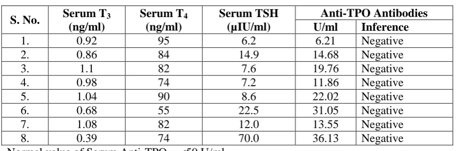
In the age groups 61-70 years, 71-80 years and >81 years, the study observed eight subjects
who had raised serum TSH. Subsequently, their blood samples were tested for anti-TPO
antibodies, which were found to be well within the normal range for laboratory and were
considered negative in all eight cases (Table 5).
Table 1: Mean haemodynamic and clinical parameters of rural and urban subjects.
Parameter
Rural area (n=100) Mean ± Standard
deviation
Urban area (n=100) Mean ± Standard
deviation
Statistical inference
Pulse rate/ minute 77.88 ± 4.54 76.90 ± 4.02 t=1.61, p=4.02*
Respiratory rate/minute 16.81 ± 1.85 16.62 ± 1.90 t=0.75, p=0.45*
SBP (mm Hg) 124.68 ± 6.17 124.12 ± 6.19 t=0.57, p=0.56*
DBP (mm Hg) 80.44 ± 4.17 80.14 ± 4.69 t=0.47, p=0.63*
*Non-significant
Table 2: Relationship of mean values of Serum T3 (ng/ml) between rural and urban subjects according to their age group.
Age group (in years)
Rural subjects (n=100) Mean ± SD
(Range)
Urban
subjects (n=100) Mean ± SD (Range)
Statistical inference
Group A (51 – 60) 1.13 ± 0.10 (0.92 – 1.32)
1.09 ± 0.13 (0.68 – 1.38)
t = 1.51 p = 0.13*
Group B (61 – 70) 1.09 ± 0.18 (0.39 – 1.3)
1.05 ± 0.08 (0.94 – 1.28)
t = 1.03 p = 0.30*
Group C (71 – 80) 1.09 ± 0.10 (0.94 – 1.28)
0.98 ± 0.07 (0.88 – 1.12)
t = 2.56 p = 0.01**
Group D (> 81) 1.09 ± 0.11 (0.92 – 1.32)
1.12 ± 0.16 (0.94 – 1.42)
Normal serum T3 value = 0.7-2.0 ng/ml *Non-significant **Significant
Table 3: Relationship of mean values of serum T4 (ng/ml) between rural and urban subjects according to their age group.
Age group (in years)
Rural subjects (N = 100) Mean ±
SD (Range)
Urban
subjects (N = 100) Mean ± SD (Range)
Statistical inference
Group A (51 – 60) 87.68 ± 8.98 (72 – 105)
87.73 ± 10.88 (55 – 110)
t = 0.02 p = 0.98*
Group B (61 – 70) 88.48 ± 16.44 (74 – 105)
88.90 ± 10.49 (70 – 110)
t = 0.12 p = 0.90*
Group C (71 – 80) 89.26 ± 9.17 (72 – 107)
90.11 ± 10.81 (70 – 104)
t = 0.21 p = 0.83*
Group D (> 81) 85.28 ± 10.11 (70 – 104)
89.85 ± 10.09 (70 – 100)
t = 0.97 p = 0.34* Normal serum T4 value = 55-135 ng/ml *Non-significant
Table 4: Relationship of mean values of Serum TSH (µIU/ml) between rural and urban
subjects according to their age group.
Age group (in years)
Rural subjects (N = 100) Mean ± SD
(Range)
Urban subjects (N = 100)
Mean ± SD (Range)
Statistical inference
Group A (51 – 60) 2.56 ± 1.39 (0.82 – 5.5)
2.56 ± 3.46 (0.54 – (22.5)
t = 0.003 p = 0.99*
Group B (61 – 70) 5.04 ± 3.47 (0.12 – 70)
2.74 ± 2.56 (0.48 – 12.0)
t = –0.696 p = 0.48*
Group C (71 – 80) 1.96 ± 0.86 (0.51 – 4.1)
2.12 ± 1.73 (0.54 – 6.2)
t = 0.317 p = 0.75*
Group D (> 81) 2.38 ± 0.98 (1.2 – 4.9)
2.10 ± 1.41 (0.54 – 4.9)
t = 0.531 p = 0.60* Normal value of Serum TSH = 0.3-5.0 µIU/ml *Non-significant
Table 5: Status of anti-TPO antibodies in subjects with raised TSH values (n = 8).
S. No. Serum T3
(ng/ml) Serum T4 (ng/ml) Serum TSH (µIU/ml) Anti-TPO Antibodies
U/ml Inference
1. 0.92 95 6.2 6.21 Negative
2. 0.86 84 14.9 14.68 Negative
3. 1.1 82 7.6 19.76 Negative
4. 0.98 74 7.2 11.86 Negative
5. 1.04 90 8.6 22.02 Negative
6. 0.68 55 22.5 31.05 Negative
7. 1.08 82 12.0 13.55 Negative
8. 0.39 74 70.0 36.13 Negative
Table 5: Status of anti-TPO antibodies in subjects with raised TSH values (n = 8).
S. No. Serum T3
(ng/ml)
Serum T4 (ng/ml)
Serum TSH (µIU/ml)
Anti-TPO Antibodies
U/ml Inference
1. 0.92 95 6.2 6.21 Negative
2. 0.86 84 14.9 14.68 Negative
3. 1.1 82 7.6 19.76 Negative
4. 0.98 74 7.2 11.86 Negative
5. 1.04 90 8.6 22.02 Negative
6. 0.68 55 22.5 31.05 Negative
7. 1.08 82 12.0 13.55 Negative
8. 0.39 74 70.0 36.13 Negative
Normal value of Serum Anti-TPO = <50 U/ml
DISCUSSION
The literature is replete with information concerning the effects of increasing age on different
aspects of thyroid hormone economy in seemingly euthyroid individuals.[8] The present study
was carried out with the objective of finding out if any age related difference exists in the
thyroid functions in normal healthy aging men of rural and urban areas of Jammu region. The
thyroid function tests viz. T3, T4, TSH were carried out by radioimmunoassay method in non
fasting subjects because fasting produces a rapid fall in serum T3 concentration.[7]
The mean age of rural subjects was 64.74 ± 12.25 years and that of urban subjects was 60.99
± 9.94 years. The difference in the mean values of age between rural and urban subjects was
statistically significant and the reason could be the unequal number of subjects in each age
group.
In the present study, the mean T3 values were higher in group A i.e. 1.13 ± 0.10 in rural
subjects and 1.09 ± 0.13 in urban subjects, declined in group B i.e. 1.09 ± 0.18 in rural
subjects and 1.05 ± 0.08 in urban subjects. The values were further low in group C both in
rural as well as in urban subjects i.e. 1.09 ± 0.10 in rural subjects and 0.98 ± 0.07 in urban
subjects.
Similar findings of progressive decrease in T3 values with advancing age in elderly was
reported by Lipson et al.,[9] Sawin et al.,[10] and Mariotti et al.[11] A possible explanation of
this decrease in serum T3 with advancing age could be decreased thyroidal production and
In the present study, the mean T3 values in the elderly rural subjects declined from 1.13 ±
0.10 in group A subjects to 1.09 ± 0.10 in group C subjects. In the elderly urban subjects, the
mean T3 values declined from 1.09 ± 0.13 in group A subjects to 0.98 ± 0.07 in group C
subjects. The difference in the mean T3 values in group C among rural and urban subjects
was statistically significant and the decline in mean T3 value was more in urban subjects as
compared to rural subjects. The explanation for this could be variations in the dietary habits
of urban and rural people. Moreover, the iodine intake of rural population has improved much
as a result of awareness being created in the rural masses due to various nutritional and
awareness programmes being launched by the government.
Even minor differences in iodine intake between populations are associated with differences
in the occurrence of thyroid disorders. Both iodine intake levels below and above the
recommended quantity are associated with an increase in the risk of disease in the
population.[12]
In the present study, the mean T3 value was found to increase slightly in age group D both in
rural and urban subjects but the difference in the mean values was statistically insignificant.
Gupta et al.,[5] failed to show any decrease in serum T3 values in older persons. The authors
observed that in studies showing opposite results, it could be the inclusion of hospitalized
patients with non-thyroidal illness, which resulted in varying reports of age related changes in
thyroid function in the literature.
In the present study, the mean T4 values were within normal range for laboratory, in all the
age groups both in rural as well as urban subjects. The mean T4 values in group A were 87.68
± 8.98 in rural subjects and 87.73 ± 10.88 in urban subjects; in group B the mean T4 values
were 88.48 ± 16.44 in rural subjects and 88.90 ± 10.49 in urban subjects; in group C the
values were 89.26 ± 9.17 in rural subjects and 90.11 ± 10.81 in urban subjects; and in group
D the values were 85.28 ± 10.11 in rural subjects and 89.85 ± 10.09. The difference in the
mean values of T4 in all age groups among rural and urban subjects was also statistically
non-significant.
Similar findings of normal range of T4 values in elderly subjects were reported by Lipson et
normal range, had been reported in few percentages of subjects in their studies on elderly
people by Kalmijn S et al.[13], Gussekloo et al.,[14] and Van den Beld et al.[15]
A similar slight increase in mean T4 values with increasing age up to group C subjects (age
group 71-80 years) followed by a decline thereafter, though the values were within normal
range for laboratory were observed in the present study also. The values were higher in group
C subjects (age group 71-80 yrs) i.e. 89.26 ± 9.17 in rural subjects and 90.11 ± 10.81 in urban
subjects, but the difference in the mean values of T4 in rural and urban subjects were
statistically non-significant.
Van den Beld et al.[15] in their study on 403 elderly men aged 73-94 years reported increased
mean values of T4, though in normal range in the subjects and they explained that the changes
in thyroid hormone concentrations may be due to a decrease in peripheral (hepatic) thyroid
hormone metabolism with aging and also probably reflecting the effect of subtle NTI
(non-thyroidal illness) and/or an increased catabolic state.
In the present study, the mean TSH values were within normal range for laboratory, in all the
age groups in rural and urban subjects. In group A, the mean TSH values were 2.56 ± 1.39 in
rural subjects and 2.56 ± 3.46 in urban subjects. In group B, the mean TSH values were 5.04
± 3.47 in rural subjects and 2.74 ± 2.56 in urban subjects. In group C, the mean TSH values
were 1.96 ± 0.86 in rural subjects and 2.12 ± 1.73 in urban subjects and in group D, the mean
TSH values were 2.38 ± 0.98 in rural subjects and 2.10 ± 1.41 in urban subjects. The
difference in the mean values of TSH in all the age groups among rural and urban subjects
was statistically non significant.
Gupta et al.,[5] in their study of subjects ranging in age from 40-70 years, observed high
values of TSH in elderly group but these were statistically insignificant. In the present study
also there was no significant difference between TSH values of rural and urban subjects.
It is possible that with increasing age there occurs a decrease in the sensitivity of the pituitary
to slight deficiencies of thyroid hormone, so that more marked deficiency than in younger
individuals would be required to elicit hypersecretion of TSH.[8]
Surks and Hollowell[16] reported positive antithyroid antibodies (ATA) in few percentages of
their subjects and the positivity of ATA was more in women than men. In the present study,
were well within the normal range for laboratory. The explanation for this could be the less
number of subjects with raised TSH and less prevalence of autoimmunity in the male subjects
of the region.
CONCLUSION
In conclusion, in the present study serum T3 levels decreased progressively with age in both
rural and urban subjects up to age group 71-80 years with a slight but insignificant increase in
age group ≥ 81 years. The mean serum T3 concentrations in rural and urban subjects were
significantly different from each other in age group 71-80 years but values did not differ
significantly in other age groups.
Serum T4 levels increased slightly in the age group 71-80 years, though in normal range and
the increase was statistically non-significant. Also a non-significant decrease in the levels
was observed after the age of 81 years. Moreover, mean serum T4 values in rural and urban
subjects did not differ significantly in any age group.
Serum TSH values increased with age up to age group 61-70 years followed by a decrease in
other groups in urban subjects and group C in rural subjects and a slight increase in the level
was observed in age group ≥81 years of rural subjects but the changes were statistically
non-significant. Moreover, mean serum TSH values in rural and urban subjects did not differ
significantly in any age group.
REFERENCES
1. Ganong, W.F. Review of Medical Physiology, 22nd edition. A Lange Medical Book,
2007: 48-49.
2. Lamberts, S.W.J., Beld A.W.V., Lely., A.J.V. The endocrinology of aging. Science, 1997:
278.
3. Lamberts, S.W.J. Endocrinology and aging. Williams Textbook of Endocrinology, 11th
Edition. Saunders Elsevier 1600, John F Kennedy Blvd Suite 1800, Philadelphia, 2008:
1185-1186.
4. Begin, M.E., Langlois, M.F., Lorrain. D., Cunnane, S.C. Thyroid function and cognition
during aging. Cur. Gerontol. Geriat. Res. Hindawi Publishing Corporation,2008.
5. Gupta, K.K., Agarwal. P.K., Roy. S.K., Agarwal, P. Thyroid functions in aging men.
Indian J. Physiol. Pharmacol, 1998; 42(4): 565-566.
7. Palmblad, J., Levi. L., Burger, A., Melander, A., Westgren, U., Schenck, H. Skuda, G.
Effects of total energy withdrawal on the levels of growth hormone, thyrotropin, cortisol,
adrenaline, nor-adrenaline, T4, T3 and rT3 in healthy males. Acta. Med. Scand, 1977; 201:
15.
8. Ingbar, S.H. The influence of aging on the human thyroid hormone economy. Geriatr.
Endocrinol, 1978; 5: 13-31.
9. Lipson, A., Elleen, L., Nickoloff, Hsu, T.H, Kasecamp, W.R., Drew, H.M., Shakir, R.,
Wagner, H.N. Jr. A study of age-dependent changes in thyroid function tests in adults. J.
Nucl. Med, 1979; 20: 1124-1130.
10.Sawin, C.T., Chopra, D., Azizi F, Mannix JE and Bacharach P. Increased prevalence of
elevated serum thyrotropin levels in the elderly. JAMA, 1979; 242(3): 247-250.
11.Mariotti, S., Barbesino, G., Caturegli, P., Bartelena, L., Sansoni, P., Fagnoni, F., Monti,
D., Fagiolo, U., Franceschi, C. Pinchera, A. Complex alteration of thyroid function in
healthy centenarians. J. Clin. Endocrinol. Metabol. 1993; 77: 1130-1134.
12.Laurberg, P. Iodine intake as a determinant of thyroid disorders in populations. Best.
Pract. Res. Clin. Endocrinol. Metab, 2010; 24(1): 13-27.
13.Kalmijn, S., Mehta, K.M, Pols, H.A., Hofman, A., Drexhage, H.A. Breteler, M.M.
Subclinical hyperthyroidism and the risk of dementia. Clin. Endocrinol, 2000; 53(6):
733-737.
14.Gussekloo, J., Van, E.E., De Craen, A.J, Meinders, A.E., Frolich, M. Westendorp RG.
Thyroid status, disability and cognitive function and survival in old age. JAMA, 2004;
292: 2591-2599.
15.Van den Beld, A.W., Visser, T.J., Feelders, R.A., Grobbee, D.E., Lamberts, S.W.J.
Thyroid hormone concentrations, disease, physical function and mortality in elderly men.
J. Clin. Endocrinol. Metabol, 2005; 90(12): 6403-6409.
16.Surks, M.I., Hollowell, J.G. Age-specific distribution of serum thyrotropin and
antithyroid antibodies in the US population: implications for the prevalence of subclinical