Exploiting the kinetic interplay between the GPIbα–VWF binding interfaces
to regulate hemostasis and thrombosis
Jianchung Chen,1 Hairu Zhou,1 Alexander Diacovo,1 X. Long Zheng,3 Jonas Emsley,4 and Thomas G. Diacovo1,2
1
Department of Pediatrics, Columbia University Medical Center, New York, New York,
10032, USA. 2Department of Pathology and Cell Biology, Columbia University Medical Center, New York, New York, 10032, USA. 3Department of Pathology and Laboratory Medicine, The Children's Hospital of Philadelphia, Philadelphia, Pennsylvania, 19104
USA.4Centre for Biomolecular Sciences, School of Pharmacy, University of Nottingham,
University Park, Nottingham, NG72RD, UK.
Correspondence
Thomas Diacovo, MD Columbia University
1130 St. Nicholas Ave, Room 924 New York, NY 10032
Tel: 212-851-4683
E-mail: [email protected]
Short title: Regulating adhesion between platelets and VWF Text word count: 4,341
Text abstract count: 200
Number of figures: 6, and 2 supplemental figures Number of tables: 1
Key points
• GPIbα–VWF-A1 bond lifetime governs platelet–VWF interactions and can be
altered to correct defects in hemostasis or prevent thrombosis.
• Targeting a distinct binding interface between GPIbα and VWF-A1 offers a
Abstract
Platelet–VWF interactions are tightly controlled to prevent vascular occlusion due to
premature aggregate formation. Although multiple mechanisms may regulate this
process, it is unclear whether the inherent properties of the bond formed the platelet
receptor GPIbα and the A1 domain of VWF can influence the site and extent to which
platelets bind VWF. Here, we demonstrate that the kinetic interplay between two distinct
regions of contact between this receptor–ligand pair not only permit maximal platelet
accrual at sites of arterial injury, but can be manipulated to correct defective hemostasis
or prevent thrombosis. This was accomplished by generating VWF-A1 knock-in mice
with mutations that enhance (I1309V) or disrupt (R1326H) GPIbα binding and by
targeting the complex with an allosteric inhibitor. Incorporation of R1326H mutation in
the major site shortened bond lifetime, yielding defects in hemostasis and thrombosis
comparable to VWF deficient animals. Similarly, pharmacologic disruption of the major
interface prevented arterial thrombosis. However, combining the R1326H mutation with
the I1309V mutation located near the minor site normalized bond kinetics and restored
the hemostatic and thrombotic properties of VWF. Hence, an important biophysical-–
biological relationship is revealed and the therapeutic benefits of modifying the GPIbα–
Introduction
The ability of blood borne cells to perform their designated biological function(s)
requires cell surface adhesion molecules to engage counter-receptors at the appropriate
time and location. For example, platelets and von Willebrand factor (VWF) co-exist in
circulating blood and must interact at sites of arterial injury in order to promote effective
hemostasis. This process is triggered upon the binding of plasma VWF to exposed
components of the injured vessel wall where it can then initiate the attachment of
platelets under shear flow conditions.1,2 VWF is a multimeric plasma glycoprotein composed of functionally distinct types of domains that are either duplicated or
triplicated in the following order from the N-terminus:
D'-D3-A1-A2-A3-D4-B1-B2-B3-C1-C2-CK.3,4 It is the interaction between VWF-A1 domain and the platelet receptor
glycoprotein Ib alpha (GPIbα) that initiates primary hemostasis by bringing platelets into
close proximity with reactive substrates generated at the site of arterial damage.5-8 The clinical significance of this interaction is underscored by the increased bleeding
tendencies of individuals who either lack the plasma protein or possess mutations within
the A1 domain of VWF. In the latter case, this includes mutations that limit binding (type
2M) or paradoxically enhance interactions (type 2B), both of which are associated with
the hereditary bleeding disorder known as von Willebrand disease (VWD).3,4
Against this background, structure analyses of native and mutant complexes have
provided insights into the nature of the interactions that support adhesion between this
receptor–ligand pair and possible mechanisms by which mutations alter this process. The
structure reveals a major and minor binding site for GPIbα on the surface of the A1
located in or directly adjacent to the larger contact surface, may disrupt electrostatic
interactions between this receptor–ligand pair or induce structural changes that reduce
GPIbα binding. In contrast, type 2B mutations are clustered in close proximity to the
minor site near the termini of the domain and may alter the conformation of a region
known as the α1-β2 loop.9-12 Consequently, this is thought to provide an essential
energetic contribution that augments binding affinity9 or reduce a steric clash that would impede adhesion.13 Clinically, this enhanced binding is believed to result in bleeding by permitting VWF multimers with the greatest hemostatic potential to aggregate
spontaneously with platelets in blood and ultimately be cleared from the circulation.3,4 Although such studies provide insight into mechanism(s) by which mutations associated
with VWD may alter binding between platelets and VWF, they cannot account for
interactions involving sites outside of the A1 and GPIbα binding domains or external
influences such as shear flow that play a role in regulating hemostasis and thrombosis.14 Indeed, several mechanisms have been identified that may prevent VWF–platelet
aggregate formation in the circulation. This includes shielding of VWF-A1 by the
adjacent D'D3 domains,15 the inhibitory effects of the β2-glycoprotein I,16 and the ability
of shear flow to activate the VWF-A1 so that it can then engage GPIbα.17 Moreover, it
has been demonstrated that forces generated by flowing blood can promote the unfolding
and elongation of VWF so that it exposes multiple A1 domains.18
An additional mechanism that has not been fully explored is the contribution that
the physicochemical properties of the bond formed between VWF-A1 and GPIbα play in
regulating platelet–VWF interactions at sites of vascular injury. Indeed, detailed analyses
into the biophysical consequences of the predicted allosteric change associated with type
2B mutations.19-22 That said, it remains unclear whether specific properties of the GPIbα– VWF-A1 bond predicted to regulate platelet–VWF interactions correspond with
biological reality.23-25
In the current study, we address this shortcoming by performing a detailed kinetic
analysis of WT and mutant murine GPIbα–VWF-A1 complexes and then validating these
observations using transgenic mice harboring modified VWF-A1 domains. We also probe
the binding interface using a novel allosteric inhibitor that prevents the formation of key
interactions between this receptor–ligand pair. Our data reveal that the physicochemical
properties of the distinct regions of contact between this receptor–ligand pair contribute
to a composite biophysical signature required for effective hemostasis and arterial
Methods
Modeling of murine GPIbα–VWF-A1 complexes
Crystal structures of the human GPIbα–VWF-A1 complex have been described for WT
and for the complex formed between the GOF mutations R1306Q and I1309V in
VWF-A1 and the platelet type GOF mutation M239V in GPIbα.9,10,13,26 The human platelet
receptor was used as a template for mouse GPIbα. Consensus rotamers with minimal
steric clashes were chosen, followed by adjustments to create reasonable van der Waals
interactions and H-bonding using COOT (http://www.biop.ox.ac.uk/coot/).
A consensus model of the human receptor–ligand pair was used to build its
murine counterpart. This included the double mutant VWF-A1 (I1309V, R1326H)
complex that was based on the human VWF-A1 (R1306Q) mutant complex using the
program SWISSMODEL (http://swissmodel.expasy.org/).10 Figures were created using PYMOL.
Mice
The generation of the VWFR1326H mutant mouse has been previously described.27 A similar targeting strategy was used to introduce the double mutation I1309V, R1326H
into exon 28 of the murine VWF gene or to substitute in the human A1 domain. VWF
deficient mice were purchased from The Jackson Laboratory. All animals were on a
C57BL/6J background and kept in a pathogen-free facility. Experiments were performed
in accordance to the guidelines set forth by Institutional Animal Care and Use Committee
Analysis of VWF expression and function
Detection and/or measurement of murine VWF A1-A2-A3 domain transcripts, plasma
VWF antigen levels, functional factor VIII levels, and multimer pattern were performed
as previously described.27-30
Platelet adhesion in flow
A parallel-plate flow chamber was used to assess platelet attachment and translocation on surface-immobilized plasma VWF or recombinant VWF-A1 protein.27 Generation and purification of recombinant proteins were performed as previously described.20,26 Citrated whole blood (150 µl) collected via cardiac puncture from anesthetized WT and mutant
VWF mice or from healthy human volunteers (venipuncture) was perfused over the
immobilized substrates at wall shear rates ranging from 100 s-1 to 1,600 s-1 for 2 min, followed by washing with Tyrode's buffer. The number of platelets attached per unit area
and translocation velocities were determined by off-line analysis. For inhibition studies,
OS1 peptide was generated and purified as previously described.31 The inhibitor was added to 1 ml of citrated whole blood 5 minutes prior to perfusing over
surface-immobilized substrates.
Platelet aggregation
The ability of ristocetin (Chronolog Corporation) to aggregate human platelets in plasma
Lumi-Aggregometer (model 540 VS, Chronolog Corporation).32 In brief, citrated whole blood obtained by cardiac puncture from anesthetized mice or collected via venipuncture from
healthy volunteers was centrifuged to obtain platelet poor plasma (PPP). Subsequently,
lyophilized human platelets (Bio/Data Corporation) reconstituted in TBS were added to
plasma at a 4:1 ratio (v/v) and a baseline obtained for 2 minutes prior to adding ristocetin
(1.25 mg/ml). Aggregation (1,200 rpm, 37°C) was allowed to proceed for 6 min. Results
are reported as maximum percent change in light transmittance from baseline with TBS
used as a reference. For inhibition studies, platelet / PPP mixture was incubated with OS1
peptide (0.1 µg/ml to 5 µg/ml) for 5 min prior to the addition of ristocetin.
In vivo thrombus formation.
Administration of anesthesia, insertion of vascular catheters, fluorescent labeling of
platelets, and surgical preparation of the cremaster muscle have been previously
described.27,32 A pulsed nitrogen dye laser was used to induce arteriole injury in the
cremaster muscle of anesthetized 8-12 week old animals. Mouse or human platelet-vessel
wall interactions were visualized by fluorescen ce microscopy. Human platelets were
administered by continuous infusion (700,000/µl at 25 µl/min) through a catheter placed
in the ipsilateral femoral artery 2 minutes before and during laser-induced injury. For
inhibition studies, 20 µg of OS1 peptide in 100 µl of saline was administered i.v. 5 min
prior to the administration of human platelets. The extent of thrombus formation was
Tail bleeding assay
Bleeding times were measured in 8-week old mice after amputating 1 cm of the tail tip
and then placing the tail in a physiological saline solution (37°C) as previously
described.27
Microsphere tethering frequency
The rate of complex formation was evaluated by measuring the frequency of tethering
events between surface-immobilized mouse platelets and microspheres coated with a low
site density of recombinant WT or mutant VWF-A1.20,21 This was determined by observing the number of beads that paused, but did not translocate, at wall shear rates
ranging from 10 s-1 to 300 s-1. This value was then normalized by dividing the number of beads that formed transient tethers by the number of non-interacting beads transported
across the field of view in the focal plane of the immobilized platelet substrate.
Estimation of the amount of VWF-A1 coupled to beads was determined using a
calibrated microbead system (Flow Cytometry Standards) following manufacturer’s
instructions. The site density of VWF-A1 on beads was estimated to be ~35 sites/µm2.
Dissociation rate constants of transient tethers
The duration of transient tethers was estimated for VWF-A1-coated microspheres
interacting with surface-immobilized platelets at wall shear stresses ranging from 0. 5 to
period. The duration of these interactions was measured by recording images at a frame
rate of 235 frames per second using 60X DIC objective (oil immersion). A total of 30–50
interact pause times for each wall shear stress was recorded. Dissociation rate constants
were determined by plotting the natural log of the number of VWF-A1–coated
microspheres that interacted as a function of time after the initiation of tethering. The
slope of the line = -koff. The force acting on the tether bond was calculated from force
balance equations (bead radius of 3.5μm) satisfied with a tether angle θ of 57.2 degrees.20,21
Statistical analyses
Details for the calculation of maximum likelihood estimates (MLEs) for the off-rates
(koff) and Bell model parameters (k0off, σ) can be found in supplemental Methods.
Statistical analysis of all other data was determined using unpaired Student’s t-test
Results
Structural and functional changes associated with I1309V and R1326H mutations
To determine the interplay between known contact surfaces and to establish the
role that the biophysical properties of the GPIbα–VWF-A1 bond play in regulating
platelet–VWF interactions, we chose mutations within the major or minor contact
interfaces of the murine A1 domain that enhance (I1309V) or disrupt (R1326H) binding.
The I1309V mutation is known to cause type 2B VWD,33 while the R1326H substitution impairs interactions with mouse GPIbα.27 Interestingly, several mammalian species
including humans have a histidine in lieu of arginine at position 1326 within the A1
domain.34 Thus, murine VWF is ideal for determining how this particular amino acid substitution alters the biophysical properties of the GPIbα–VWF-A1 bond and its ability
to support hemostasis and thrombosis. Models of WT and mutant murine complexes were
built to better understand the effect of the mutations on binding (Figure 1A). Although
crystal structure analyses have yet to identify the exact mechanism by which the I1309V
mutation augments adhesion,9,13 it is located in the α1-β2 loop adjacent to the type 2B
mutation R1306Q.9,10 The latter mutation is believed to result in a local conformational change with a re-positioning of residues Q1311 and R1334 that reinforce interactions
with human GPIbα.9 Figure 1B illustrates the position of residue 1309 in the context of
the complex formed with murine GPIbα and the augmentation in binding that occurs
upon re-positioning of residues Q1311 and R1334.
To understand how these mutations affect adhesion under flow conditions, we
and I1309V substitutions and determined their ability to support mouse platelet
interactions when surface-immobilized in a parallel plate flow system.27 The R1326H mutation impaired mouse platelet accumulation and increased translocation velocity
~13-fold as compared to WT rVWF-A1 (Figure 1C-D). In contrast, the I1309V mutation
decreased translocation velocity ~5-fold and greatly augmented platelet accumulation.
Strikingly, rVWF–A1 containing both amino acid substitutions yielded an adhesive
phenotype comparable to WT.
I1309V and R1326H mutations differentially alter bond formation and dissociation
To determine whether the biophysical properties of rVWF-A1 expressing both
mutations truly reflect that of the WT complex, we analyzed the formation and
dissociation of transient adhesive events, known as tether bonds, that occur when flowing
rVWF-A1–coated microspheres transiently interact with GPIbα expressed on
surface-immobilized mouse platelets.20,21 On-rate was determined by evaluating the frequency with which microspheres interacted with surface-immobilized mouse platelets at various
wall shear rates. Whereas the I1309V mutation enhanced the rate of association ~3.5–fold,
this was abolished when combined with the R1326H substitution (Figure 2A). In fact,
murine rVWF-A1 possessing the double mutation displayed a rate of association that
more closely resembled the WT complex with a maximal tethering frequency at a wall
shear rate of 85 s-1.
The effects of the mutations on the kinetics of dissociation (koff) were determined
previously showed the utility of this system for assessing the biophysical properties of the
interaction between human GPIbα and rVWF-A1. Moreover, the use of microspheres
with a uniform size and shape permits the calculation of the amount of shear force acting
on the tether bond. As previously observed for the human complex, the distribution of
interaction times indicated that the majority of WT and mutant tether bonds fit a straight
line, the regressed slope of which corresponded to -koff (Figure 2B-E). In addition, the rate
of dissociation behaved in accordance with the equation of Bell, which states that koff =
k0off exp(σFb/kT), where Fb is the applied force, k0off is the dissociation rate constant in
the absence of force, kT is the thermal energy, and σ (reactive compliance) relates the sensitivity of bond off-rate to an applied force.35 The larger the values for k0off and σ, the
shorter the bond lifetime (1/ k0off) and the more prone the receptor–ligand interaction is to
force-driven dissociation, respectively. Maximum likelihood estimates (MLE) of the
kinetics of GPIbα–VWF-A1 interactions and their subsequent fit to the Bell equation
were performed.36 Regression analyses of these simulations fit the data at bond forces ranging from 36 pN to 217 pN and thus permitted the determination of k0off and σ (Figure
2F and Table 1). Whereas the major effect of the I1309V substitution was to prolong the
lifetime of the tether bond (6-fold), R1326H shortened its duration (2.5-fold). However,
the kinetic (k0off) and mechanical (σ) properties of rVWF-A1 bearing both mutations were
nearly identical to that of the native complex.
To determine whether the predictions based on biophysical studies truly have biological
relevance, we generated knock-in mice that express either the R1326H (VWF R1326H) mutation alone27 or in combination with I1309V (VWF I1309V, R1326H) (supplemental Figure 1A-C). VWF gene transcription, multimer pattern, antigen levels as well as factor
VIII function and platelet counts were similar to WT littermates (supplemental Figure
1D-H). Consistent with the reduction in bond lifetime and impaired adhesion, animals
expressing the R1326H mutation had prolonged tail bleeding times and a diminished
capacity to form arterial thrombi (Figure 3A-B). In fact, VWF R1326H mice possessed a phenotype similar to that observed for mice lacking this plasma protein (VWF KO). Remarkably, the additional incorporation of the I1309V mutation corrected the observed
perturbations in platelet–VWF interactions. Not only did VWF-A1 double mutant
animals have hemostatic properties similar to WT littermates, but also laser-induced
thrombi were comparable in size to mice possessing the WT plasma protein (Figure
3A-B). Direct evidence that plasma VWF I1309V, R1326H has adhesive properties equivalent to its WT counterpart is demonstrated by its ability to support mouse platelet accumulation
and translocation velocities at similar levels (Figure 3C-D). This was not the case for
plasma VWF R1326H which impaired platelet adhesion and yielded a ~3-fold increase in translocation velocity. These data support a close association between the lifetime of the
GPIbα–VWF-A1 bond and the ability of these hemostatic elements to support critical
biological processes.
Given the importance of the major contact site in supporting mouse platelet–VWF
interactions in vivo, we next explored the therapeutic utility of targeting this region in the
human complex. This was accomplished by using a novel cyclic peptide, termed OS1,
which was isolated from a cysteine-constrained phage display library.31 It interacts with the curved concave face of the GPIbα leucine-rich repeat,37 stabilizing a conformation of a regulatory loop known as the β-switch10 so that it prevents the formation of electrostatic
interactions essential for binding to the VWF-A1 domain (Figure 4A). However, the
effect of OS1 is limited to human GPIbα as demonstrated by its inability to reduce mouse
platelet–VWF interactions in vitro and in vivo (Figure 4B-C). In contrast, incubation of
whole blood from healthy volunteers with the inhibitor not only limited platelet
accumulation on surface-immobilized human rVWF-A1 protein, but also impaired
ristocetin-induced platelet aggregation in a dose dependent manner (Figure 4D-E).
To directly test the ability of OS1 to prevent human platelet-mediated arterial
thrombosis, we generated an animal in which the majority of the murine A1 domain was
replaced with its human counterpart (supplemental Figure 2A-B). VWF gene
transcription, multimer pattern, antigen levels and platelet counts in VWF HA1 mice were similar to WT littermate controls (supplemental Figure 2C-F). In addition, animals
containing the human VWF-A1 domain manifested impaired platelet–vessel wall
interactions that resulted in a prolongation in tail bleeding time as well as a reduction in
thrombus size in laser-injured arterioles (Figure 5A-B). Consistent with these
VWF HA1 mice formed large and often occlusive thrombi in laser-injured arterioles of nearly identical size to that observed in their VWF R1326H counterparts (Figure 5D). Accumulation of human platelets on surface-immobilized plasma VWF from either
animal was also comparable (Figure 5E). Yet, only plasma VWF from mice possessing
the human VWF-A1 domain supported ristocetin-induced platelet agglutination /
aggregation of human platelets demonstrating that the R1326H mutation in murine
VWF-A1 was not sufficient to support this process (Figure 5F). Importantly, the OS1 peptide
was able to impair human platelet accumulation on surface-immobilized plasma VWF
HA1
and inhibited ristocetin-induced platelet agglutination / aggregation (Figure 6A-B).
Moreover, human platelet-mediated thrombus formation in arterioles of VWF HA1 mice was reduced by >80% in the presence of the inhibitor (Figure 6C). The data demonstrate
the potential therapeutic utility in targeting the major contact interface formed between
Discussion
Detailed structural and biophysical analyses of small regions of contact between GPIbα
and VWF-A1 have yielded tremendous insight into the relationship between
force-lifetime-and-chemistry of an interaction critical for supporting hemostasis and thrombosis.
Here, we have taken a unique approach to better understand the role that the kinetic
properties of the contact surfaces formed between this receptor–ligand pair play in
supporting these biologically relevant processes. By generating animals with mutations
contained within the major and minor binding sites of murine VWF-A1 that either reduce
or enhance attachment to GPIbα, we provide the first in vivo evidence that bond lifetime
does play an important role in regulating platelet–VWF interactions at sites of arterial
injury.
To determine whether data obtained using a murine-based system were applicable
to the human receptor–ligand pair, we first measured the kinetic properties of the murine
GPIbα–VWF-A1 bond and its response to an applied force. Values for the intrinsic
off-rate were similar between the species (k0off values of 3.05 ± 0.55 s-1 and 3.45 ± 0.37 s-1 for
the mouse and human complex, respectively). However, the reactive compliance (σ) for
the WT murine complex was ~1.5-fold higher than for its human counterpart (0.028 ±
0.001 nm versus 0.018 ± 0.002 nm, respectively), suggesting that the interaction between
the murine receptor–ligand pair may be slightly more prone to dissociate upon the
application of hydrodynamic force. The effects of the I1309V substitution mirrored that
Surprisingly, the observed alterations in adhesion did not persist when this mutation was
combined with R1326H. In fact, the biophysical properties of the double mutant bond
more closely resembled that of the native complex. These observations led us to predict
that murine plasma VWF I1309V, R1326H should possess hemostatic and thrombotic properties similar to the native plasma protein if bond lifetime plays an important role in
these processes. This was borne out by the fact that I1309V, R1326H double knock-in
mice had tail bleeding times and formed arterial thrombi of similar size to that observed
for WT littermates. Based on these observations we conclude that the major and minor
contact interfaces not only contribute to a biophysical signature well suited for supporting
platelet–VWF interactions under the hydrodynamic conditions encountered in the arterial
circulation, but also that they must be maintained within strict limits in order to optimize
platelet accumulation at sites of vascular injury.
Although the precise mechanism(s) by which the I1309V mutation augments
binding remains controversial,9,13 its pronounced effects on the biophysical properties of
the murine GPIbα–VWF-A1 bond made it suitable for gaining insight into the
relationship between bond lifetime and the requirement for shear flow to initiate platelet
attachment to VWF. It is well known that the application of a specific level of shear is
essential to achieve optimal adhesion between GPIbα and VWF-A1 (termed the shear
threshold effect). 20 Shear-induced forces are thought to influence the GPIbα–VWF-A1
bond in two ways. As the applied force increases from low levels, the bond lifetime
increases until it is of sufficient duration to promote effective interactions between
accordance with Bell’s model.35 However, this concept has been challenged in a study suggesting that the shear threshold effect may be due to a transition of a slip bond from a
low to high affinity state in response to a force-induced conformation change in the
complex.41 Despite these conflicting views, our data suggest that the ability of the
I1309V mutation to prolong the lifetime of the GPIbα–VWF-A1 bond beyond that of the
native complex is essential for promoting platelet–VWF interactions at low flow states.
Moreover, the process is reliant on the biophysical properties of the major contact
interface. In the presence of the R1326H substitution, the I1039V mutation could no
longer abolish the requirement for shear flow to support the tethering of murine
rVWF-A1 coated beads to mouse GPIbα, nor slow mouse platelet translocation velocities and
augment platelet attachment to surface immobilized VWF-A1 containing the double
mutation.
It is important to note that the ability to prolong the lifetime of the GPIbα
–VWF-A1 bond is not unique to type 2B mutations as it has also been reported for snake venoms.
This is best exemplified by the ability of botrocetin, a component of Bothrops jararaca
venom, to promote platelet–VWF aggregation in plasma as well as contribute to the loss
of VWF multimers and platelets from the blood.42 Interestingly, we have shown that the major effect of botrocetin is to increase the bond lifetime two-fold for both human and
mouse complexes without affecting the on-rate.26 This did not involve allosteric changes
in GPIbα or VWF-A1, but relied on the ability of the venom protein to form a biological
clasp that prolonged their embrace. These results further support our current observations
that the lifetime of the GPIbα–VWF-A1 bond contributes significantly to promoting
There has been much speculation on whether one or both of the identified binding
surfaces between GPIbα and VWF-A1 could be targets for the prevention or treatment of
thrombosis.43 Our current study helps clarify this issue. We demonstrate that a cyclic
peptide (OS1) proven to alter the conformation of GPIbα so that is incapable of forming
several key interactions with the major binding interface of VWF-A1 significantly
reduced human platelet-mediated thrombus formation in laser-injured arterioles of VWF
HA1
mice. Thus, the development of such agents that can alter specific interactions
between this receptor–ligand pair offers a powerful approach to reducing arterial
thrombosis.
In summary, animals bearing specific mutations in a protein critical to promoting
hemostasis and thrombosis help to establish an essential link between kinetic
measurements of a receptor-ligand bond and their overall significance to a biologically
relevant process. In addition, our data shed new light on the cooperation between the two
distinct GPIbα binding sites within VWF-A1 in generating a biophysical signature
ideally suited for supporting platelet–VWF interactions to sites of arterial injury. Insights
into how the interaction between a receptor–ligand pair can be altered by manipulating
the kinetics of a receptor-ligand interaction are relevant to the development of therapies
Acknowledgements
We thank Aychyn C. Huang and Jeffrey S. Jhang for performing factor VIII function
analysis, Gray Shaw for supplying the OS1 peptide, and Ian Laurenzi for performing the
analysis of tether bond formation. We are grateful to Barry Coller and Michael Rosen for
critical review of the manuscript. This research is supported by National Institute of
Health grants HL103989 and HL097971. The authors have no competing financial
interests.
Authorship contributions
T.G.D. conceived the study, supervised the project, conducted experiments, and wrote the
manuscript. J.C. generated VWF mutant mice and conducted experiments. H.Z. and
X.L.Z. performed experiments and J.E. generated atomic models of the murine GPIbα–
VWF complex. I.J.L. conducted MC simulations and statistical analysis of the
References
1. Ruggeri ZM, Orje JN, Habermann R, Federici AB, Reininger AJ.
Activation-independent platelet adhesion and aggregation under elevated shear stress. Blood.
2006;108(6):1903-1910.
2. Sakariassen KS, Bolhuis PA, Sixma JJ. Human blood platelet adhesion to artery
subendothelium is mediated by factor VIII-Von Willebrand factor bound to the
subendothelium. Nature. 1979;279(5714):636-638.
3. Nichols WC, Ginsburg D. von Willebrand disease. Medicine (Baltimore).
1997;76(1):1-20.
4. Sadler JE. New concepts in von Willebrand disease. Annu. Rev. Med. 2005;56:173-191.
5. Cruz MA, Diacovo TG, Emsley J, Liddington R, Handin RI. Mapping the glycoprotein
Ib-binding site in the von Willebrand factor A1 domain. J. Biol. Chem.
2000;275(25):19098-19105.
6. Piétu G, Meulien P, Cherel G, et al. Production in Escherichia coli of a biologically
active subfragment of von Willebrand factor corresponding to the platelet glycoprotein Ib,
collagen and heparin binding domains. Biochem. Biophys. Res. Commun.
1989;164(3):1339-1347.
7. Savage B, Saldivar E, Ruggeri ZM. Initiation of platelet adhesion by arrest onto
fibrinogen or translocation on von Willebrand factor. Cell. 1996;84(2):289-297.
glycoprotein Ib binding domain of von Willebrand factor expressed in Escherichia coli.
Biochemistry. 1991;30(21):5202-5209.
9. Dumas JJ, Kumar R, McDonagh T, et al. Crystal structure of the wild-type von
Willebrand factor A1-glycoprotein Ibalpha complex reveals conformation differences
with a complex bearing von Willebrand disease mutations. J. Biol. Chem.
2004;279(22):23327-23334.
10. Huizinga EG, Tsuji S, Romijn RA, et al. Structures of glycoprotein Ibalpha and its
complex with von Willebrand factor A1 domain. Science. 2002;297(5584):1176-1179.
11. Emsley J, Cruz M, Handin R, Liddington R. Crystal structure of the von Willebrand
Factor A1 domain and implications for the binding of platelet glycoprotein Ib. J. Biol.
Chem. 1998;273(17):10396-10401.
12. Fukuda K, Doggett TA, Bankston LA, Cruz MA, Diacovo TG, Liddington RC.
Structural basis of von Willebrand factor activation by the snake toxin botrocetin.
Structure. 2002;10(7):943-950.
13. Blenner MA, Dong X, Springer TA. Towards the Structural Basis of Regulation of
von Willebrand Factor Binding to Glycoprotein Ib. J. Biol. Chem.
2014;289(9):5565-5579
14. Lenting PJ, Pegon JN, Groot E, de Groot PG. Regulation of von Willebrand
factor-platelet interactions. Thromb Haemost. 2010;104(3):449-455.
15. Ulrichts H, Udvardy M, Lenting PJ, et al. Shielding of the A1 domain by the D'D3
domains of von Willebrand factor modulates its interaction with platelet glycoprotein
16.Hulstein JJ, Lenting PJ, de Laat B, Derksen RH, Fijnheer R, de Groot PG.
beta2-Glycoprotein I inhibits von Willebrand factor dependent platelet adhesion and
aggregation. Blood. 2007; 110(5):1483–1491.
17. Dong JF, Berndt MC, Schade A, McIntire LV, Andrews RK, Lopez JA.
Ristocetin-dependent, but not botrocetin-Ristocetin-dependent, binding of von Willebrand factor to the platelet
glycoprotein Ib-IX-V complex correlates with shear-dependent interactions. Blood.
2001;97(1):162-168.
18. Schneider SW, Nuschele S, Wixforth A, et al. Shear-induced unfolding triggers
adhesion of von Willebrand factor fibers. Proc Natl Acad Sci USA 2007; 104(19):7899–
7903.
19. Arya M, Kolomeisky AB, Romo GM, Cruz MA, Lopez JA, Anvari B. Dynamic force
spectroscopy of glycoprotein Ib-IX and von Willebrand factor. Biophys J.
2005;88(6):4391-4401.
20. Doggett TA, Girdhar G, Lawshé A, et al. Selectin-like kinetics and biomechanics
promote rapid platelet adhesion in flow: the GPIbα-vWF tether bond. Biophys J. 2002;83(1):194-205.
21. Doggett TA, Girdhar G, Lawshe A, et al. Alterations in the intrinsic properties of
the GPIb alpha – vWF tether bond define the kinetics of the platelet-type von
Willebrand disease mutation, G233V. Blood. 2003;102(1):152-160.
22. Kumar RA, Dong JF, Thaggard JA, Cruz MA, Lopez JA, McIntire LV. Kinetics of
23. Dufrêne YF, Evans E, Engel A, Helenius J, Gaub HE, Müller DJ. Five challenges to
bringing single-molecule force spectroscopy into living cells. Nat Methods.
2011;8(2):123-127.
24. Neuman KC, Nagy A. Single-molecule force spectroscopy: optical tweezers,
magnetic tweezers and atomic force microscopy. Nat Methods. 2008;5(6):491-505.
25. Weisel JW, Shuman H, Litvinov RI. Protein-protein unbinding induced by force:
single-molecule studies. Curr Opin Struct Biol. 2003;13(2):227-235.
26. Fukuda K, Doggett T, Laurenzi IJ, Liddington RC, Diacovo TG. The snake venom
protein botrocetin acts as a biological brace to promote dysfunctional platelet aggregation.
Nat Struct Mol Biol. 2005;12(2):152-159.
27. Chen J, Tan K, Zhou H, et al. Modifying murine von Willebrand factor A1 domain
for in vivo assessment of human platelet therapies. Nat Biotechnol. 2008;26(1):114-119.
28. Cao WJ, Krishnaswamy S, Camire RM, Lenting PJ, Zheng XL. Factor VIII
accelerates proteolytic cleavage of von Willebrand factor by ADAMTS13. Proc Natl
Acad Sci U S A. 2008;105(21):7416-7421.
29. Laje P, Shang D, Cao W, et al. Correction of murine ADAMTS13 deficiency by
hematopoietic progenitor cell-mediated gene therapy. Blood. 2009;113(10):2172-2180.
30. Niiya M, Endo M, Shang D, et al. Correction of ADAMTS13 deficiency by in utero
gene transfer of lentiviral vector encoding ADAMTS13 genes. Mol Ther.
31. Benard SA, Smith TM, Cunningham K, et al. Identification of peptide antagonists
to glycoprotein Ibalpha that selectively inhibit von Willebrand factor dependent
platelet aggregation. Biochemistry. 2008;47(16):4674-4682.
32. Magallon J, Tan K, Zhao H, Tronik-Le Roux D, Liddington RC, Diacovo TG.
Humanized mouse model of thrombosis is predictive of the clinical efficacy of
antiplatelet agents. Circulation. 2011;123(3):319-326.
33. Federici AB, Mannucci PM, Stabile F, et al. A type 2b von Willebrand disease
mutation (Ile546-->Val) associated with an unusual phenotype. Thromb Haemost.
1997;78(3):1132-1137.
34. Jenkins PV, Pasi KJ, Perkins SJ. Molecular modeling of ligand and mutation sites of
the type A domains of human von Willebrand factor and their relevance to von
Willebrand's disease. Blood. 1998;91(6):2032-2044.
35. Bell GI. Models for the specific adhesion of cells to cells. Science.
1978;200(4342):618-627.
36. Uz B, Arslan E, Laurenzi IJ. Maximum likelihood estimation of the kinetics of
receptor-mediated adhesion. J. Theo. Bio. 2010;262 (3):478-448.
37. McEwan PA, Andrews RK, Emsley J. Glycoprotein Ibα inhibitor complex structure
reveals a combined steric and allosteric mechanism of von Willebrand factor antagonism.
Blood. 2009;114(23):4883-4885.
38. Marshall BT, Long M, Piper JW, Yago T, McEver RP, Zhu C. Direct observation of
39. Thomas WE. Mechanochemistry of receptor-ligand bonds. Curr Opin Struct Biol.
2009;19(1):50-55.
40. Yago T, Lou J, Wu T, et al. Platelet glycoprotein Ibalpha forms catch bonds with
human WT vWF but not with type 2B von Willebrand disease vWF. J Clin Invest.
2008;118(9):3195-3207.
41. Kim J, Zhang CZ, Zhang X, Springer TA. A mechanically stabilized receptor-ligand
flex-bond important in the vasculature. Nature. 2010;466(7309):992-995.
42. Sanders WE, Read MS, Reddick RL, Garris JB, Brinkhous KM. Thrombotic
thrombocytopenia with von Willebrand factor deficiency induced by botrocetin. An
animal model. Lab Invest. 1988;59(4):443−452.
43. Sadler JE. Biomedicine. Contact--how platelets touch von Willebrand factor. Science.
Figure legends
Figure 1. I1309V mutation rescues the defect in platelet adhesion associated with the R1326H mutation. (A and B) Proposed models for the WT (A) and double mutant
(I1306V, R1326H) (B) murine VWF-A1 domains (gray) in complex with murine GPIbα
(cyan). The β-switch hairpin loop of GPIbαis shown in green and key side chains involved in forming the interface are shown in stick. Zooms reveal details of the
electrostatic interactions that may be altered by the mutations R1326H and I1309V,
respectively. (C and D) Accumulation (C) and translocation velocity (wall shear rate of
1,600 s-1) (D) of mouse platelets on surface-immobilized recombinant murine VWF-A1 proteins. Platelet attachment and their subsequent motion were digitally recorded on an inverted Nikon microscope (Eclipse TE2000) with a plan 10X or 20X objective, respectively. The number of platelets attached per unit area and translocation velocities were determined by off-line analysis (Image-Pro Plus, Media Cybernetics). Data are
representative of the mean of three separate experiments performed in triplicate (±
standard deviation).
Figure 2. Kinetics of tether bond formation and dissociation. (A) Frequency of
transient adhesive interactions between microspheres coated with recombinant VWF-A1
protein and surface-immobilized mouse platelets (wall shear rates 10 s-1 to 300 s-1). (B–E) Representative graphs depicting the distribution of pause times for more than 30
stresses. The dissociation rate constant (koff) is the negative slope of the linear regression
through the experimental data. (F) Effect of force on the GPIbα–VWF-A1 tether bond as
a function of koff. pN, piconewtons. Tethering frequency and estimation of koff values for
VWF-A1 coated microspheres transiently interacting with surface-immobilized platelets
were determined by recording digital images with a Nikon 10X plan or 60X DIC
objective (oil immersion) at frame rates of 30fps or 235 fps, respectively (Speed Vision
Technologies, San Diego, CA). Results represent the mean ± standard deviation.
Figure 3. Hemostatic and thrombotic properties of plasma VWF I1309V, R1326H. (A) Tail bleeding times for WT, VWF KO, VWF R1326H, and VWF I1309V, R1326H mice. Each point represents one individual animal; lines show the mean of each group. (B) Thrombus
formation in laser-injured arterioles of WT and VWF mutant mice (n = 7 mice per
genotype; 1 arteriole per mouse). Platelet-vessel wall interactions were visualized
through a 20X water-immersion objective (Olympus LUMPlanFl, 0.5 NA) using a Zeiss
Axiotech Vario microscope equipped with a Yokogawa CSU-22 spinning disk confocal
scanner, iXON EM camera, and 561 nm laser line to detect rhodamine-labeled mouse
platelets (Revolution XD, Andor™ Technology). The extent of thrombus formation was assessed for 2 min post injury and the area (µm2) of coverage determined by off-line analysis (Image IQ, Andor™ Technology and Image-Pro Plus, Media Cybernetics). (C and D) Accumulation (C) and translocation velocities (wall shear rate 1,600 s-1) (D) of mouse platelets on surface-immobilized WT or mutant plasma VWF at the indicated wall
shear rates (n = 3 separate experiments). Images were obtained using a CCD camera and
significant (P > 0.05). *P < 0.05 and **P < 0.0001 relative to control.
Figure 4. Disrupting the major contact interface with the allosteric inhibitor OS1 limits human platelet–VWF interactions. (A) Ribbon representation of the human
GPIbα–VWF-A1–OS1 ternary complex (upper panel). The change in R-loop
conformation imposed upon OS1 cyclic peptide (purple) binding to GPIbα is depicted in
the panel on the right. (B and C) OS1 peptide is specific for human GPIbα. (B)
Accumulation of mouse platelets on surface-immobilized murine rVWF-A1 protein (wall
shear rate of 1,600 s-1) in the absence of presence of the inhibitor (n=3). Images were obtained with a CCD camera and analyzed using Image-Pro Plus (C) Thrombus
formation in laser injured arterioles of WT mice animals that received an infusion of
vehicle control or OS1 peptide (n = 7 mice per treatment condition; 1 arteriole per
mouse). Fluorescent imagines were obtained using a 20X water-immersion objective, a
Yokogawa CSU-22 spinning disk confocal scanner, and a 561 nm laser line to detect
rhodamine-labeled mouse platelets. (D) Accumulation of human platelets on
surface-immobilized human VWF-A1 protein (wall shear rate of 1,600 s-1) in the absence or presence of the inhibitor (n=3). Images were obtained using a CCD camera and analyzed
using Image-Pro Plus. (E) Ristocetin-induced aggregation of human platelets in the
presence of human plasma. Data are the mean ± standard deviation, ns = not significant
Figure 5. Hemostatic and thrombotic properties of plasma VWF HA1. (A) Tail bleeding times for WT, VWF R1326H, and VWF HA1 mice. Each point represents one individual animal; lines show the mean of each group. (B) Thrombus formation in
laser-injured arterioles of WT and VWF mutant mice (n = 7 mice per genotype; 1 arteriole per
animal). Fluorescent imagines were obtained using a 20X water-immersion objective, a
Yokogawa CSU-22 spinning disk confocal scanner, and a 561 nm laser line to detect
rhodamine-labeled mouse platelets. (C) Accumulation of mouse platelets on
surface-immobilized WT or mutant plasma VWF at a wall shear rate of 1,600 s-1 (n = 3). Images were obtained using a CCD camera and analyzed using Image-Pro Plus. (D) Human
platelet mediated thrombus formation in laser injured arterioles of WT or VWF mutant
animals (n = 7 mice per genotype; 1 arteriole per animal). A 488 nm laser line was used
to detect BCECF-labeled human platelets. (E) Accumulation of human platelets on
surface-immobilized WT or mutant plasma VWF at a wall shear rate of 1,600 s-1 (n = 3). (F) Representative plot depicting the extent of ristocetin-induced agglutination /
aggregation of lyophilized human platelets in plasma from human volunteers or VWF
mutant animals (n = 2 experiments performed in duplicate). Data are the mean ± standard
deviation, ns = not significant (P > 0.05).
Figure 6. Disrupting the major contact interface between human GPIbα and VWF
HA1
reduces arterial thrombus formation. (A and B) Effect of the OS1 peptide on
analyzed using Image-Pro Plus. (C) Effect of OS1 peptide on human platelet-mediated
thrombus formation in arterioles of VWF HA1 mice (n = 7 mice per genotype; 1 arteriole per animal). A 488 nm laser line was used to detect BCECF-labeled human platelets.
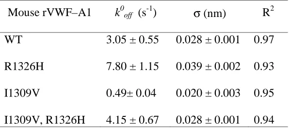
Table 1. Intrinsic dissociation rates constants (k0off) and reactive compliance (σ) values
for murine GPIbα–VWF-A1 tether bond
Values were determined by using maximal likelihood statistical estimates of Bell
parameters.
Mouse rVWF–A1 k0off (s-1) σ (nm) R2
WT
R1326H
I1309V
I1309V, R1326H
3.05 ± 0.55
7.80 ± 1.15
0.49± 0.04
4.15 ± 0.67
0.028 ± 0.001
0.039 ± 0.002
0.020 ± 0.003
0.028 ± 0.001
0.97
0.93
0.95