ISSN Online: 2157-9415 ISSN Print: 2157-9407
Percutaneous Cholecystostomy in High Risk
Patients with Acute Cholecystitis
Mujahid Ahmad Mir, Sheikh Viqar Manzoor, Farooq Ahmad Reshi, Waheed Ahmad Zargar,
Shaukat Jeelani, Faraidon Faiq Ahmad, Aung Zar Ko, Balvinder Singh
Department of General Surgery, Government Medical College, Srinagar, India
Abstract
Aims and Objectives: To assess efficacy and safety of percutaneous cholecys-tostomy (PC) in high risk patients with acute cholecystitis. Materials and Methods: The study was carried out in high risk patients with acute calculous or acalculous cholecystitis. Patients qualifying for the study were subjected to PC under ultrasound (USG) guidance. A cholecystogram was done postopera-tively, to help establish satisfactory catheter position. Results: 24 (70.59%) pa-tients had empyema-gallbladder, 8 (23.53%) had acute calcular cholecystitis and 2 (5.9%) patients were diagnosed as acalcular cholecystitis. None of the patients was fit for general anesthesia at the time of admission. Median hospi-tal-stay after performing procedure was 4 days. Clinical success rate was re-ported 100% in our study. Bile cultures yielded growth of E Coli in 10 (29.41%), klebsela in 8 (23.53%), pseudomonas aeruginosa in 6 (17.65%) and Proteus mirabilis in 4 (11.8%) of patients. 6 (17.65%) patients did not grow any organism in their bile. Growth noted was sensitive to imipenem 29.41% (10), ciprofloxacin 17.65% (6), levofloxacin 17.65% % (6) and cefuroxime 11.76% (4). No major complication was recorded in our study. No procedure related death was observed. Tube displacement occurred in one patient and minor bleeding was reported in 2 patients. Catheter was removed after a mean of 25.25 days. All patients underwent definitive surgical intervention during the follow up period of 3 months. Conclusion: USG guided PC is a safe and effective procedure for treating high-risk patients who present with acute cholecystitis. Once the acute symptoms diminish or resolve, it should be fol-lowed by elective surgery.
Keywords
Percutaneous Cholecystostomy, Cholecystitis, Ultrasound, High Risk, Cholecystogram
How to cite this paper: Mir, M.A., Man-zoor, S.V., Reshi, F.A., Zargar, W.A., Jeela-ni, S., Ahmad, F.F., Ko, A.Z. and Singh, B. (2017) Percutaneous Cholecystostomy in High Risk Patients with Acute Cholecysti-tis. Surgical Science, 8, 154-161.
https://doi.org/10.4236/ss.2017.83017
Received: January 30, 2017 Accepted: March 10, 2017 Published: March 13, 2017
Copyright © 2017 by authors and Scientific Research Publishing Inc. This work is licensed under the Creative Commons Attribution International License (CC BY 4.0).
1. Introduction
Cholecystitis is a common condition that in selected patients, carries significant risk for morbidity and mortality. The treatment of severe acute biliary inflam-mation/infection still results in fatalities and increased hospital costs. The treat-ment of acute cholecystitis is early cholecystectomy [1]. Laparoscopic cholecys-tectomy is now the Gold standard for the management of symptomatic choleli-thiasis and acute cholecystities [2]. Although cholecystectomy is generally safe, the mortality rate of cholecystectomy in patients at high risk for surgery from comorbid conditions ranges between 14% and 30% [3] [4]. Factors contributing to the high surgical mortality in this setting include the presence of sepsis, poor general condition of the patient, immunosuppression and diminished liver func-tion. As a temporizing measure, high-risk patients are treated with a regimen consisting of decompression of the gallbladder combined with broad-spectrum antibiotics.
Percutaneous cholecystostomy (PC) is a technique that consists of percutane-ous placement of a catheter under imaging guidance in the gallbladder lumen.
[5] [6] PC is indicated in poor surgical candidate/high risk patients with acute calculous or acalculous cholecystitis, unexplained sepsis in critically ill patients (diagnostic for cholecystitis as etiology of sepsis if clinical improvement after cholecystostomy), cholangitis, biliary obstruction for drainage of biliary tree fol-lowing failed endoscopic retrograde cholangiopancreaticography (ERCP) and percutaneous transhepatic cholangiography (PTC). This minimally invasive procedure can aid stabilization of a patient to enable a more measured surgical approach with time for therapeutic planning.
2. Aims and Objectives
To assess efficacy and safety of percutaneous cholecystostomy in high risk pa-tients with acute cholecystitis in terms of clinical improvement and complica-tions respectively following the procedure.
3. Materials and Methods
The study was conducted in Post-graduate Department of General Surgery, Govt Medical College Srinagar, over a period of three years (2013-2015). The study was prospective, observational and was carried out in high risk patients, not fit for general anaesthesia because of underlying co-morbidities with acute calcul-ous or acalculcalcul-ous cholecystitis. Patients having un-optimised coagulopathies, suspected gallbladder malignancy, gallbladder packed with calculi preventing catheter insertion or patients with massive ascites were excluded from the study.
procedure in every patient: Complete blood count, complete urine examination, Serum urea and creatinine, Blood sugar (Random/Fasting), Serum Sodium and Potassium, Liver function tests, X-Ray Chest (P/A View), ECG-12 leads, PT/ INR, HIV and Hepatitis serology.
4. Operative Procedure
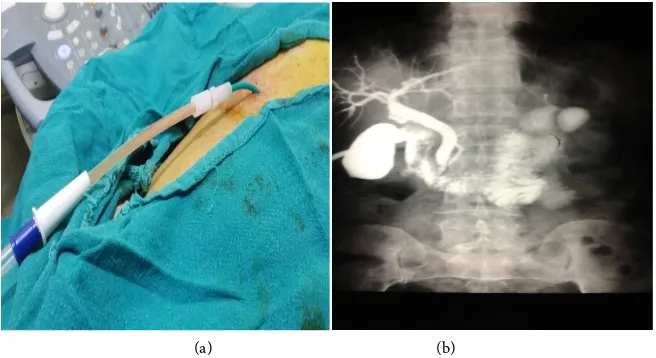
All the cases were done under local anesthesia. The procedure was performed with the patient in supine position after per urethral catheterization. Regular monitoring of the vital signs by a suitably trained staff member was done during the procedure. Procedure was performed using ultrasound guidance and 14-Fre- nch locking pigtail catheter with trocar, under all aseptic precautions. Catheter was secured to skin after attaching a gravity drainage bag. (Figure 1(a)). Bile was sent for gram staining, culture and/or cell count.
Bed rest (for 2 - 4 hours) with regular monitoring vital signs, provision of adequate analgesia was routinely indicated in the first few hours following the procedure. Catheter was flushed and aspirated regularly with saline (6 to 8 hourly). A cholecystogram (injection of contrast into the indwelling catheter under fluoroscopy), was performed when the patient was stable, to help establish satisfactory catheter position and the state of the gallbladder (Figure 1(b)).
5. Results
The study included 34 patients with mean age of 70.11 years, minimum and maximum age was 40 and 98 years respectively. The male: female ratio was 2.4 in our study. The mean BMI of the patients in our study was 31.28. (SD = 3.001), shown in Table 1.
In our study most of the patients presented with chief complaints of pain in upper abdomen and/or right upper quadrant abdomen (88.2%), vomiting (82.3 %), and abdominal swelling/lump (88.2%). Fever and jaundice were present in 70.5% and 8.82% of patients respectively.
[image:3.595.210.538.524.703.2](a) (b)
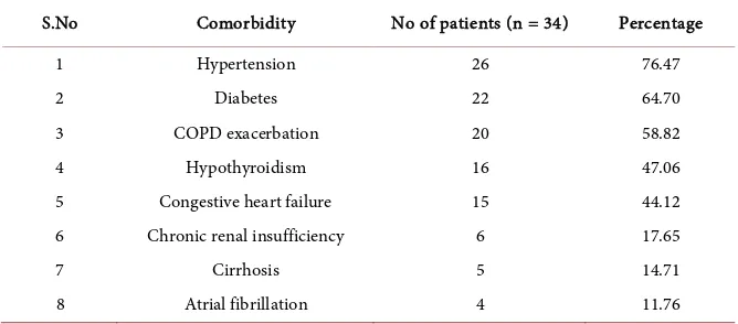
Majority of the patients enrolled in our study were having complicated chole-cystitis. 24 (70.59%) had empyema-gallbladder, 8 (23.53%) had acute calcular cholecystitis and 2 (5.9%) patients were diagnosed as acalcular cholecystitis. All the patients had at-least one comorbidiy. The comorbidity profile of patients is shown in Table 2.
Median hospital stay after performing procedure was 4 days. None of the pa-tients in our study required to stay admitted for more than a week after the pro-cedure was performed. Maximum and minimum hospital-stay following proce-dures was 6 and 3 days respectively.
Clinical success is defined by the French Society of Interventional Radiology as the disappearance of fever and pain as well as reduction of leucocytosis. Visu-al anVisu-alogue score was used to assess pain preoperatively and postoperatively (Table 3).
Pain relief after 48 hours following procedure was statistically significant (P < 0.0001) There was statistically significant reduction in total leukocyte count within 48 hours of procedure.
[image:4.595.206.540.375.551.2]All of the procedures were done via trans-peritoneal route using tracer tech-nique. Bile cultures yielded growth of E Coli in 10 (29.41%) of patients, klebsela
Table 1. Socio-demographic profile of patients.
S No variables No. of patients (n = 34) Percentage
1 Age (years)
40 - 49 4 11.76
50 - 59 2 5.9
60 - 69 6 17.65
70 - 79 17 50.0
80 & above 5 14.7
2 SEX Male 10 29.41
Female 24 70.59
3 BMI (kg/m2)
<30 8 14.7
30 - 34.99 20 58.82
35 and above 8 23.53
Table 2. Showing comorbidity profile of patients.
S.No Comorbidity No of patients (n = 34) Percentage
1 Hypertension 26 76.47
2 Diabetes 22 64.70
3 COPD exacerbation 20 58.82
4 Hypothyroidism 16 47.06
5 Congestive heart failure 15 44.12
6 Chronic renal insufficiency 6 17.65
7 Cirrhosis 5 14.71
[image:4.595.206.540.585.732.2]in 8 (23.53%), pseudomonas aeruginosa in 6 (17.65%) and Proteus mirabilis in 4 (11.8%) of patients. 6 (17.65%) patients did not grow any organism in their bile even after 48 hours of incubation. Most of the growth noted was sensitive to imepenem 29.41% (10), ciprofloxin 17.65% (6), levofloxin 17.65% (6) and cefu-roxime 11.76% (4). Culture and sensitivity of two patients was not available and 6 bile cultures were sterile.
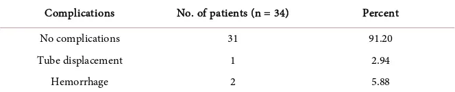
No major complication was recorded in any of the patients enrolled in our study. Tube displacement occurred in one patient which was removed on 4th day
after the procedure and procedure was repeated. Minor bleeding was reported in 2 patients (Table 4).
Catheter was removed after a mean of 25.25 days (range 17 - 35 days). 28 (82.35%) patients underwent laparoscopic cholecystectomy, 2 (5.88%) open cho- lecystectomy and CBD exploration, 4 (11.76%) open cholecystectomy.
6. Discussion
All 34 patients enrolled in our study who underwent percutaneous cholecys-tostomy showed clinical improvement within 48 hours. This level of response suggests that this procedure is an effective alternative to surgery, whether used as a stopgap measure until the patient is clinically fit for an operation or as defini-tive management for those with serious comorbidity or terminal disease. If a pa-tient is not well enough to be transferred to the radiology suite, this procedure can even be done at bedside in the intensive care unit under local anesthesia.
[image:5.595.207.540.528.633.2]In our series of 34 patients the mean age of patients was 70.11 years. This was comparable to that in studies conducted by Gordon B. Werbel et al. [6], Van Steenbergen et al. [7] and Ozgur Bafiaran et al. [8]. Patients presentation was consistent with study conducted by Ozgur Bafiaran, et al. [8]. Majority of the pa-tients enrolled in our study were having complicated cholecystitis. 24 (70.59%) had empyema-gallbladder, 8 (23.53%) had acute calcular cholecystitis and 2
Table 3. Showing improvement in pain after the procedure.
VAS Score of pain Number of patients
At admission 48 hours after procedure
Absent (0) 0 15
Mild (1 - 3) 0 19
Moderate (4 - 6) 8 0
Severe (7 - 10) 26 0
Table 4. Showing complication profile of procedure.
Complications No. of patients (n = 34) Percent
No complications 31 91.20
Tube displacement 1 2.94
[image:5.595.207.539.666.733.2](5.9%) patients were diagnosed as acalcular cholecystitis. In study by Alexander M. Eggermont et al. [9] the procedure was performed in six critically ill patients who had acute acalculous cholecystitis. In study by Gordon B. Werbel et al. [6]
and Van Steenbergen et al. [7], critically ill patients who had acute cholecystitis complicated by empyema formation were chosen as sample population. In study by Shaista Afzal Saeed et al. [10], 25 patients had acute calculus cholecystitis, 10 acalculous cholecystitis, 04 empyema and 2 patients had gallbladder perforation.
All of the patients (n = 34; 100%) had at least 1 comorbidity, with a mean number of 3.2 comorbidities (median 1). The maximum number of comorbidi-ties was 5 (n = 2). Hypertension was most common 26 (76.50%), followed by diabetes mellitus 22 (64.70%) and exacerbation of COPD 20 (58.82%). The com- orbidity profile of our patients was comparable with that of the patients studied by Nicole Cherng et al. [11].
The procedure was done under local anesthesia using trans-peritoneal ap-proach in all patients. The procedure was technically successful in all (34) the patients studied, consistent with that in the study by Griniatsos John et al. [12]. The culture/sensitivity findings of drained fluid/bile were comparable with that reported by Ahmed Farouk Abdulaal et al. [13]. Clinical improvement was no-ticed in all patients within 48 hours. Statistically significant reduction in the val-ues of white blood cells, axillary body temperature and visual analogue score of pain were observed within 48 hours. This is at par with studies by Asgaut Viste
et al. [14], C. Codina et al. [15]. Median hospital stay after performing procedure
was 4 days (range 3 - 6). Follow-up after drainage was with a median of 3 mon- ths (range 2 - 4 months). During that time definitive surgical intervention was done in all the patients after proper optimization.
No complication was seen during or after procedure in 31 (91.20%) patients. Complications occurred in 3 (8.82%) patients including hemorrhage in 2 pa-tients which settled of its own after 2 days with no blood transfusion require-ment and tube displacerequire-ment in 1 patient which required removal and replace-ment of catheter 2 days after the initial procedure In literature complication rate of around 10% [16]: mainly bile leaks, nearly always after trans-peritoneal drai-nage [16], or bleeding requiring transfusion or not [16], drain migration (8.6%), and more rarely, digestive tract perforations or pneumothorax have been docu-mented. There was no direct procedure-related mortality in our study.
All the cases were done via transperitoneal route using trocar technique in our study Complication rate may decrease if the procedure is done via transhepatic route and/or using Saldinger technique.
7. Conclusion
References
[1] Nahrwold, D.L. (1997) Acute Cholecystitis. In: Sabiston, D., Ed., Textbook of Sur-gery, 15th Edition, Saunders, Philadelphia, 1126-1131.
[2] Legorreta, A.P., Silber, J.H., et al. (1993) Increased Cholecystectomy Rate after the Introduction of Laparoscopic Cholecystectomy. The Journal of American Medical Association, 270, 1429-1432.https://doi.org/10.1001/jama.1993.03510120051029 [3] Houghton, P.W.G., Jenkinson, L.R. and Donaldson, L.A. (1985) Cholecystectomy in
the Elderly: A Prospective Study. British Journal of Surgery, 72, 220-222.
https://doi.org/10.1002/bjs.1800720327
[4] Frazee, R.C., Nagorney, D.M. and Mucha, P. (1989) Acute Calculus Cholecystitis.
Mayo Clinic Proceedings, 64, 163-167.
https://doi.org/10.1016/S0025-6196(12)65670-5
[5] Volgelzang, R.L. and Nemcek, A.A. (1988) Percutaneous Cholecystostomy: Diag-nostic and Therapeutic Efficacy. Radiology, 168, 29-34.
https://doi.org/10.1148/radiology.168.1.3289094
[6] Werbel, G.B., Nahrwold, D.L., Joehl, R.J., Volgelzang, R.L. and Rege, R.V. (1989) Percutaneous Cholecystostomy in the Diagnosis and Treatment of Acute Cholecys-titis in the High-Risk Patient. Archives of Surgery, 124, 782-786.
https://doi.org/10.1001/archsurg.1989.01410070032007
[7] Van Steenbergen, W. (1993) Percutaneous Transhepatic Cholecystostomy for Acute Complicated Calculous Cholecystitis in Elderly Patients. Journal of the American Geriatrics Society, 41, 157-162.https://doi.org/10.1111/j.1532-5415.1993.tb02051.x [8] Özgür, B., Nazl, Y., Haldun, S., et al. (2005) Ultrasound-Guided Percutaneous
Cholecystostomy for Acute Cholecystitis in Critically Ill Patients: One Center’s Ex-perience. The Turkish Journal of Gastroenterology, 16, 134-137.
[9] Eggermont, A.M., Laméris, J.S. and Jeekel, J. (1985) Ultrasound-Guided Percuta-neous Transhepatic Cholecystostomy for Acute Acalculous Cholecystitis. Archives of Surgery, 120, 1354-1356.https://doi.org/10.1001/archsurg.1985.01390360020005 [10] Saeed, S.A. and Masroor, I. (2010) Percutaneous Cholecystostomy (PC) in the
Management of Acute Cholecystitis in High Risk Patients. Journal of the College of Physicians and Surgeons Pakistan, 20, 612-615.
[11] Cherng, N., Witkowski, E.T., Sneider, E.B., et al. (2012) Use of Cholecystostomy Tubes in the Management of Patients with Primary Diagnosis of Acute Cholecysti-tis. Journal of the American College of Surgeons, 214, 196-201.
https://doi.org/10.1016/j.jamcollsurg.2011.11.005
[12] Griniatsos, J., Petrou, A., Pappas, P., et al. (2008) Percutaneous Cholecystostomy without Interval Cholecystectomy as Definitive Treatment of Acute Cholecystitis in Elderly and Critically Ill Patients. Southern Medical Journal, 101, 586-590.
[13] Abdulaal, A.F., Sharouda, S.K., et al. (2014) Percutaneous Cholecystostomy Treat-ment for Acute Cholecystitis in High Risk Patients. The Egyptian Journal of Radi-ology and Nuclear Medicine, 45, 1133-1139.
https://doi.org/10.1016/j.ejrnm.2014.07.001
[14] Asgaut, V., Dag, J., et al. (2015) Percutaneous Cholecystostomy in Acute Cholecys-titis; A Retrospective Analysis of a Large Series of 104 Patients. BMC Surgery, 15, 17.https://doi.org/10.1186/s12893-015-0002-8
[16] Pandanaboyana, S., Mittapalli, D., Marioud, A., et al. (2013) Clinical Outcomes of a Percutaneous Cholecystostomy for Acute Cholecystitis: A Multicentre Analysis.
HPB, 15, 511-516.https://doi.org/10.1111/j.1477-2574.2012.00610.x
Submit or recommend next manuscript to SCIRP and we will provide best service for you:
Accepting pre-submission inquiries through Email, Facebook, LinkedIn, Twitter, etc. A wide selection of journals (inclusive of 9 subjects, more than 200 journals)
Providing 24-hour high-quality service User-friendly online submission system Fair and swift peer-review system
Efficient typesetting and proofreading procedure
Display of the result of downloads and visits, as well as the number of cited articles Maximum dissemination of your research work
Submit your manuscript at: http://papersubmission.scirp.org/