SYNCHRONISATION IN THE PREFRONTAL-STRIATAL CIRCUIT TRACKS BEHAVIOURAL CHOICE IN A GO NO-GO TASK IN RATS
Christine Stubbendorff1,2, Manuel Molano-Mazon3,4, Andrew MJ Young1, Todor V Gerdjikov1
1Department of Neuroscience, Psychology and Behaviour, University of Leicester,
Leicester, LE1 9HN, United Kingdom, 2Faculty of Science, University of Nottingham, Sutton Bonington, LE12 5RD, United Kingdom, 3Centre for Systems Neuroscience, University of Leicester, Leicester LE1 7QR, United Kingdom, 4Laboratory of Neural Computation, Istituto Italiano di Tecnologia, 38068 Rovereto (TN), Italy
Number of pages: 28 Number of figures: 6 Number of tables: 5
Number of words in the abstract: 206 Number of words in Introduction: 502 Number of words in Discussion: 1300
Keywords: striatum, prelimbic cortex, choice behaviour, reward, discrimination
All correspondence to: Dr Todor V. Gerdjikov
Department of Neuroscience Psychology and Behaviour University of Leicester, Leicester, LE1 9HN, United Kingdom Phone: +44 (0)116 229 7190
Abstract
Rodent striatum is involved in sensory-motor transformations and reward-related learning.
Lesion studies suggest dorsolateral striatum, dorsomedial striatum, and nucleus accumbens
underlie stimulus-response transformations, goal-directed behaviour and reward expectation
respectively. In addition, prefrontal inputs likely control these functions. Here we set out to
study how reward-driven behaviour is mediated by the coordinated activity of these structures
in the intact brain. We implemented a discrimination task requiring rats to either respond or
suppress responding on a lever after the presentation of auditory cues in order to obtain
rewards. Single unit activity in the striatal subregions and prelimbic cortex was recorded
using tetrode arrays.Striatal units showed strong onset responses to auditory cues paired with
an opportunity to obtain reward. Cue onset responses in both striatum and cortex were
significantly modulated by previous errors suggesting a role of these structures in maintaining
appropriate motivation or action selection during ongoing behaviour. Furthermore, failure to
respond to the reward-paired tones was associated with higher pre-trial coherence among
striatal subregions and between cortex and striatum suggesting a task-negative corticostriatal
network whose activity may be suppressed to enable processing of reward-predictive cues.
Our findings highlight that coordinated activity in a distributed network including both
Introduction
Adaptive behaviour requires the ability to associate multiple cues with a variety of possible
outcomes and behavioural strategies. Striatum is the main input structure to the basal ganglia
and is associated with cognitive and motivational processing as well as with the execution of
motor responses and is considered a key brain region for the regulation of stimulus-driven
behaviour (Hamid et al., 2016, Haber, 2003, Ito and Doya, 2015, Yin et al., 2008).
Region-specific lesions suggest that dorsolateral striatum (DLS), dorsomedial striatum (DMS) and
nucleus accumbens (NAc) contribute differently to specific components of reward-directed
behaviour (Yin et al., 2006, Yin et al., 2005, Hart et al., 2014). Whereas DMS is implicated in
the updating of stimulus-response-outcome contingencies, DLS is primarily associated with
automated stimulus-response behaviour and NAc is thought to mainly integrate motivational
aspects of learning (Yin et al., 2006, Haber, 2003, Yin et al., 2005). Activity between these
regions however is likely to be highly coordinated during reward-related behaviour. Within
striatum, axons and dendrites in each subregion often cross into other subregions (Haber,
2003). Successful behaviour necessitates integration of reward processing, associative
learning and motor planning suggesting that interaction between brain regions maintains
these processes (Haber and Knutson, 2010, Liljeholm and O'Doherty, 2012).
Striatal-dependent reward-related behaviour is modulated by prefrontal input. Prelimbic
cortex (PrL) sends strong projections to both core and shell of the NAc as part of the limbic
cortico-striatal-thalamic circuit and to DMS as part of the associative cortico-striatal-thalamic
circuit (Heidbreder and Groenewegen, 2003, Gabbott et al., 2005, Hart et al., 2014). PrL is
involved in goal-directed behaviour and complex behaviour that requires flexible switching
between different context-dependent strategies (Riga et al., 2014, Heidbreder and
aspects of reward-seeking behaviour, including the updating of response-outcome
contingencies (Eagle and Robbins, 2003, Yin et al., 2008, Van Waes et al., 2012). While DLS
does not receive direct prefrontal input, multiple stimulus-response-outcome contingencies
require a level of executive control over DMS vs. DLS behavioural function such as habitual
vs. goal-directed processes (Riga et al., 2014, Moorman and Aston-Jones, 2015).
How reward-related behaviours guided by multiple cues are encoded in the coordinated
activity of the prefrontal-striatal network is not well understood. Cue responses may reflect
upcoming behavioural choice (Nicola et al., 2004a) and/or previous trial experience (Kim et
al., 2009). To investigate this we assessed the activity of striatal subregions and PrL
simultaneously in a modified go/no-go cue-discrimination task; this task is unlikely to be
isolated to a single subregion (involving a classical component, operant discrimination, etc.)
thus enabling us to study the activity of the subregions in combination. We found that in both
striatum and PrL previous errors resulted in higher cue onset excitatory responses for go cues
and that while cue-onset inhibition was not modulated significantly by previous errors it was
higher on cues preceding errors in the current trial. We also identified that pre-trial
intrastriatal and corticostriatal coherence was significantly higher preceding failures to
respond to reward-predicting cues.
Methods
Animals
Male Lister Hooded rats (n = 4; Charles River, Cambridge, UK) weighing 225-250g on
arrival were kept on reversed light/dark cycle (12:12h; lights on 19.00h). Animals had access
to water ad libitum and access to food (LabDiet 5LF5, PMI Nutrition Intl, Brentwood, MO)
by the University of Leicester Animal Welfare and Ethics Review Body (AWERB) and under
project and personal licences issued by the UK Home Office under the UK Animals
(Scientific Procedures) Act 1986.
Apparatus
Rats were pre-trained in standard operant chambers [Med Associates, Fairfield, VT; 30 x 31 x
24 cm (height x width x depth); prod. no. ENV-008] placed in sound attenuated, ventilated
cubicles and fitted with a magazine (Med Associates prod. nr. ENV-200R2M) for delivery of
sugar pellets (45 mg Dustless Precision Pellets, Bio Serv, Sheffield UK; Product No F0021)
and a retractable lever (Med Associates prod. nr. ENV-112CM) positioned to the left of the
magazine. A stimulus light (Med Associates prod. nr. ENV-221M) was positioned
immediately above the food magazine and the lever. A speaker was positioned above the
magazine just below the ceiling of the box and a house light was positioned at the top of the
opposite wall of the chamber. For electrophysiological recordings, the wall-fitted magazine
was replaced by a custom made square receptacle [2 x 5 x 3 cm (height x width x depth)]
attached to the grid floor 3.5 cm from the wall to allow access to the reward in animals with a
tetrode implant. Auditory stimuli were applied using custom-made tone generators based on
an NE555 integrated circuit (Texas Instruments, Dallas, TX).
Discrimination task
Rats were handled for 1-2 days and exposed to the sugar pellets in their home cage before the
start of the behavioural training. Rats were initially trained to press a lever for sugar pellets
using standard shaping techniques. Subsequently rats were trained on a continuous
reinforcement schedule, which continued until the rat performed 100 lever presses within 30
minutes in two consecutive sessions (all animals achieved this within 2-4 sessions). The
trials) to auditory cues of different frequencies (1 vs. 10 kHz (75dB): counterbalanced). Each
trial started with the presentation of either the go or no-go tone. Four seconds after tone onset
the lever was presented allowing the rat a 4 second response interval to press the lever. Upon
lever press or, if the rat did not press the lever, at the end of the 4 second response interval,
the lever retracted and the tone was switched off. Rats were rewarded with a sugar pellet on
both correct lever press (hit) and correct omission of lever press (correct rejection: CR) trials
whereas incorrect lever press (false alarm: FA) resulted in a 60 second time-out with the
house light and lever light switched off. Incorrect omission of lever press (miss) had no
programmed consequence (Fig. 1A). Each trial was followed by a 60 second inter-trial
interval (ITI). Implantation surgeries were carried out when animals were fully trained.
Tetrode implantation surgery
Rats were anaesthetised with 4% v/v isofluorane (Schering-Plough) in O2, and maintained between 2-3%. Immediately post induction, an injection of glycopyronnium bromide was
administered (6-8µg/kg; i.m.; Anpharm, Warsaw, Poland) to reduce respiratory tract
secretions. The animal was mounted in a stereotaxic frame and the head was adjusted so that
lambda and bregma were aligned on the same horizontal plane. To prevent corneal
desiccation, Lacri-Lube Eye Ointment (Allergan, Westport, Ireland) was applied to the eyes.
A homeothermic heat pad (Harvard Apparatus, Boston, Massachusetts, USA) was used to
maintain body temperature between 36oC and 37oC. Glucose (5%, 3ml/hr, s.c.) was given via an infusion pump (Intec, K.D, Scientific, Holliston, Massachusetts, USA) for the duration of
the surgery. A scalp incision was made along the midline, the periosteum was retracted and
10 stainless steel anchoring screws (Morris Co., Southbridge, Massachusetts, USA, part
number 0X 1/8 flat) were affixed to the skull. A right-side craniotomy was then performed
above mPFC and striatum. Implantation co-ordinates were: +0.8 to +0,4 mm AP; 3.6 to 4.0
mm DV for DMS; +1.2 to +1.6 mm AP; 1.1 to 2.3 mm ML; -6.4 to -7.0 mm DV for NAc,
and +3.2 mm AP; 1.1 mm ML; -2.6 mm DV for PrL (Paxinos and Watson, 2007). The dura
was incised and the tetrode array was advanced into the target structures. The medial of each
tetrodes per structure was targeted at these locations and distance between tetrode tips was
minimal (~ 200 micron). Two tetrodes were implanted in DLS and in DMS and three in NAc.
Each tetrode was made of four 12 μm tungsten wires (H-Formvar insulation with Butyral
bond coat, California Fine Wire Company, Brover Beach, CA) twisted together and heated to
form a bundle. The tip of each wire was gold plated to reduce impedance to 150 - 400 kΩ.
The tetrodes were threaded through a 0.17 mm outer diameter silica tube (SGE Analytical
Science; Milton Keynes, UK) to increase stability and loaded into a microdrive (Versadrive,
Neuralynx, Bozeman; Montana, USA). Within the drive, each tetrode was glued to a delrin
shuttle which was threaded onto an adjustment screw, allowing the shuttle and tetrode to be
moved independently by manually turning the screw. The tetrodes were sealed with paraffin
wax and the implant was built up using layers of light curing dental cement (Flowable
Composite, Henry Schein; Gillingham, UK). Antibiotic ointment (Fuciderm; Uldum,
Denmark) was applied to the wound and the skin was sutured. A non-steroidal
anti-inflammatory analgesic (Carprieve, 5mg/kg; S.C; Norbrook Laboratories Ltd; Corby, UK)
was given in jelly for a minimum 3 days post-surgery or as advised by the University of
Leicester named veterinary surgeon, based on post-op monitoring. Oral antibiotics (Baytril,
2.5%, 0.2ml/kg; S.C., Bayer; Leverkusen, Germany) were given in jelly twice daily
(Harley’s, UK) for 5 days after surgery. The animals were given a week to recover from the
surgery before behavioural testing and remained individually housed for the remainder of the
Electrophysiological Recordings and Data Analysis
The tetrodes were advanced ~0.125mm approximately 20 minutes before each recording
session. During the discrimination task, rats were recorded through a metal coil-wrapped
headstage cable. An op-amp based 32 channel head-stage amplifier (HST/8o50-G1-GR, 1x
gain, Plexon Inc., Dallas, TX, USA) was plugged directly into the head implant and the signal
was passed through a preamplifier (PBX2, 1000x gain; Plexon Inc., Dallas, TX, USA) and
digitized at 25 kHz. For spike sorting the raw signal was band-pass filtered 300-3,000Hz and
spikes were sorted using the Matlab-based Wave_clus software to yield single-unit spike
trains (Quiroga et al., 2004). Single units were detected by applying a threshold of 5 x signal
noise. Signal noise was estimated as the median absolute deviation of the band-passed signal
(Rey et al., 2015). Spike sorting was achieved with super-paramagnetic clustering using a
single parameter (‘temperature’) where in the super-paramagnetic regime clusters of a
relatively large size, corresponding to the different single units, are captured (Fig. 1D). All
automatic detection thresholds and sorting solutions were examined individually and adjusted
if needed. In addition to this we inspected cross-correlograms and autocorrelograms of units
obtained on the same wire as well as average cluster waveforms and ISI intervals for
violations of a refractory period. To examine how synchrony between structures is modulated
in this task, cross-spectrum based spike coherence among regions was calculated during
baseline (-3 to 0 sec before cue onset) and in the cue response phase (0 to 3 seconds after cue
onset) (Halliday, 2015) (Matlab code available online at http://www.neurospec.org). The
total product moment correlation between two spike trains denoted as R2 was obtained by integration of the coherence, defined as the ratio of the magnitude squared cross spectrum
between the two signals to the product of their auto spectra. Minimum Mean Square Error
(MMSE) pre-whitening was applied to the two signals prior to spectral analysis. Behavioural
analysed with permutation tests conducted using the statcond function of the EEGLAB
toolbox in Matlab
(https://uk.mathworks.com/matlabcentral/fileexchange/27960-resampling-statistical-toolkit) (Delorme and Makeig, 2004).
At the end of the experiments, rats were terminally anaesthetized with ketamine (100mg/kg,
i.p.) and tetrode tip locations were lesioned (15 sec of 30µA) to allow visual verification of
recording sites. Following this, rats were killed with a sodium pentobarbital overdose (200mg
in 1ml, i.p.) and perfused transcardially with saline and 4% paraformaldehyde. After
perfusion, brains were refrigerated (5oC) for 24 hours and transferred to 30% sucrose solution for a further 2-3 days after which they were rapidly frozen and stored at -20o C. Tetrode placement was verified visually while cutting the frozen brains in 30μm slices on a cryostat
(Fig. 1E,F and Fig. 5C). In one rat the position of the tetrodes targeting NAc and PrL could
not be verified and single unit responses from these tetrodes were excluded from the analysis.
Results
Discrimination task
Response rates to go and no-go tones as well as lever press latency in hit and FA trials were
calculated from all 49 sessions included in electrophysiological analyses. All rats successfully
learned to discriminate between go and no-go tones and maintained a high average level of
discrimination, i.e. go trial hit rate (number of hits divided by total number of go trials) above
0.75 and no-go trial FA rate (number of FA divided by total number of no-go trials) below
0.25, until the end of the experiment (Fig. 1B). Consistent with prior studies we noted that
lever-press latency was longer in FA trials than hit trials [t(1190) = 5.53, p = 0.005,
Striatal neurons show onset responses to cues predicting upcoming reward availability Medium spiny neurons (MSN) represent more than 90% of rat striatal and accumbal neurons,
and unlike GABAergic interneurons are characterized by relatively low firing rates. We
recorded units with low baseline activity (< 6 Hz), and the firing rates we observed are
consistent with previous studies (Barnes et al., 2005, Sharott et al., 2009). We recorded from
a total of 99 (DLS), 80 (DMS) and 105 (NAc) putative MSN cells; based on each neuron’s
mean modulation across trial types (below) 56, 15 and 51, respectively, of these neurons were
inhibited by cue onset and 43, 65 and 54, respectively, were excited. Average firing rates
were 1.82 ± 0.19, 1.64 ± 0.17 and 1.94 ± 0.14 Hz (mean ± SEM).
To determine an appropriate analysis window, we looked for the interval yielding the highest
number of neurons whose activity was significantly modulated (either excited or inhibited) by
the stimulus cue. We tested analysis windows of increasing duration (0.1-4sec) in 50ms
increments and found that the highest number of neurons were modulated significantly
immediately after cue onset (relative to 3 sec baseline; Wilcoxon rank sum test, p<0.05) (Fig.
2A-B). Single unit responses to cue onset were visualised by calculating a sliding-window
area under the receiver operating characteristics curve (auROC) by comparing the
distribution of firing rates in a 100 ms window against the distribution of baseline firing rates
across all trials (2C), as done previously (Tian et al., 2016). Neurons showing a significant
modulation in at least one trial type (hit, miss, CR, or FA) were selected for further analysis;
82 (DLS), 66 (DMS) and 93 (NAc) (average baseline firing rates: 1.79 ± 0.16, 1.50 ± 0.18
and 1.85 ± 0.16 Hz). Spike responses in excited and inhibited neurons were analysed
separately.
Previous work shows that whether or not an animal made an error on a previous trial affects
neural activity on subsequent trials, possibly related to the role of striatum in updating
behavioural strategy as a function of experience (Kim et al., 2007, Kim et al., 2009).
Therefore, we broke down striatal cue onset responses by previous trial outcome: correct, i.e.,
rewarded (hit or CR) vs. incorrect (miss or FA) trials. Baseline-subtracted striatal activity in
excited units was higher after previous errors [Fig 3A; F(1,272) = 25.95, p = 0.003,
permutation 2-way ANOVA] with a significant interaction between previous trial outcome x
current trial response [F(3,272) = 3.06, p =0.012, permutation 2-way ANOVA]. This
previous-trial outcome effect was similar across the striatal subregions [structure x previous
trial outcome interaction: F(2,384) = 0.0463, p = 0.8632, permutation 2-way ANOVA]. We
noted a main effect of structure [F(2,384) = 4.64, p=0.0373], however the difference in
excitation between subregions was very small (DLS: 2.12 Hz, NAc: 2.11 Hz, DMS: 2.97 Hz)
and the slight apparent increase in DMS was not significant using pairwise comparisons (ps >
.09). Further, current trial outcome (hit, miss, CR or FA) related to cue onset activity only
after previous errors with higher onset activity to go cues (hit and miss) than to FA (Fig 3B;
Table 1). Inhibited neurons on the other hand were not significantly modulated by previous
trial outcome [F(1,560 = 1.16, p =0.211, permutation 2-way ANOVA]. Overall, inhibition
was stronger preceding errors (miss and FA) than correct choices (hit and CR) [Fig. 3C;
F(3,600) = 13.20, p = 0.003; permutation 2-way ANOVA; for pairwise comparisons see
Table 2]. Again we noted a main effect of structure [F(2,600) = 5.26, p = 0.022] with DMS
showing the least inhibition (DLS: -1.07 Hz, NAc: -1.23 Hz, DMS: -0.91 Hz), however the
pairwise differences were small and only significant between DMS and NAc (t(350),
p=0.005). We conclude that go-cue onset excitation in putative MSNs across striatal
subregions is enhanced after previous errors. Inhibition on the other hand is associated with
Errors are associated with higher prestimulus striatal coherence
To investigate how striatal subregion synchronisation relates to the animal’s decisions, we
calculated prestimulus (3 sec before cue onset) spike coherence between neuronal pairs (1010
NAc-DMS, 742 NAc-DLS and 343 DMS-DLS neuron pairs) [Fig. 4; F(3, 600) = 252.01, p =
0.003; F(2,600) = 2.07, p = 0.485; and F(6, 600) = 23.82, p = 0.003; trial, structure and
interaction effects respectively; two-way permutation ANOVA]. Remarkably for all structure
pairs, incorrect choices (miss and FA) were associated with higher pre-stimulus coherence
than correct choices (Table 3). In addition, pre-stimulus coherence between NAc-DMS and
DMS-DLS was higher in misses than FAs (Table 3) and NAc-DMS pre-stimulus coherence
was lower in hit than CR (Table 3).
Cue-evoked (3 sec after cue onset; prestimulus-subtracted) striatal coherence was not
affected by trial or subregions [F(3,3360) = 4.12, p = 0.211; F(2,3360) = 5.44, p = 0.157;
F(6,3360) = 4.19, p = 0.202, trial, structure and interaction effects, two-way permutation
ANOVA].
Cue-related PrL activity is higher after previous error trials, however spike responses do not encode the animal’s upcoming choice
Previous work implicates PrL in the encoding of stimulus-response-outcome associations and
behavioural flexibility in reward-related tasks (Halladay and Blair, 2015, Hosking et al.,
2015, Moorman and Aston-Jones, 2015). We recorded activity in PrL neurons in parallel with
striatum recordings to determine how corticolimbic connectivity is affected in this task. PrL
firing rates were consistent with the cells being pyramidal cells (Bruno and Simons, 2002).
We recorded from a total of 36 putative pyramidal neurons in PrL with baseline firing rate of
the neuron’s mean modulation across trial types and 30 were significantly modulated by cue
onset (Fig. 5A,D, compare Fig. 2B).
Repeating the effect found in striatum, cue-onset activity was modulated by previous error
trials in excited but not inhibited neurons [Fig. 5C; F(1,16) = 4.00, p = 0.017; F(1,80) = 2.72,
p = 0.142 respectively, two-way permutation ANOVA]. Unlike striatum, PrL spike activity at
cue onset did not relate to subsequent trial outcome [F(3,68) = 1.98, p = 0.410 and F(3,112) =
0.80, p = 0.366; excited and inhibited neurons, respectively; one-way permutation ANOVA].
We conclude that reward-cue activity in PrL tracks previous errors as found in striatum
however unlike striatum it does not appear to encode the animal’s imminent choice on the
current trial.
Misses are associated with higher prefrontal-striatal coherence
Previous lesion work suggests interactions between mPFC and striatal subregions may
underlie action selection in reward-related tasks (Baker and Ragozzino, 2014, Christakou et
al., 2004). To determine whether neurophysiological interactions in the intact brain support
these conclusions, we calculated spike coherence between PrL and the three striatal
subregions (900 PrL-NAc, 246 PrL-DMS and 330 PrL-DLS neuron pairs) before cue onset (3
sec) and on presentation of the cue (3 sec after onset) (Fig. 6A). The prestimulus results
repeated the prominent effect on misses reported in striatum [compare Fig. 6A and Fig. 4;
F(3,3132) = 433.11, p < 0.003; F(2, 3132) = 51.11, p = 0.008; F(6, 3132) = 9.03, p = 0.291;
trial, structure and interaction effects respectively; two-way permutation ANOVA].
Specifically Prl-NAc coherence was highest preceding misses (Table 4) but also higher
preceding FA errors than correct choices (Table 4). In the PrL-DMS pair, pre-cue coherence
was highest preceding misses (Table 4), and lowest preceding hits but note that hit-FA was
not significant (Table 4). In the PrL-DLS pair, coherence was highest preceding misses
CR and FA trials were preceded by intermediate coherence values. To summarize a correct
go response was preceded by relatively low coherence whereas failure to respond was
preceded by relatively high coherence. This effect replicated across all striatum subregions.
Finally we observed that Prl-NAc and Prl-DMS but not Prl-DLS cue-evoked
coherence was higher on miss trials [Fig. 6B; F(3,2856) = 69.37, p = 0.003; F(2, 2856) =
15.05, p = 0.042; F(6, 2856) = 42.52, p = 0.003; trial, structure and interaction effects
respectively; two-way permutation ANOVA; Table 5). In addition, Prl-NAc cue-evoked
coherence was lower on CR trials (Table 5). This dissociation maps directly onto the direct
PrL to NAc and DMS but not DLS projections and suggests that cue-triggered
synchronisation preceding miss and CR trials is specific to direct projections (Heidbreder and
Groenewegen, 2003, Gabbott et al., 2005, Hart et al., 2014, Voorn et al., 2004). This mapping
onto anatomical connection after stimulus onset (Fig. 6B) was not apparent in the baseline
synchronization (Fig 6A). This suggests that behaviourally relevant sensory stimulation may
produce coherence patterns that are more directly related to anatomical connections than
those observed in the adapted network state (at baseline). This possibility requires further
exploration. Overall we conclude that high synchronisation between PrL and the striatal
subregions is associated with failures to respond to the reward predicting cue.
Discussion
This work investigated the representation of reward-related decisions in a distributed circuit
encompassing multiple striatal subregions and prelimbic cortex. Striatum showed robust
excitatory and inhibitory responses to cue onset. Excitatory responses were higher following
previous errors and for go cues. On the other hand, inhibitory cue-onset responses were
higher preceding error choices. We also investigated how synchronisation in the
and structures was that corticostriatal and intra-striatal spike synchronisation was higher
preceding failures to respond to the reward-paired tones. This finding suggests a network
whose activity may be suppressed to enable processing of reward-predictive cues.
Whereas some previous work report activity in dorsal striatum to be unaffected by cue onset
(Root et al., 2010, Berke, 2008, Kimchi et al., 2009), other work suggest cue-triggered
activity in NAc depends on the subsequent behaviour or outcome of the trial (Nicola et al.,
2004a, Roitman and Loriaux, 2014). In addition, dorsal striatal and NAc neurons have been
previously shown to modulate their activity according to the rat’s actions in previous trials
(Kim et al., 2009, Oyama et al., 2015). Consistent with this here we found that cue-related
excitatory activity was increased after previous errors (Fig. 3A). This observation suggests
that striatal spiking activity may serve to maintain appropriate action selection during
ongoing behaviour. However, we also found that cue onset activity related to the structure of
the task (higher for go cues than no-go cues) rather than the animal’s behavioural choice (hit,
miss, etc.) (Fig. 3B). It would appear therefore that this onset activity is not related to
attention to specific cues, but may perhaps reflect fluctuations in motivation after negative
feedback. This was in contrast with cue-onset inhibition which did relate to the animal’s
behavioural choice: inhibition was significantly more pronounced when the animal was about
to commit an error, independent of any possible motor preparation component (i.e., preceding
both FA and miss errors; Fig. 3C). It is unclear what local or modulatory networks may
underlie this distinction. In particular striatal cholinergic interneurons signal the occurrence
of motivationally salient stimuli, provide an inhibitory signal to medium spiny projection
neurons (MSNs), and may mediate reward-guided behaviour (English et al., 2011). It is thus
possible that cholinergic inhibition of subsets of MSNs may account for the current
tempting to refer to recent work implicating direct and indirect pathway MSNs in
reward-driven behaviours. Direct pathway MSNs may support the execution of desired actions
whereas indirect pathway MSNs may be related to the inhibition of competing responses and
whose trial-to-trial activity may thus relate to cue attention and correct behavioural choice
(Vicente et al., 2016). Previous lesion work ascribe different roles for DLS, DMS and NAc in
reward-directed responding (Yin et al., 2006, Yin et al., 2005, Hart et al., 2014). Here we
found no significant differences in cue-related responses between striatal subregions. It must
be pointed out however that the task used in the current study is unlikely to be isolated to a
single subregion due to the engagement of multiple reward-related processes (classical
conditioning, discrimination, operant responding, etc.) and is likely to involve contributions
from all three subregions (Haber and Knutson, 2010, Liljeholm and O'Doherty, 2012). Thus
the present results are not inconsistent with previous lesion work.
Our second major finding which was repeated across striatal subregions and
prefrontal-striatal synchrony analyses was that failure to respond to reward-predicting cues (i.e., miss
trials) was associated with higher intra- and inter-region spike coherence (Fig. 4). Increased
coherence preceding miss trials may relate to low levels of attention to external stimuli.
Previous work has linked fluctuation in cortical activity and network connectivity to
attentional state (Melloni et al., 2007, Forstmann et al., 2010, Sadaghiani et al., 2010, Herzog
et al., 2014) but our results are the first to suggest that a high spike synchronisation network
state in the prefrontal-striatal circuit may impair task performance, potentially being
associated with low levels of attention to external stimuli causing the rat to miss the cue. Here
we also observed that higher intrastriatal synchronisation was associated with an increased
likelihood of false alarms (Fig 4). Changes in NAc activity has been linked to
that high baseline synchronisation between NAc and the dorsal striatal subregions may
produce a ‘go’ bias regardless of the cue value. Interestingly given the lack of a NAc-DLS
direct projection this synchronisation-produced bias may involve an extended ‘task positive’
circuit encompassing multiple brain regions which biases the activity of both structures
(Sadaghiani et al., 2010). Further causal studies using perturbation of target circuits using
recent viral approaches are needed to further investigate this issue.
Previous work implicates PrL in the encoding of stimulus-response-outcome
associations and in successful switching between behavioural strategies depending on context
(Halladay and Blair, 2015, Hosking et al., 2015, Moorman and Aston-Jones, 2015). We
therefore examined how PrL cue-evoked neuronal activity relates to reward-directed
decisions. The number of PrL neurons we were able to record was not high (36) although
power estimates are not readily available for spike data. Bearing this caveat in mind, the
following was observed. Whereas cue-induced striatal responses related to the animal’s
choices, this effect was not apparent in PrL. It should be noted however that unlike previous
experiments where rats were trained to make a choice immediately upon the presentation of
the cue, here cue onset signalled a delayed opportunity to make a behavioural choice (4 sec;
Fig. 1A). Thus reported firing rate increases on cue presentation in other studies may relate to
action initiation (compare PrL projections to motor and premotor cortices (Bedwell et al.,
2014)). PrL cue onset excitation was increased after previous errors, regardless of
behavioural choice on the current trial (compare Fig 5C). Because this onset activity was not
related to behavioural choice it likely reflects global variables such as fluctuations in
motivation after negative feedback. The significance of these PrL cue-onset firing rate
fluctuations must be distinguished from PrL-striatum network activation effects. For
example, previously mPFC-NAc disruption has been shown to interfere with the planning of
updating of response-outcome contingencies (Christakou et al., 2004). Our
neurophysiological data extends this to show that pre-stimulus synchronisation between PrL
and striatum profoundly affects behavioural choice. Repeating the results we report with
striatum subregions, increased baseline PrL-striatum coherence was associated with misses.
This finding is especially robust as it replicated across multiple subregions and with
within-subregion results (compare Fig. 6A with Fig. 4). There is in fact an extensive human
literature implicating pre-stimulus inter-cortical coherence in stimulus detection (Melloni et
al., 2007, Forstmann et al., 2010). Further in rats, increased prefrontal-parietal coherence
preceded detection failures in an auditory detection task (Herzog et al., 2014). Low detection
rates following high coherence may represent functional inhibition within an underlying
cortical network diverting attention away from external stimuli to focus attention on internal
representations such as working memory (Hanslmayr et al., 2007, van Dijk et al., 2008,
Mazaheri et al., 2009). Through the associative, sensory-motor and limbic
cortico-striatal-thalamic circuits, PrL and striatal subregions are intricately connected to both task-positive
and task-negative networks implicated in the regulation of attention to external stimuli
(Sadaghiani et al., 2010, Van Waes et al., 2012). Here we identify neurophysiologically a
‘task-negative’ network encompassing PrL and striatum whose activity signals a failure to
respond to a reward-predicting cue.
In summary, here we investigated prefrontal-striatal spike network activity in the context of
reward-related decisions in rats. We show that activity in this system relates to previous and
upcoming behavioural choices in a way that supports the coordinated role of striatal
subregions in maintaining appropriate action selection. We also identify for the first time a
reward-predictive cues. Thus our findings highlight the significance of coordinated
prefrontal-striatal activity in underlying reward-related decisions.
Acknowledgement
MMM was funded by a European Union's Horizon 2020 research and innovation programme
under the Marie Sklodowska-Curie grant agreement No 699829. CS was in receipt of a
University of Leicester postgraduate studentship. We thank the University of Leicester
Biomedical Workshop for excellent technical support. We also thank Dr Gonzalo Urcelay
and Dr David Halliday for feedback during the preparation of the manuscript.
Author contribution
Experimental design: C Stubbendorff, AMJ Young and TV Gerdjikov. Execution of
experiment: C Stubbendorff. Data analysis: C Stubbendorff, M Molano, TV Gerdjikov.
Manuscript: C Stubbendorff, TV Gerdjikov.
Conflict of interest statement Authors state no competing interests.
Data accessibility
The data associated with this manuscript can be obtained upon request by contacting the
References
BAKER, P. M. & RAGOZZINO, M. E. 2014. Contralateral disconnection of the rat prelimbic cortex and dorsomedial striatum impairs cue-guided behavioral switching. Learn Mem, 21, 368-79. BARNES, T. D., KUBOTA, Y., HU, D., JIN, D. Z. & GRAYBIEL, A. M. 2005. Activity of striatal neurons
reflects dynamic encoding and recoding of procedural memories. Nature, 437, 1158-61. BEDWELL, S. A., BILLETT, E. E., CROFTS, J. J. & TINSLEY, C. J. 2014. The topology of connections
between rat prefrontal, motor and sensory cortices. Front Syst Neurosci, 8, 177.
BERKE, J. D. 2008. Uncoordinated firing rate changes of striatal fast-spiking interneurons during behavioral task performance. J Neurosci, 28, 10075-80.
BRUNO, R. M. & SIMONS, D. J. 2002. Feedforward mechanisms of excitatory and inhibitory cortical receptive fields. J Neurosci, 22, 10966-75.
CHRISTAKOU, A., ROBBINS, T. W. & EVERITT, B. J. 2004. Prefrontal cortical-ventral striatal interactions involved in affective modulation of attentional performance: implications for corticostriatal circuit function. J Neurosci, 24, 773-80.
CURZON, P., BANNON, A. W. & DECKER, M. W. 1999. Effect of 192IgG-saporin injection into the
nucleus basalis magnocellularis on acquisition and performance of a go/no-go procedure in the rat. Psychobiology, 27, 114-122.
DELORME, A. & MAKEIG, S. 2004. EEGLAB: an open source toolbox for analysis of single-trial EEG dynamics including independent component analysis. J Neurosci Methods, 134, 9-21.
EAGLE, D. M. & ROBBINS, T. W. 2003. Lesions of the medial prefrontal cortex or nucleus accumbens core do not impair inhibitory control in rats performing a stop-signal reaction time task. Behav Brain Res, 146, 131-144.
ENGLISH, D. F., IBANEZ-SANDOVAL, O., STARK, E., TECUAPETLA, F., BUZSAKI, G., DEISSEROTH, K., TEPPER, J. M. & KOOS, T. 2011. GABAergic circuits mediate the reinforcement-related signals of striatal cholinergic interneurons. Nat Neurosci, 15, 123-30.
FORSTMANN, B. U., ANWARDER, A., SCHAFER, A., NEUMANN, J., BROWN, S., WAGENMAKERS, E., BOGACZ, R. & TURNER, R. 2010. Cortico-striatal connections predict control over speed and accuracy in perceptual decision making. PNAS, 107, 15916-15920.
FUNAMIZU, A., ITO, M., DOYA, K., KANZAKI, R. & TAKAHASHI, H. 2015. Condition interference in rats performing a choice task with switched variable- and fixed-reward conditions. Front Neurosci, 9, 27.
GABBOTT, P. L., WARNER, T. A., JAYS, P. R., SALWAY, P. & BUSBY, S. J. 2005. Prefrontal cortex in the rat: projections to subcortical autonomic, motor, and limbic centers. J Comp Neurol, 492,
145-77.
HABER, S. N. 2003. The primate basal ganglia: parallel and integrative networks. Journal of Chemical Neuroanatomy, 26, 317-330.
HABER, S. N. & KNUTSON, B. 2010. The reward circuit: linking primate anatomy and human imaging. Neuropsychopharmacology, 35, 4-26.
HALLADAY, L. R. & BLAIR, H. T. 2015. Distinct ensembles of medial prefrontal cortex neurons are activated by threatening stimuli that elicit excitation versus inhibition of movement. J Neurophysiol, 114, 793-807.
HALLIDAY, D. M. 2015. Nonparametric directionality measures for time series and point process data. J Integr Neurosci, 14, 253-77.
HAMID, A. A., PETTIBONE, J. R., MABROUK, O. S., HETRICK, V. L., SCHMIDT, R., VANDER WEELE, C. M., KENNEDY, R. T., ARAGONA, B. J. & BERKE, J. D. 2016. Mesolimbic dopamine signals the value of work. Nat Neurosci, 19, 117-26.
HARDING, E. J., PAUL, E. S. & MENDL, M. 2004. Animal behaviour: Cognitive bias and affective state. Nature, 22, 312.
HART, G., LEUNG, B. K. & BALLEINE, B. W. 2014. Dorsal and ventral streams: the distinct role of striatal subregions in the acquisition and performance of goal-directed actions. Neurobiol Learn Mem, 108, 104-18.
HEIDBREDER, C. A. & GROENEWEGEN, H. J. 2003. The medial prefrontal cortex in the rat: evidence for a dorso-ventral distinction based upon functional and anatomical characteristics. Neurosci Biobehav Rev, 27, 555-579.
HERZOG, L., SALEHI, K., BOHON, K. S. & WIEST, M. C. 2014. Prestimulus frontal-parietal coherence predicts auditory detection performance in rats. J Neurophysiol, 111, 1986-2000.
HOSKING, J. G., COCKER, P. J. & WINSTANLEY, C. A. 2015. Prefrontal Cortical Inactivations Decrease Willingness to Expend Cognitive Effort on a Rodent Cost/Benefit Decision-Making Task. Cereb Cortex, 26, 1529-38.
ITO, M. & DOYA, K. 2015. Distinct neural representation in the dorsolateral, dorsomedial, and ventral parts of the striatum during fixed- and free-choice tasks. J Neurosci, 35, 3499-514.
KIM, H., SUL, J. H., HUH, N., LEE, D. & JUNG, M. W. 2009. Role of striatum in updating values of chosen actions. J Neurosci, 29, 14701-12.
KIM, Y. B., HUH, N., LEE, H., BAEG, E. H., LEE, D. & JUNG, M. W. 2007. Encoding of action history in the rat ventral striatum. J Neurophysiol, 98, 3548-56.
KIMCHI, E. Y., TORREGROSSA, M. M., TAYLOR, J. R. & LAUBACH, M. 2009. Neuronal correlates of instrumental learning in the dorsal striatum. J Neurophysiol, 102, 475-89.
LILJEHOLM, M. & O'DOHERTY, J. P. 2012. Contributions of the striatum to learning, motivation, and performance: an associative account. Trends Cogn Sci, 16, 467-75.
MAZAHERI, A., NIEUWENHUIS, I. L., VAN DIJK, H. & JENSEN, O. 2009. Prestimulus alpha and mu activity predicts failure to inhibit motor responses. Hum Brain Mapp, 30, 1791-800.
MELLONI, L., MOLINA, C., PENA, M., TORRES, D., SINGER, W. & RODRIGUEZ, E. 2007. Synchronization of neural activity across cortical areas correlates with conscious perception. J Neurosci, 27,
2858-65.
MOORMAN, D. E. & ASTON-JONES, G. 2015. Prefrontal neurons encode context-based response execution and inhibition in reward seeking and extinction. PNAS, 112, 9472-9477.
NICOLA, S. M., YUN, I. A., WAKABAYASHI, K. T. & FIELDS, H. L. 2004a. Cue-evoked firing of nucleus accumbens neurons encodes motivational significance during a discriminative stimulus task. j Neurophysiol, 91, 1840-1865.
NICOLA, S. M., YUN, I. A., WAKABAYASHI, K. T. & FIELDS, H. L. 2004b. Firing of Nucleus Accumbens Neurons During the Consummatory Phase of a Discriminative Stimulus Tak Depends on Previous Reward Predictive Cues. J Neurophysiol, 91, 1866-82.
OYAMA, K., TATEYAMA, Y., HERNADI, I., TOBLER, P. N., IIJIMA, T. & TSUTSUI, K. 2015. Discrete coding of stimulus value, reward expectation, and reward prediction error in the dorsal striatum. J Neurophysiol, 114, 2600-15.
PAXINOS, G. & WATSON, C. 2007. The Rat Brain In Stereotaxic Coordinates. 6th Edition ed.: Academic Press.
QUIROGA, R. Q., NADASDY, Z. & BEN-SHAUL, Y. 2004. Unsupervised Spike Detection and Sorting with Wavelets and Superparamagnetic Clustering. Neural Comput, 16, 1661-1687.
REY, H. G., PEDREIRA, C. & QUIAN QUIROGA, R. 2015. Past, present and future of spike sorting techniques. Brain Res Bull, 119, 106-17.
RIGA, D., MATOS, M. R., GLAS, A., SMIT, A. B., SPIJKER, S. & VAN DEN OEVER, M. C. 2014. Optogenetic dissection of medial prefrontal cortex circuitry. Front Syst Neurosci, 8, 230. ROITMAN, J. D. & LORIAUX, A. L. 2014. Nucleus accumbens responses differentiate execution and
ROITMAN, M. F., WHEELER, R. A. & CARELLI, R. M. 2005. Nucleus accumbens neurons are innately tuned for rewarding and aversive taste stimuli, encode their predictors, and are linked to motor output. Neuron, 45, 587-97.
ROOT, D. H., TANG, C. C., MA, S., PAWLAK, A. P. & WEST, M. O. 2010. Absence of cue-evoked firing in rat dorsolateral striatum neurons. Behav Brain Res, 211, 23-32.
SADAGHIANI, S., HESSELMANN, G., FRISTON, K. J. & KLEINSCHMIDT, A. 2010. The relation of ongoing brain activity, evoked neural responses, and cognition. Front Syst Neurosci, 4, 20.
SHAROTT, A., MOLL, C. K., ENGLER, G., DENKER, M., GRUN, S. & ENGEL, A. K. 2009. Different subtypes of striatal neurons are selectively modulated by cortical oscillations. J Neurosci, 29,
4571-85.
TIAN, J., HUANG, R., COHEN, J. Y., OSAKADA, F., KOBAK, D., MACHENS, C. K., CALLAWAY, E. M., UCHIDA, N. & WATABE-UCHIDA, M. 2016. Distributed and Mixed Information in Monosynaptic Inputs to Dopamine Neurons. Neuron, 91, 1374-89.
VAN DIJK, H., SCHOFFELEN, J. M., OOSTENVELD, R. & JENSEN, O. 2008. Prestimulus oscillatory activity in the alpha band predicts visual discrimination ability. J Neurosci, 28, 1816-23. VAN WAES, V., BEVERLEY, J. A., SIMAN, H., TSENG, K. Y. & STEINER, H. 2012. CB1 Cannabinoid
Receptor Expression in the Striatum: Association with Corticostriatal Circuits and Developmental Regulation. Front Pharmacol, 3, 21.
VICENTE, A. M., GALVAO-FERREIRA, P., TECUAPETLA, F. & COSTA, R. M. 2016. Direct and indirect dorsolateral striatum pathways reinforce different action strategies. Curr Biol, 26, R267-9. VOORN, P., VANDERSCHUREN, L. J., GROENEWEGEN, H. J., ROBBINS, T. W. & PENNARTZ, C. M. 2004.
Putting a spin on the dorsal-ventral divide of the striatum. Trends Neurosci, 27, 468-74. YIN, H. H., KNOWLTON, B. J. & BALLEINE, B. W. 2006. Inactivation of dorsolateral striatum enhances
sensitivity to changes in the action-outcome contingency in instrumental conditioning. Behav Brain Res, 166, 189-96.
YIN, H. H., OSTLUND, S. B. & BALLEINE, B. W. 2008. Reward-guided learning beyond dopamine in the nucleus accumbens: the integrative functions of cortico-basal ganglia networks. Eur J Neurosci, 28, 1437-48.
Figure Captions
Figure 1. A. Behavioural paradigm. Rats were trained to either respond (go trials) or supress responding (no-go trials) to discrete auditory cues of different frequencies (1 or 10 kHz,
counter-balanced). B. Mean response rates (no. hits/total no. go trials; no. FA/total no. no-go
trials) for discrimination sessions included in the single unit analyses (49 sessions from 4
rats). The dashed line represents chance level. Inserted pie charts depict the proportion of hit,
correct rejection (CR), miss and false alarm (FA) trials. C. Latency to lever press was
significantly higher in FA trials compared with Hit trials, *** p < 0.001, error bars indicate
+/- SEM. D. Example waveforms from two neurons recorded in DLS by four tetrode wires. E
& F. Verification of tetrode placement in dorsal striatum (E) and NAc (F) based on histology.
Figure 2. Striatal subregion activity triggered by reward-predicting cues. A. Striatal single unit responses to cue onset in Hit trials in excited (top) and inhibited (bottom) neurons. B.
Number of neurons significantly modulated by upcoming trial outcome (p < 0.05) for
intervals of varying duration. Dashed lines indicate the upper limit of chance levels estimated
using the inverse binomial formula with p = 0.05 (Matlab function binoinv). C. Mean
z-transformed firing rates of DLS, DMS and NAc excited (top) and inhibited (bottom) neurons
using trials that elicited the greatest significant response to trial onset (time bin: 100ms,
against baseline). Shaded area indicates bootstrapped 99% confidence intervals.
Figure 3. Striatal cue-onset activity relative to baseline in the first 100ms following cue onset is associated with upcoming behavioural choice. Responses from the striatal subregions were
pooled together due to the lack of a subregion x trial interaction. A. Cue onset activity in
excited neurons in relation to correct (hit & CR) or incorrect (miss & FA) behavioural
previous incorrect responses. C. Effect of trial outcome in inhibited neurons. *p < 0.05; **p
< 0.01.
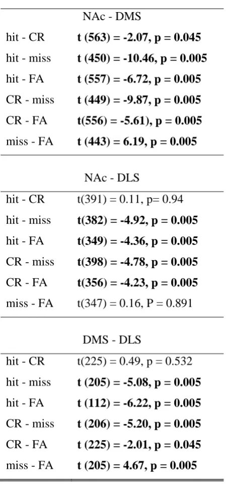
Figure 4: Association between striatal synchronisation and behavioural choice. A strong association between pre-stimulus coherence (3 sec before cue onset) and behavioural choice
was present between all three striatal subregions. *p < 0.05; **p < 0.01.
Figure 5: PrL activity triggered by reward-predicting cues A. Example PrL single unit response on cue onset in hit trials. B. Number of neurons significantly modulated by
upcoming trial outcome (p < 0.05) for intervals of varying duration. Dashed line indicates
chance levels as in Fig. 2B. C. Average single unit responses in excited neurons in relation to
correct (hit & CR) or incorrect (miss & FA) behavioural choice in the previous trial. *p <
0.05. D. Mean z-transformed firing rates of PrL excited (top) and inhibited (bottom) neurons
using trials that elicited the greatest significant response to trial onset (time bin: 100ms,
against baseline). Shaded area indicates bootstrapped 99% confidence intervals. E.
Verification of tetrode placement in PrL based on histology.
Figure 6: Association between PrL-striatal synchronisation and behavioural choice. A. A strong association between pre-stimulus coherence and behavioural choice was present
between PrL and all three striatal subregions. B. Cue-triggered PrL-striatal synchronisation.
Tables
Table 1: Pairwise comparisons between trial types in cue-excited neurons following error trials. Significant comparisons are listed in bold (permutation t-test, p > 0.05).
Effect of previous error
hit - CR t(59) = 1.35, p = 0.244 hit - miss t(51) = -0.05, p = 0.871
hit - FA t(49) = 2.81, p = 0.005 CR - miss t(70) = -1.58, p = 0.124
CR - FA t(68) = 1.51, p= 0.134
[image:25.595.67.301.424.560.2]miss - FA t (60) = 3.01, p = 0.005
Table 2: Pairwise comparisons between trial types in cue-inhibited neurons. Significant comparisons are listed in bold (permutation t-test, p > 0.05).
Effect of current trial
hit - CR t(246) = -0.02, p = 0.901
Table 3: Pairwise comparisons of prestimulus coherence between trial types in striatal subregion pairs. Significant comparisons are listed in bold (permutation t-test, p > 0.05).
Prestimulus coherence NAc - DMS
hit - CR t (563) = -2.07, p = 0.045 hit - miss t (450) = -10.46, p = 0.005 hit - FA t (557) = -6.72, p = 0.005 CR - miss t (449) = -9.87, p = 0.005 CR - FA t(556) = -5.61), p = 0.005 miss - FA t (443) = 6.19, p = 0.005
NAc - DLS
hit - CR t(391) = 0.11, p= 0.94
hit - miss t(382) = -4.92, p = 0.005 hit - FA t(349) = -4.36, p = 0.005 CR - miss t(398) = -4.78, p = 0.005 CR - FA t(356) = -4.23, p = 0.005 miss - FA t(347) = 0.16, P = 0.891
DMS - DLS
hit - CR t(225) = 0.49, p = 0.532
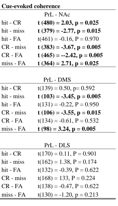
Table 4: Pairwise comparisons of prestimulus coherence between trial types for PrL- striatal subregion pairs. Significant comparisons are listed in bold (permutation t-test, p > 0.05).
Prestimulus coherence PrL - NAc
hit - CR t(332) = -1.23 p= 0.2043
hit - miss t (409) = -9.26, p = 0.005 hit - FA t (495) = -5.89, p = 0.005 CR - miss t (219) = -4.66, p = 0.005 CR - FA t (305) = -2.60, p = 0.005 miss - FA t (382) = 5.92, p = 0.005
PrL - DMS
hit - CR t (71) = -3.81, p = 0.005 hit - miss t (106) = -6.36, p = 0.005 hit - FA t (136) = -0.83, p = 0.503
CR - miss t (106) = -5.58, p = 0.005 CR - FA t(136) = 0.10, p= 0.950
miss - FA t (100) = 4.91, p = 0.005
PrL - DLS
hit - CR t(183) = -3.01, p = 0.005 hit - miss t (183) = -5.11, p = 0.005 hit - FA t(144) = -6.38, p = 0.005 CR - miss t (91) = -6.10, p = 0.005 CR - FA t(143)= -0.94, P = 0.373
Table 5: Pairwise comparisons of cue-evoked coherence between trial types for PrL- striatal subregion pairs. Significant comparisons are listed in bold (permutation t-test, p > 0.05).
Cue-evoked coherence PrL - NAc
hit - CR t (480) = 2.03, p = 0.025 hit - miss t (379) = -2.77, p = 0.015 hit - FA t(461) = -0.16, P = 0.970 CR - miss t (383) = -3.67, p = 0.005 CR - FA t (465) = --2.42, p = 0.005 miss - FA t (364) = 2.71, p = 0.025
PrL - DMS
hit - CR t(139) = 0.50, p= 0.592 hit - miss t (103) = -3.45, p = 0.005 hit - FA t(131) = -0.22, P = 0.950 CR - miss t (106) = -3.55, p = 0.015 CR - FA t(134) = -0.61, P = 0.532 miss - FA t (98) = 3.24, p = 0.005
PrL - DLS
8 8bV5 8b5 8b75 re s p o n s e Tr a te la te n c y TC s e c E xxx Duration: Go LeverTpress No Reward
leverTpress consequenceNo
Nogo LeverTpress No TimeToutTC68sE leverTpress Reward ToneTon: LeverTout: 4s 4s
D:TExampleTtetrodeTwaveforms E:TDorsalTstratumTtetrodeTplacements F:TNAcTtetrodeTplacements
3 bregmaT3b9Vmm bregmaT3b56mm bregmaT3bV8mm bregmaT3b88mm bregmaT8b3Vmm bregmaTVb84mm bregmaTVbV8mm bregmaTVb5Vmm bregmaTVb76mm hit FA 8 3 V 3 4 go nogo h it 8 6 3
m is s 3 3 6 C R 8 3 7
3 3 8
3ms
388T
μ
hit miss CR FA spikeszsec 4b 2b b time/3sec8
g1 b 1
time/3sec8
g1 b 1
2b 1b b
time/3sec8
g1 b 1
b 6 3 1b b time/3sec8
g1 b 1
spikeszsec
2b b
time/3sec8
g1 b 1
time/3sec8
g1 b 1
1b b DMS b 2b 4b 6b 8b
intervals/3sec81 2 3 4 b noy/of/neuron s b 2b 4b 6b 8b
intervals/3sec81 2 3 4 b NAc b 2b 4b 6b 8b
intervals/3sec81 2 3 4 b noy/of/neuron s Cy/Population/cuegtriggered/activity time/3sec8 time/3sec8/ DLS/excited/neurons g2 g1 b 1 2 zscore/3sp ikeszs8 time/3sec8 DLS/inhibited/neurons g2 g1 b 1 2 zscore/3sp ikeszs8 g2 g1 b 1 zscore/3sp ikeszs8 2 DMS/excited/neurons DMS/inhibited/neurons NAc/excited/neurons NAc/inhibited/neurons g2 g1 b 1 2 zscore/3sp ikeszs8 g2 g1 b 1 zscore/3sp ikeszs8 2
g2 g1 b 1 2 3 4
g2 g1 b 1 2 3 4
g2 g1 b 1 2 3 4
g2 g1 b 1 2 3 4
g2 g1 b 1 2 3 4
g2 g1 b 1 2 3 4
*
spikes/sec
0 4 8 12 16 20
hit CR miss FA
spikes/sec
-8 -6 -4 -2
0 hit CR miss FA
**
0 5 10 15 20 25 30
spikes/sec
**
after correct
NAc-DMS
hit CR miss FA
NAc-DLS DMS-DLS
**
hit CR miss FA
R2
** * *
*
0 0.2 0.4 0.6 0.8
0 0.2 0.4 0.6
hit CR miss FA
0 0.2 0.4 0.6 0.8 1
timeF7sec4
61 z 1 2z 1z z spikesysec miss CR FA noDFofFneuron 5 z z 1z 15 2z EDFPrLFtetrodeFplacements bregmaF3D72mm bregmaF3D24mm intervalF7sec41 2 3 4
spikesysec after correct after error v CDFEffectFofFpreviousFerror z 4 8 12 16 2z 62 62 61 z 2 zscoreF7sp ikesys4 timeF7sec4FrelativeFtoFcueFonset PrLFinhibitedFneurons 62 61 z 1 zscoreF7sp ikesys4 2 1
62 61 z 1 2 3 4
hit CR miss FA R2 PrL-NAc B.1Cue-triggered1coherence1 PrL-NAc R2
hit CR miss FA
PrL-DMS
hit CR miss FA
PrL-DMS
hit CR miss FA
PrL-DLS
hit CR miss FA
PrL-DLS ** ** ** ** ** ** 0 0.1 0.2 0.3 0.4 0 0.1 0.2 0.3 0 0.1 0.2 0.3 0.4
hit CR miss FA