CUSTOM SCLERAL PROSTHESIS
*Dr. Avinash Kumar
Department of Prosthodontics, KVG
ARTICLE INFO ABSTRACT
The eye is a vital organ and an essential component of facial expression. from tumor, congenital anomaly and external
a crippling effect
followed by fabrication of an ocular prosthesis to improve good option when reconstruction
possible nor desirable. There are different materials and techniques u same. Resin is proved to be the better among the a
prostheses. Through our clinical report, we have stock iris and
prosthesis providing functionally and esthetically satisfactory
Copyright©2017 Dr. Avinash Kumar and Dr. Deviprasad Nooji
which permits unrestricted use, distribution, and reproduction in any medium, provided the original work is properly cited.
INTRODUCTION
Loss of eye has a psychological effect on patient and their families. Immediate replacement of the lost eye is necessary to aid in physical and psychological healing for the patient and to improve social acceptance. (Mishra and Choudhary
Surgical procedures adopted for the removal of an eye classified by Peyman, Saunders and Goldberg (1987) into three general categories: enucleation, evisceration and exenteration. According to Scoll (1982) Enucleation is a surgical procedure in which the globe and the attached portion of the optic nerve are excised from the orbit. Evisceration is removal of the contents of globe while leaving the sclera and extra ocular muscles intact. Exenteration is the most radical of the three procedures and involves removal of the eye, adnexa, and the part of the bony orbit. (Mishra and Choudhary
Frequently, an ocular implant is placed in the tissue bed to facilitate the construction of an ocular prosthe
apart from its cost the chief drawback associated with ocular implants is the erosion of the overlying tissue, leads to exposure of the implant or contamination of the implants at the time of insertion. (Beumer and Zlotolow, 1996
for the replacement of missing eye was obtained from the Egypt dynasty, used precious stones, earthenware, copper, and gold. Materials such as vulcanite and celluloid were used during 19th century. In the early part of 20th century, Muller
*Corresponding author:Dr. Avinash Kumar,
Department of Prosthodontics, KVG Dental College and Hospital, Sullia
ISSN: 0975-833X
Article History:
Received 28th February, 2017
Received in revised form
15th March, 2017
Accepted 24th April, 2017
Published online 31st May, 2017
Citation: Dr. Avinash Kumar and Dr. Deviprasad Nooji,
51467. Key words:
Custom scleral prosthesis, Scleral wax pattern, Iris, Ocular implants.
CASE STUDY
CUSTOM SCLERAL PROSTHESIS
Dr. Avinash Kumar and Dr. Deviprasad Nooji
of Prosthodontics, KVG Dental College and Hospital,
ABSTRACT
The eye is a vital organ and an essential component of facial expression.
from tumor, congenital anomaly and external injury requiring surgical intervention. Loss of an eye has a crippling effect on the psychology of the patient. Enucleation of the eye is therefore
followed by fabrication of an ocular prosthesis to improve esthetics. ood option when reconstruction by plastic surgery or the use of osseo possible nor desirable. There are different materials and techniques u same. Resin is proved to be the better among the available materials for prostheses. Through our clinical report, we have fabricated a semi
stock iris and customized sclera. This prosthesis had the advantages of both stock and thesis providing functionally and esthetically satisfactory result.
Dr. Avinash Kumar and Dr. Deviprasad Nooji. This is an open access article distributed under the Creative Commons Att use, distribution, and reproduction in any medium, provided the original work is properly cited.
Loss of eye has a psychological effect on patient and their families. Immediate replacement of the lost eye is necessary to aid in physical and psychological healing for the patient and to Choudhary, 2009) Surgical procedures adopted for the removal of an eye are classified by Peyman, Saunders and Goldberg (1987) into three general categories: enucleation, evisceration and exenteration. ll (1982) Enucleation is a surgical procedure in which the globe and the attached portion of the optic nerve are excised from the orbit. Evisceration is removal of the contents of globe while leaving the sclera and extra ocular is the most radical of the three procedures and involves removal of the eye, adnexa, and the Choudhary, 2009) Frequently, an ocular implant is placed in the tissue bed to facilitate the construction of an ocular prosthesis. However, apart from its cost the chief drawback associated with ocular implants is the erosion of the overlying tissue, leads to exposure of the implant or contamination of the implants at the 1996)First evidence for the replacement of missing eye was obtained from the Egypt dynasty, used precious stones, earthenware, copper, and gold. Materials such as vulcanite and celluloid were used
century, Muller-
Department of Prosthodontics, KVG Dental College and Hospital,
Uri family fabricated glass eye using sand with low iron oxide content. In 1944, by the combined efforts of the individuals of the armed forces of the United States, methyl
resin was successfully used for the fabrication of the ocular prosthesis. (Benson, 1977; Putanikar
usage of resin gained popularity because of its light weight, translucency, better fracture resistance, ease of fabrication, easy adjustability, and its capability for intrinsic and extrinsic coloring. (Benson, 1977) In the majority of patients satisfaction depends on how the prosthetic eye (and its components) resembles the contralateral side.
Dugad et al., 2014; Long and
2013; Worrell, 2014) There are several techniques documented in the literature for fitting and fabricating the artificial eye. It includes fitting a stock eye, modifying a stock eye on the positive replica of the ocular defect and the fabrication of the custom eye prosthesis (Benson
Taicher et al., 1985). In custom ocular prosthesis, both sclera and iris are custom made. First two techniques are less time consuming but often have the disadvantages like compromised esthetics and unreliable fit. Custom ocular prosthesis provides improved esthetics, and fit but usually more time
and complicated. (Benson, 1977
Taicher et al., 1985) This clinical report demonstrates a technique for fabricating ocular prosthesis with stock iris and custom made sclera to provide func
satisfactory result.
International Journal of Current Research Vol. 9, Issue, 05, pp.51463-51467, May, 2017
Dr. Avinash Kumar and Dr. Deviprasad Nooji, 2017. “Custom scleral prosthesis”, International Journal of Current Research
Available online at http://www.journalcra.com
and Hospital, Sullia
The eye is a vital organ and an essential component of facial expression. Ocular defects might result requiring surgical intervention. Loss of an eye has on the psychology of the patient. Enucleation of the eye is therefore normally esthetics. A custom ocular prosthesis is a by plastic surgery or the use of osseo-integrated implants is neither possible nor desirable. There are different materials and techniques used for the fabrication of the vailable materials for fabrication of ocular fabricated a semi-customized scleral prosthesis with customized sclera. This prosthesis had the advantages of both stock and custom ocular
result.
is an open access article distributed under the Creative Commons Attribution License, use, distribution, and reproduction in any medium, provided the original work is properly cited.
Uri family fabricated glass eye using sand with low iron oxide content. In 1944, by the combined efforts of the individuals of of the United States, methyl-methacrylate resin was successfully used for the fabrication of the ocular Putanikar et al., 2015) Since then usage of resin gained popularity because of its light weight, translucency, better fracture resistance, ease of fabrication, easy adjustability, and its capability for intrinsic and extrinsic In the majority of patients, how the prosthetic eye (and its contralateral side. (Bi et al., 2013; and Gutta, 2013; Pruthi and Jain, There are several techniques documented literature for fitting and fabricating the artificial eye. It includes fitting a stock eye, modifying a stock eye on the positive replica of the ocular defect and the fabrication of the Benson, 1977; Putanikar et al., 2015; . In custom ocular prosthesis, both sclera and iris are custom made. First two techniques are less time-consuming but often have the disadvantages like compromised esthetics and unreliable fit. Custom ocular prosthesis provides cs, and fit but usually more time-consuming 1977; Putanikar et al., 2015; This clinical report demonstrates a technique for fabricating ocular prosthesis with stock iris and custom made sclera to provide functionally and esthetically
INTERNATIONAL JOURNAL OF CURRENT RESEARCH
51463-Case report
A 28-year-old female patient reported to The Department of Prosthodontics KVG Dental college & Hospital, Sullia, Karnataka, India in November 2016 with evisceration (Naveen
et al., 2010) of (removal of the eye's contents, leaving the scleral shell and extra-ocular muscles intact, performed to reduce pain or improve cosmesis in a blind eye) right eye (Fig- 1), since she was 14 years old. A stock prosthesis was given at the age of 20 years. Since then she had changed her prosthesis thrice and the one which she was wearing at the time of presenting to our Department was 1 year old which was ill-fitting. On examination of the eye socket revealed a healthy conjunctiva with no signs of infection or inflammation of the enophthalmic eye. According to the treatment based classification system given by Himanshi et al., the patient was categorized under Class III phthisis bulbi, i.e., includes ocular defects that produce moderate enophthalmos with disfigured sclera. (Aggarwal et al., 2014)Hence, it was decided to replace the deficient eye with a semi-customized ocular prosthesis with stock iris and custom made scleral shell, with the scleral and iris shade matched with that of the contralateral eye of the patient. Entire procedure was explained to the patient and her consent was obtained.
Clinical Procedures
Impression of the eye socket
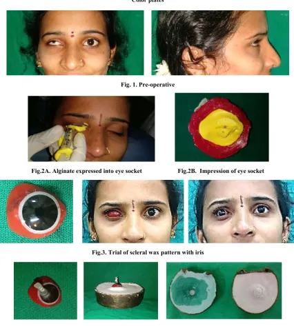
Patient was made into supine position for free flow of alginate in the socket to make the impression. The patient’s eye socket and eyebrows were coated with a thin layer of Petroleum Jelly (Vaseline) for comfortable separation of the impression. Alginate was mixed and loaded in a disposable plastic syringe and expressed into the eye socket under the eyelids (fig- 2A).13Beading and boxing was done around the eye to record the extra ocular area and a thin mix of alginate is poured over the previous impression of the socket with syringe. Gauze was placed over the alginate while it was setting for anchorage of the dental plaster. Dental plaster was mixed in thick consistency and placed over the gauze as backing for support of the impression. The impression was removed from the socket after both alginate and plaster was set taking care to remove the impression from the lower, shallower eyelid sulcus first, and then rotated out from the deeper upper eyelid sulcus to prevent distortion of the impression (Fig- 2B).
Pouring of the cast
Boxing of the impression was done and poured in Die stone (Kalastone, Kalabhai Pvt., Ltd., Mumbai, India) to obtain a split cast mold. (Mishra and Choudhary, 2009)The impression was poured upto the height of contour to obtain the lower half of the split cast mold. After the die stone was set, four orientation indices were made and separating medium was applied and second half or remainder of the impression was poured covering the entire impression. After the die stone was set, the two halves of the cast were separated and the impression was retrieved to obtain a mold space for fabrication of scleral wax pattern.
Scleral wax pattern fabrication
The inner surface of the split cast was coated with separating medium. The cast was re-assembled and the mold cavity was
filled with molten base plate wax (Modeling wax, Dental products of India Ltd.). The two halves of the cast were separated to retrieve the wax pattern. The pattern was highly polished and free from dust and debris before placing it in the eye socket.
Scleral wax pattern try- in
Scleral wax pattern was tried in the eye socket (Fig- 3). It was evaluated for fullness of the both palpabre, and under- extensions or overextensions. Areas of under- extensions were corrected by adding base plate wax. Any overextensions were reduced. The contour of the wax pattern was evaluated with patient’s eyes open and then by manual palpation with the eyes close. The contours of the eyelids with wax pattern were made to resemble the adjacent natural eye.
Placement of Iris
Iris of the patient’s old stock eye was used, as it had the same shade and size of the natural eye. Scleral part of the stock eye was trimmed off using an acrylic trimmer. The position of the natural iris was determined by instructing the patient to look straight ahead to a distant object. (Poonnanna et al., 2012)The distance of iris of the natural eye was measured using a divider opposing the iris from the inner canthus to the center of iris and the same distance was marked and engraved on the scleral wax pattern. Then similarly, distance from the outer canthus to the center of the iris was measured using the divider and transferred to the scleral wax pattern. The pattern was taken out and the selected iris was placed and adjusted to the horizontal and vertical axis according to the marking engraved on the scleral wax pattern (Fig- 3). Scleral wax pattern with the iris was tried in the eye socket and checked for symmetry and function by asking the patient to perform various movements.
Processing
After the trial of wax pattern a handle was attached to the iris which was made of cold cure acrylic so as to prevent the dislodgement of the iris during dewaxing as well as packing. Wax pattern was invested as in the case of complete denture laboratory procedures. The second pour was poured in such a way that the handle attached to the iris was embedded into the plaster of the counter flasking. Then the dewaxing was done after the final set, taking care so that there was complete wax elimination from the mold space (Fig- 4).
Packing
Tooth colored heat cured PMMA (DPI-Heat cure, Dental Products of India Ltd.) of appropriate shade, matching with the scleral color of normal eye of the patient was mixed in a ceramic jar in the ratio of 3:1 and packed in the mold in dough stage and kept for bench curing to enable complete polymerization and prevention of any excess unreacted monomer. This enables the minimization of porosities and gives a good finish to the prosthesis. We used long curing cycle of 8 hours so as to prevent the presence of any residual monomer in the prosthesis which is very essential. It prevents any unanticipated irritation or sensitivity and thereby rejection of the prosthesis by the patient. After curing, the mold was allowed to cool down to room temperature. Resin sclera with the iris attached over it was obtained after deflasking.
Color plates
[image:3.595.85.512.63.537.2]
Fig. 1. Pre-operative
Fig.2A. Alginate expressed into eye socket Fig.2B. Impression of eye socket
Fig.3. Trial of scleral wax pattern with iris
Fig.4. Photograph showing wax pettern invested with handle attachment, and mold space after dewaxing
[image:3.595.73.520.556.798.2]
Fig.5. Finished ocular prosthesis without characterization Fig.6. Try in for the plane sclera with stock iris and modified sclera
Acrylic resin extension from the iris was trimmed off using an acrylic trimmer, followed by polishing with Buff and pumice to obtain a smooth glossy surface (Fig- 5).
Try- in of final prosthesis
The scleral blank with iris was tried in the eye socket (Fig- 5). Stability of the prosthesis, contour of the sclera, and the position of the iris was reconfirmed. Under- supported areas in the eyelids due to polymerization shrinkage of acrylic resin were modified by adding a layer of modeling wax on the scleral blank and verified for the movements of ocular prosthesis in all directions. It was modified till the prosthesis showed adequate support and fullness in the eyelid in harmony with the adjacent natural eye (Fig- 6).
Characterization of the prosthesis
After the try-in of the prosthesis, the sclera was characterized to match the natural eye of the patient to give life like appearance. A close- up view photograph of the patient eye was taken to observe the scleral pattern. It was seen that there was a slight yellowish hue present on the medial side of the natural eye, greyish hue around the iris and few blood vessels laterally and medially. Modified sclera with iris was invested in a flask followed by separating the two compartments of the flask after dewaxing (Fig-7). It produced an even space for packing clear heat polymerizing acrylic resin after painting to give the conjunctival effect. Acrylic resin forming the sclera was trimmed uniformly to a depth of around 0.5 mm around the iris carefully. Over the surface of the sclera painting was done using the soft color tones of brown, blue, white and yellow (Favicryl, Pedilite Industries Ltd., Mumbai, India) to match the sclera of the contralateral natural eye. Red nylon fibers were placed along the outer periphery to simulate the blood vessels. Clear heat polymerizing acrylic resin was mixed and trial packing was done to remove the excess acrylic followed by bench curing, long curing cycle. The ocular prosthesis was carefully retrieved from the flask, trimmed and finely polished to obtain a glass like finish (Fig- 8).
Placement of final prosthesis
The properly finished and polished prosthesis was inserted in the socket after it was washed with soap solution and cleaned thoroughly with water (Fig- 9). Harmonious movement of the prosthesis was examined by instructing the patient to perform movements in various directions. Necessary adjustments were made and final finishing and polishing was carried out. Post insertion instructions were given to the patient, regarding the usage, cleaning with soap solution and recall was done in 1,3 days, 1 week and 1 month. The fit of the prosthesis was to be evaluated every 6 months. (Taylor 2nd ed)
DISCUSSION
Pre-fabricated prosthesis carries potential disadvantages of poor fit (which endangers the eye to granuloma formation), poor esthetics and poor eye movements. (Cain, 1982) According to Beumer et al. (1996)intimate contact between the ocular prosthesis and the tissue bed is needed to distribute even pressure, so a prefabricated prosthesis should be avoided. Moreover, the voids in the prefabricated prosthesis collect mucus and debris, which can irritate mucosa and act as a potential source of infection, which are minimized in custom-made prosthesis. (Cain,, 1982; Grisius and Robert, 1993) Customized ocular prosthesis has the advantages over stock eyes like, better contouring, color matching, and coordinated movements with the contralateral eye. (Benson, 1977; Taicher
et al., 1985)Customizing the iris demands extra skill and time from the operator. (Putanikar et al., 2015)This can be avoided if stock iris matching with the contralateral natural eye is available. Semi-customizing the prosthesis using the stock iris and customized sclera will have advantages of both stock and custom prosthesis. The close adaptation of the custom-made ocular prosthesis to the tissue bed provides maximum comfort and restores full physiologic function. Limitations of the technique are that the clinician is dependent on the availability of a prefabricated eye with properly matching iris. Also, the long-term color stability of the heat-cured acrylic and the strength of its union with the stock eye will have to be closely evaluated.
Fig.8. Definitive ocular prosthesis after characterization
Fig.9. Post-operative view showing the rehabilitation of the ocular defect with custom scleral prosthesis
Conclusion
Success of the ocular prosthesis largely depends on the precise laboratory technique and artistic skills of the operator. Through this technique, the demand for the artistic skill and consumption of time are reduced by the use of precisely selected stock iris, yet esthetic and functional requirements are met by the semi-customized sclera.
REFERENCES
Aggarwal H, Singh RD, Kumar P, Gupta SK, Alvi HA. 2014. Prosthetic guidelines for ocular rehabilitation in patients with phthisis bulbi: A treatment-based classification system. J Prosthet Dent., 111(6):525-8.
Benson P. 1977. The fitting and fabrication of a custom resin artificial eye. J Prosthet Dent., 38(5):532-8.
Beumer J, Zlotolow I. 1996. Restoration of facial defects. In: Beumer J, editor. maxillofacial rehabilitation - prosthodontic and surgical considerations 1st ed. St. Louis: C. V. Mosby publishers; p. 350-64.
Beumer J, Zlotolow I. 1996. Restoration of facial defects. In: Beumer J, editor. maxillofacial rehabilitation - prosthodontic and surgical considerations 1st ed. St. Louis: C. V. Mosby publishers; p. 350-64.
Bi Y, Wu S, Zhao Y, Bai S. 2013. A new method for fabricating orbital prosthesis with a CAD/CAM negative mold. J Prosthet Dent., 110:424-8.
Cain JR. 1982. Custom ocular prosthesis. J Prosthet Dent., 48:690-4.
Dugad JA, Dholam KP, Chougule AT. 2014. Vacuum form sheet as a guide for fabrication of orbital prosthesis. J Prosthet Dent., 112:390-2.
Grisius MM, Robert L. 1993. Treatment of lagophthalmos of the eye witha custom prosthesis. J Prosthet Dent.,70:333-5. Long JA. and Gutta R. 2013. Orbital, periorbital, and ocular reconstruction. Oral MaxillofacSurgClin North Am., 25:151-66.
Mishra SK. and Choudhary R. 2009. Reproduction of custom made eye prosthesis maneuver: a case report. J Dentist Oral Hygiene, Dec;1(5):59-63.
Naveen HC, Porwal A, Nelogi S. 2010. Prosthetic rehabilitation of phthisis bulbi by digital imaging technique. A case report. Cont Lens Anterior Eye, 33: 231-4.
Poonnanna AA, Tripathi G, Porwal A, Patel M. 2012. Art and science behind esthetic ocular prosthesis: a case report. Int J Dent Case Reports, 2(5):103-109.
Pruthi G. and Jain V. 2013. Light weight prosthesis for a patient with bilateral orbital exenteration-a clinical report. J Prosthodont Res., 57:135-9.
Putanikar NY, Patil AG, Shetty PK, Nagaral S, Mithaiwala HI. 2015. Prosthetic rehabilitation of a patient with ocular defect using semicustomized prosthesis: A case report. J Int Oral Health, 7(4):81-84.
Taicher S, Steinberg HM, Tubiana I, Sela M. 1985. Modified stock-eye ocular prosthesis. J Prosthet Dent, 54(1):95-8. Taylor TD. Clinical maxillofacial prosthetics.Quintessence
Publication Co, Inc; 2nd ed. P.266-276.
Worrell E. 2014. Ocular prosthetic obturator: an innovative medical device. Br J Ophthalmol., 98:862-4.