Original Article
Effect of prostaglandin E1 in patients with advanced
lung cancer treated with chemotherapy
Hai-Xia Ren1, Lian-Tian Tian2, Liang Zhang3, Bo Yang3, Lei Li3, Ming-Jiang Li3, Wei-Dong Zhang3, Xiao-Ping Li3
1Department of pharmacy, Tianjin First Central Hospital, Tianjin 300192, China; 2Occupational Respiratory Disease Research Center, West China No. 4 Hospital/West China School of Public Health, Sichuan University, Chengdu 610041, China; 3Department of Thoracic Surgery, Tianjin First Central Hospital, Tianjin 300192, China
Received February 17, 2017; Accepted January 25, 2018; Epub March 15, 2018; Published March 30, 2018
Abstract: To examine potential VTE risk reducing effect of prostaglandin E1 (PGE1) as supplemental therapy in patients with advanced lung cancer treated by chemotherapy.68 patients were recruited in this study: 46 males with age range from 58-85 and the average age of 67.48 ± 6.06; 22 females with age range from 55-85 and the average age of 68.05 ± 7.72. All patients were randomly assigned into three groups. Patients in the therapy group (33 patients) only received chemotherapy. On the other hand, patients in experimental group (25 patients) received combination therapy with chemotherapy and intravenous injection of PGE1 (Alprostadil, produced by Beijing Tide Pharmaceutical company) for 14 days. While in the control group (10 patients), only the supportive therapy was given. All patients were followed up till the end of their lives, with the exact time of death recorded, among which 51 died in our hospital, the rest were informed by phone calls to confirm the exact time of death. The median survival time was 61 days in the experimental group, which was not statistically significant to those of control group (52.5 days, p=0.237) and therapy group (56 days, p=0.656). In the therapy group, 9 cases showed small amount of VTE during treatment, one patient died because of fatal pulmonary embolism. In experimental group, only 2 cases experienced non-fatal VTE. The main adverse reaction of PGE1 was nodular vasculitis in 3 cases which was well tolerated. Concomitant application of PGE1 with chemotherapy significantly reduced the risk of VTE in patients with advanced lung cancer while it did not extend the patients’ survival time.
Keywords: Prostaglandin E1, advanced lung cancer, survival time
Introduction
Prostaglandins (PG) are unstable bioactive sub-stances, to work by synthesis of arachidonic acid in reaction to external stimuli (such as soft tissue injuries) [1]. Depending on structure and function, prostaglandins are divided into
sever-al types with different or even conflicted func -tions. Aberrantly high level of PG was reported in breast and gynecological cancer, colon can-cer, lung cancer and other types of malignan-cies [2]. In addition, prostaglandin E (PGE) lev-els were higher in patients with bone me- tastases than in those without metastasis [3]. And this aberrant high level of PGE was believed to facilitate bone metastases [4]. prostaglandin E1 (PGE1) and Prostaglandin B2 (PGB2) were not involved in cyclic adenosine monophos-phate (cAMP) synthesis which can be interpret-ed as inhibition of cell proliferation, suggesting potential anti-tumor effects of PG [5].
Widely expressed in peripheral and coronary blood vessels, PGE1 leads to relaxation of smooth muscle, and inhibits platelet aggrega-tion as well as atherosclerotic lipid plaque
for-mation [6]. Also, PGE1 has significant anti-inflammatory and anti-oxidative stress effect
[7]. Ishikawa found that PGE1 inhibits secretion of tumor necrosis factor (TNF) and interleu-kin-10 (IL-10) in mononuclear cells [8]. Fang dis-covered that PGE1 inhibits endothelial cell acti-vated oxygen (reactive oxygen species, ROS), enhances endothelial nitric oxide synthase (eNOS) expression, and promotes NO release [9].
coagu-lation facilitates cancer cells attachment,
inva-sion and transfer which may in turn influence
biology of the tumor, resulting in a poorer out-come of chemotherapy [11]. Patients treated with long-term chemotherapy may experience small pulmonary artery spasms, increase in pulmonary vascular resistance, pulmonary hypertension and increase of right heart load, which may lead to right heart failure or heart failure [6, 12, 13]. Nowadays studies show that humoral factors such as prostaglandins play an important role in hypoxic pulmonary vasocon-striction [14]. There are two types of prosta-glandins derivatives which are thromboxanes and prostacyclins. Among them, thromboxane A2 (TXA2) causes vasoconstriction, prostaglan-din F2a (PGF2A) cable and vasodilation of pros-tacyclin (PGI2), alprostadil (PGE1) in two cate-gories. Hypoxic pulmonary vasoconstriction largely depends on local vasoconstrictive sub-stances and vasodilative subsub-stances [15]. In addition, hypoxia, hypercapnia, stimulation of aortic body and carotid body chemoreceptors, and sympathetic activity lead to increased cat-echolamine secretion, pulmonary vasoconstric-tion, and pulmonary arterial hypertension [16]. Chronic hypoxia can also generate secondary polycythemia, increase blood viscosity and hematocrit, increase platelet adhesion effect of microcirculation perfusion, leading to throm-bosis in pulmonary circulation and coronary cir-culation, and potential heart failure [17]. PGE1 can dilate bronchial and artery vein blood vessels, and thus increase myocardial contrac-tility [18]. By exciting mucosa of the respiratory tract, PGE1 receptor exerts its action directly on bronchial smooth muscle. PGE1 can decrease section of reacting substance and
other inflammatory mediators, release inhibito -ry overactive sympathetic neurotransmitter, decrease airway hyper responsiveness during heart failure and bronchial dilation [19]. Therefore ventilation and hypoxia are improved. Furthermore, PGE1 dilates arteries and veins, reduces pre- and after- load of the heart and pulmonary artery pressure, enhances myocar-dial contractility and renal vascular expansion, and increases urine output [19]. Microcirculation and macrocirculation of the heart are improved with strong anti-platelet aggregation effect of PGE1. In this paper, we used PGE1 as supple-mental therapy to examine its effect in patients treated with chemotherapy [20].
Materials and methods Materials
Clinical data of inpatients were collected from January 2013 to December 2015. Protocol of the study was approved by review board
com-mittee of Tianjin first central hospital. Written
informed consent was obtained from each patient. 68 patients with advanced lung
can-cer were diagnosed by pathological confirma -tion or by distant metastases without patho-logical diagnosis. Inclusion criteria: (1) for non-small cell lung cancer (NSCLC) cases, TNM staging of T3-T4 are required; for small cell lung cancer (SCLC) cases, extensive stage are required. (2) ECOG score is 3 or 4. (3) patients were not under chemotherapy or targeted ther-apy 3 months before enrollment, with only sup-portive care. The histological grading and TNM
classification of the patients’ cases were per -formed according to the recommendations of the International Union Against Cancer (2009). Exclusion criteria: (1) patients with medical his-tory that affects blood coagulation (such as severe liver and kidney disease, thromboem-bolic disease, diabetes, surgery trauma and severe infections within 1 month, receiving
anticoagulant and antifibrinolytic or haemostat -ic treatment within two weeks). (2) Platelet <100,000/l. (3) pre-existing hemoptysis. (4) Patients in critical condition, with unstable vital signs or clinical expected survival time less than 15 days.
68 cases were enrolled in this study: 46 males with the age range from 58-85 years old and average age of 72.35 ± 5.73 (years ± SD); 22 females with the age range from 55-85 years old and average age of 67.65 ± 5.90 (years ± SD). According to pathology reports, 43 cases were NSCLC, 13 cases were SCLC, and the rest 12 cases were with no clear pathological
clas-sification. ECOG scores were 3 points in 30
cases and 4 points in 38 cases. At baseline, all patients received a panel of tests including rou-tine blood test, liver and kidney function tests, complete blood cell count with differential leu-kocyte count, serum biochemistry, tumor mark-ers panel examination, blood gas analysis, DIC check; and received physical examination, bronchoscopy, computed tomography (CT) scan of the chest and upper abdomen, and
electro-cardiogram. After clinical staging confirmed,
group (10 cases), therapy group (33 cases) and experimental group (25 cases) based on their ECOG score by coin throwing random method. Clinic data of the three groups (gender, age, lesion location and pathological results) are
comparable and not statistically significant.
Treatment
Pathology is confirmed by transbronchial biop -sy, sputum biopsy from percutaneous lung
puncture or pleural fluid exfoliative cells. The
therapy group (33 cases) only received chemo-therapy. Experimental group (25 cases) re- ceived chemotherapy combined with intrave-nous injection of PGE1 (Alprostadil, produced by Beijing Tide Pharmaceutical company) for 14 days. Supportive therapy was given to patients in the control group (10 cases). PGE1 was administered at the standard dosage recom-mended by medication instruction which is 10 ug in 10 ml 5% glucose or 0.9% NS once daily for 14 days. The exact time of death was recorded for each patient. 51 died in our hospi-tal and their time of death was recorded by doc-tor. For the rest of patients, they were followed up once a week by phone and their time of
death was confirmed by phone call to their
close family members at home. Response and toxicity criteria
Throughout the study, use of any other
antico-agulants or fibrinolytic drugs is prohibited, but
use of non-steroidal anti-inflammatory drugs is
allowed.
VTE diagnostic criteria: VTE includes deep venous thrombosis of lower extremity, non-fatal pulmonary embolism or death-related venous thromboembolism (fatal pulmonary embolism and death of unknown causes).
The adverse reaction of PGE1 being observed in this study is any clinical related hemorrhage
occurred starting from the first day of drug
administration throughout the course of study to no later than 3rd day after the last
administra-tion. Hemorrhage is defined as any of the fol -lowing conditions: (1) hemorrhage occurred in organs such as skull, vertebral tube, eyes, peri-toneum, joint, pericardium, or intramuscle, (2) hemoglobin levels reduced to 20 g/L or less, or (3) infusion of 2 or more units of whole blood or red blood cell suspension.
The clinical stages of deep venous thrombosis:
acute stage defined as occurrence less than 7 days after onset; sub-acute stage defined as
between 9th and 13th day of onset; chronic
stage defined as after 30th day of onset.
Statistical analysis
The statistical analysis was performed using SPSS software, version 19.0 (IBM SPSS Inc., Chicago, IL, USA). The data were evaluated sta-tistically using an independent sample t-test when a simple comparison between two groups was required, analysis of variance (ANOVA) was used to compare the data in multiple groups,
and least significant difference and
Student-Newman-Keuls processes were used to com-pare the differences between each pair of
groups. Finally, χ2 tests were used to examine
the statistical significance of differences in the
expression rate. Results
Demographic data of patients
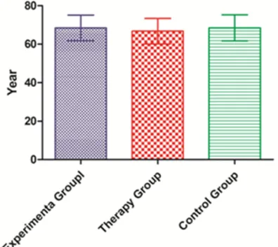
[image:3.612.90.288.73.249.2]Experiment group was consisted of 18 male patients and 7 female patients with the aver-age aver-age of 68.48 ± 6.64 (years ± SD); therapy group included 22 male patients and 11 female patients with the average age of 66.79 ± 6.59 (years ± SD); control group had 6 male patients and 4 female patients with the average age of 68.50 ± 6.77 (years ± SD) (Figure 1). ECOG scores were comparable among all three
groups, with 11 cases of 3 points and 14 cases of 4 points in experiment groups, 15 cases of 3
[image:4.612.93.360.85.216.2]points and 18 cases of 4 points in therapy group, and 4 cases of 3 points and 6 cases of 4 point in control group. As to the disease type, the experiment group had 18 cases of NSCLC, 4 cases of SCLC, and 3 cases of undiag-nosed lung cancer; the therapy group had 21 cases of NSCLC, 6 cases of SCLC, and 6 cases of undiagnosed lung cancer; the control group had 4 cases of NSCLC, 3 cases of SCLC, and 3 cases of undiagnosed lung can-cer. The distribution of clinical Table 1. Demographic data of the three groups
Experimental
group n=25 Therapy group n=33 Control Group n=10
Average Age 68.48 ± 6.64 66.79 ± 6.59 68.50 ± 6.77 P=0.32 ECOG
3 point 11 15 4 P =0.43
4 point 14 18 6 P =0.26
Tumor Type
NSCLC 18 21 4 P=0.33
SCLC 4 6 3 P=0.78
Undiagnosed 3 6 3 P =0.83
[image:4.612.90.288.249.419.2]ECOG: Eastern Cooperative Oncology Group.
Figure 2. Survival time of all three groups. The aver-age survival time was 61.6 ± 13.3 days in the experi-mental group. Average survival time was 59.8 ± 16.4 days in the therapy group. In control group, it was 55.3 ± 15.7 days. There was no significant differ -ences between the experimental and therapy group (p=0.656).
Figure 3. Survival curves of all three groups through the study.
characteristics among the three groups showed
no significant differences (Table 1).
Comparison of survival time
The median survival time was 61 days in the experimental group with average survival time of 61.6 ± 13.3, whereas the median survival time was 56 days in the therapy group with average survival time of 59.8 ± 16.4, which was similar to that of the control group (52.5 days,
55.3 ± 15.7). There was no significant differ -ences in survival time between the experimen-tal and therapy group (p=0.656) (Figures 2, 3). VTE and adverse reactions
During PGE1 treatment, 3 patients in experi-mental group developed nodular vasculitis. In therapy group (n=33), 9 cases showed small amount of VTE during treatment with 1 patient died due to fatal pulmonary embolism. In the experimental group (n=25) only one patient experienced non-fatal VTE. On the other hand, in the control group, 3 patients developed VTE. The results showed that the incidence rate of
VTE was significantly lower in the experimental
group compared with the therapy group (Tables 2, 3).
Discussion
[image:4.612.89.287.527.656.2]effect on bone marrow and leads to reduced levels of granulocyte and platelet [23]. Th- rombosis formation is easier in cancer patients than in healthy people due to their hypercoagu-lable state and thrombin produced by tumor cell membrane [24]. Patients with advanced lung cancer usually have disorder of coagula-tion; therefore they are more likely to develop VTE with chemotherapy treatment [25] . In this study, it was found that administration of PGE1
concurrently with chemotherapy significantly
decreased the incidence rate of VTE and reduced the risk of fatal embolism. In terms of survival time, the present study showed that the survival time of patients treated with
che-motherapy alone was not significantly different
from that of the supportive treatment group
[26]. And there was no significant difference in
the mean survival time among all three groups, which is different from previous reports where it was shown that chemotherapy combined with anticoagulant can prolong survival time of patients [27]. The possible reason may be that the sample sizes of this study were too small to show statistical differences.
It had been more than 10 years since the first report claiming that PGE may be beneficial in
the treatment of ischaemic peripheral vascular disease [28]. PGE1 administered
intra-arterial-ly increased calf blood flow at doses between 1
and 10 ng/kg/min [29]. However, therapeutic
benefit has been controversial for PGE adminis -tered intravenously. PGE1 adminis-tered intra-venously at a dose of 10 ug per day was
report-ed to produce clinical benefit for patients with
advanced lung cancer [30]. However, a large double-blind controlled trial of PGE1 showed no
beneficial effect in patients suffering from
ulcers secondary to advanced lung cancer. The healing rate in PGE1 treated group was not
higher than that of the placebo group which was approximately 50%.
Our study showed that PGE1 significantly
reduced the incidence of VTE during chemo-therapy in patients with advanced lung cancer. The underlying mechanisms could be that
PGE1 can enhance fiber dissolution activity,
reduce pathological changes during ischemic injury, and promote the VTE thrombolysis. The
beneficial effects may be related to the multi -ple biological activities of PGE1. ①The effects
on coagulation and fibrinolytic system: activa -tion of platelet prostaglandin receptor by PGE1 inhibits the release of TXA2. Therefore the TXA2 induced strong release and aggregation of platelet was inhibited and vasoconstrictive effect of platelet was reduced. This could con-tribute to the prevention of VTE, especially in the hypercoagulation and microvascular occlu-sion state of patients with advanced lung can-cer under chemotherapy. Previous investiga-tions showed that PGE1 can enhance local thrombolytic infusion effect of thrombolytic drugs (tPA, urokinase, streptokinase), which was seen in animal models of porcine coronary artery thrombosis, rabbits inferior vena cava thrombus and jugular vein thrombosis and in vitro thrombus model [31]. Moreover, PGE1 can inhibit the activity of plasminogen activator inhibitor -1 (PA-1) in vivo. In addition, PGE1 has a direct protective effect on vascular
endothe-lial cells, which is beneficial to the production of
tPA by endothelial cells and the enhancement
[image:5.612.88.515.85.134.2]of local fibrinolytic activity. ②The effects of PGE1 on impaired microcirculation: PGE1 is a powerful vasodilator by relaxing vascular sm- ooth muscle for the whole body. The application of PGE1 in patients with advanced lung cancer, especially in those under chemotherapy, facili-tates the establishment of collateral circulation Table 2. The VTE and adverse reactions of experimental and therapy group
Experimental group n=25 Therapy group n=33
VTE rate 4.0% (1/25) 27.3% (9/33) χ2=3.89 P=0.049
Adverse reaction (nodular vasculitis) 12.0 (3/25) 0.0% (0/33) χ2=2.09 P=0.148
VTE: Vein thrombosis embolism syndrome.
Table 3. The VTE and adverse reactions of experimental and control group
Experimental group n=25 Control group n=10
VTE rate 4.0% (1/25) 30.0% (3/10) χ2=2.55 P=0.061
Adverse reactions (nodular vasculitis) 12.0 (3/25) 0.0% (0/10) χ2=0.23 P=0.542
[image:5.612.89.523.172.219.2]and improves local microcirculation, thus pre-venting the pathological process of ischemic injury. Moreover, PGE1 can inhibit platelet aggregation, reduce blood viscosity, reduce the aggregation of red blood cells and improve its deformability, thereby improving the perfusion and repair of damaged tissue [32, 33]. Aside
from its beneficial effect on prevention and
treatment of VTE, the only adverse reaction of PGE1 is vasculitis which is mild and well toler-ated. Taken together, the application of PGE1
has significant benefits to patients with
ad-vanced lung cancer. Conclusion
In this study, we discovered that PGE1 signifi -cantly reduced the incidence rate of VTE in patients with advanced lung cancer. However
we did not find significant effect on overall sur -vival time of patients among all three groups. Therefore, it is still uncertain whether PGE1 administration could lead to therapeutic im- provement in patients treated by chemothera-py. Further study needs to be carried out to test other infusion regimes with different doses and even different routes of administration.
Acknowledgements
This work was supported by the Foundation of Tianjin Municipal Bureau of Health (No. 2013ky09).
Disclosure of conflict of interest
None.
Address correspondence to: Dr. Xiao-Ping Li, De- partment of Thoracic Surgery, Tianjin First Central Hospital, Fukang Road, Nankai District, Tianjin 300192, China. Tel: 86-22-23627068; E-mail: eon-elxp@163.com
References
[1] Zhang SH, Zhao JL. [Prostaglandins and optic papilla blood flow]. Zhonghua Yan Ke Za Zhi 2017; 53: 73-76.
[2] Tastekin D, Tas F, Karabulut S, Duranyildiz D, Serilmez M, Guveli M, Vatansever S. Clinical significance of serum tenascin-C levels in breast cancer. Tumour Biol 2014; 35: 6619-25.
[3] Barlow M, Edelman M, Glick RD, Steinberg BM, Soffer SZ. Celecoxib inhibits invasion and me-tastasis via a cyclooxygenase 2-independent
mechanism in an in vitro model of Ewing sar-coma. J Pediatr Surg 2012; 47: 1223-7. [4] Bassalyk LS, Kushlinskiĭ NE, IuN S, Siniukov
PA, Fedenko AN. [The relationship between the prostaglandin E level in the tumor and the times of the metastasis of primary osteogenic sarcoma]. Vopr Onkol 1989; 35: 1301-5. [5] Arai H, Nomura Y, Kinoshita M, Shimizu H, Ono
K, Goto H, Takigawa M, Nishimura F, Washio N, Kurihara H, Murayama Y. Response of human gingival fibroblasts to prostaglandins. J Peri -odontal Res 1995; 30: 303-11.
[6] Fitzsimmons C, Proudfoot D, Bowyer DE. Mono-cyte prostaglandins inhibit procollagen secre-tion by human vascular smooth muscle cells: implications for plaque stability. Atherosclero-sis 1999; 142: 287-93.
[7] Uemura T, Takamatsu H, Kawasaki T, Uemura T, Takamatsu H, Kawasaki T, Taniguchi M, Ya-mamoto E, Tomura Y, Uchida W, Miyata K. Ef-fect of YM-254890, a specific Galphaq/11 in -hibitor, on experimental peripheral arterial disease in rats. Eur J Pharmacol 2006; 536: 154-61.
[8] Noguchi K, Iwasaki K, Endo H, Kondo H, Shi-tashige M, Ishikawa I. Prostaglandins E2 and I2 downregulate tumor necrosis factor alpha-induced intercellular adhesion molecule-1 ex-pression in human oral gingival epithelial cells. Oral Microbiol Immunol 2000; 15: 299-304. [9] Fang W, Li H, Zhou L, Su L, Liang Y, Mu Y. Effect
of prostaglandin E1 on TNF-induced vascular inflammation in human umbilical vein endo -thelial cells. Can J Physiol Pharmacol 2010; 88: 576-83.
[10] Walker AJ, Baldwin DR, Card TR, Powell HA, Hubbard RB, Grainge MJ. Risk of venous thromboembolism in people with lung cancer: a cohort study using linked UK healthcare data. Br J Cancer 2016; 115: 115-21.
[11] Yoshii Y, Numata T, Ishitobi W, Ohnishi M, Horie M. Lung adenocarcinoma complicated by Trousseau’s syndrome successfully treated by a combination of anticoagulant therapy and chemotherapy. Intern Med 2014; 53: 1835-9. [12] Grossman E, Messerli FH. Calcium
antago-nists. Prog Cardiovasc Dis 2004; 47: 34-57. [13] Burger CD, D’Albini L, Raspa S, Pruett JA. The
evolution of prostacyclins in pulmonary arterial hypertension: from classical treatment to mod-ern management. Am J Manag Care 2016; 22: S3-15.
[14] Cahill PA, Redmond EM, Sitzmann JV. Endothe-lial dysfunction in cirrhosis and portal hyper-tension. Pharmacol Ther 2001; 89: 273-93. [15] Leuchte HH, Michalek J, Soenmez O, Meis T,
[16] Yamamoto T, Wada A, Tsutamoto T, Ohnishi M, Horie M. Long-term treatment with a phospho-diesterase type 5 inhibitor improves pulmo-nary hypertension secondary to heart failure through enhancing the natriuretic peptides-cGMP pathway. J Cardiovasc Pharmacol 2004; 44: 596-600.
[17] Kang J, Li Y, Hu K, Lu W, Zhou X, Yu S, Xu L. Chronic intermittent hypoxia versus continu-ous hypoxia: Same effects on hemorheology. Clin Hemorheol Microcirc 2016; 63: 245-55. [18] Riise J, Ørstavik Ø, Qvigstad E, Dahl CP, Osnes
JB, Skomedal T, Levy FO, Krobert KA. Prosta-glandin E1 facilitates inotropic effects of 5-HT4 serotonin receptors and β-adrenoceptors in failing human heart. Basic Res Cardiol 2012; 107: 295.
[19] Buckley J, Birrell MA, Maher SA, Nials AT, Clarke DL, Belvisi MG. EP4 receptor as a new target for bronchodilator therapy. Thorax 2011; 66: 1029-35.
[20] Teixeira MM, Williams TJ, Hellewell PG. Role of prostaglandins and nitric oxide in acute inflam -matory reactions in guinea-pig skin. Br J Phar-macol 1993; 110: 1515-21.
[21] Yin DP, Sankary HN, Chong AS, Ma LL, Shen J, Foster P, Williams JW. Protective effect of isch-emic preconditioning on liver preservation-re-perfusion injury in rats. Transplantation 1998; 66: 152-7.
[22] Takahashi Y. Real-time intraoperative diagno-sis of lung adenocarcinoma high risk histologi-cal features: a necessity for minimally invasive sublobar resection. Minim Invasive Surg Oncol 2017; 1: 12-19.
[23] Schiller JH, Harrington D, Belani CP, Langer C, Sandler A, Krook J, Zhu J, Johnson DH. Eastern Cooperative Oncology Group. Comparison of four chemotherapy regimens for advanced non-small-cell lung cancer. N Engl J Med 2002; 346: 92-8.
[24] Fuentes HE, Tafur AJ, Caprini JA. Cancer-asso-ciated thrombosis. Dis Mon 2016; 62: 121-58. [25] Khorana AA, Dalal M, Lin J, Connolly GC. Inci-dence and predictors of venous thromboem-bolism (VTE) among ambulatory high-risk can-cer patients undergoing chemotherapy in the United States. Cancer 2013; 119: 648-55.
[26] Abu AW. Video-assisted thoracoscopic surgery for non-small cell lung cancer. Minim Invasive Surg Oncol 2017; 1: 1-11.
[27] Robert F, Busby E, Marques MB, Reynolds RE, Carey DE. Phase II study of docetaxel plus enoxaparin in chemotherapy-naive patients with metastatic non-small cell lung cancer: preliminary results. Lung Cancer 2003; 42: 237-45.
[28] Reiter M, Bucek RA, Stümpflen A, Dirisamer A, Minar E. Prostanoids in the treatment of inter-mittent claudication--a meta-analysis. Vasa 2002; 31: 219-24.
[29] Eo S, Kwon C, Lee H, Cho S, Kim J, Baek G, Yeo J, Lim C. Quantification of the effect of Lipo-PGE1 on angiogenesis. J Plast Reconstr Aes-thet Surg 2015; 68: 104-12.
[30] Hughes D, Otani T, Yang P, Newman RA, Yantiss RK, Altorki NK, Port JL, Yan M, Markowitz SD, Mazumdar M, Tai HH, Subbaramaiah K, Dan-nenberg AJ. NAD+-dependent 15-hydroxypros-taglandin dehydrogenase regulates levels of bioactive lipids in non-small cell lung cancer. Cancer Prev Res (Phila) 2008; 1: 241-9. [31] Lee S, Lee J, Choi YW. Design and evaluation of
prostaglandin E1 (PGE1) intraurethral liquid formulation employing self-microemulsifying drug delivery system (SMEDDS) for erectile dysfunction treatment. Biol Pharm Bull 2008; 31: 668-72.
[32] Kuznetsov MR, Koshkin VM, Karalkin AV, Bold-in B. V, Rodionov SV, Sergeeva NA, Petukhov EB, Golosnitskii PIu. [Preoperative care of mi-crocirculatory vessels in patients with lower limb arteriosclerosis obliterans]. Angiol Sosud Khir 2005; 11: 19-24.