Abstract—Biothermomechanics of skin tissue is highly
interdisciplinary, involving bioheat transfer, burn damage, biomechanics and physiology. Characterization of the thermomechanical behaviour of skin tissue is of great importance and can contribute to a variety of medical applications. However, few quantitative studies have been conducted on the temperature dependent mechanical properties of skin tissue. This paper reports the tensile and compressive behaviours of skin tissue under different temperatures and discusses the effect of temperature and corresponding dermal collagen denaturation on the mechanical properties of skin tissue. The results show that temperature has great influence on both the tensile and compressive properties of skin tissue, but the mechanisms are different. The variation of skin tensile properties is caused by the thermal denaturation of skin collagen with increasing temperature, while the variation of skin compressive behaviour of skin tissue is maybe due to the hydration change with thermal denaturation.
Index Terms—Compressive behaviour, skin tissue, tensile
behaviour, thermomechanical behaviour, thermal denaturation.
I. INTRODUCTION
Skin, generally consists of three layers: epidermis, dermis, and subcutaneous tissue. It is the largest single organ of the body and plays a variety of important roles including sensory, thermoregulation and host defense etc. Advances in laser, microwave and similar technologies have led to recent developments in thermal treatments of diseased and injured skin tissue such as skin cancer and skin burn. The aim is to induce thermal injury precisely within skin tissue several millimeters below the surface but without affecting the
Manuscript received March 13, 2007. This work is supported by the Overseas Research Studentship (ORS) and Overseas Trust Scholarship of Cambridge University, the National Natural Science Foundation of China (10328203, 10572111, 10632060), the National 111 Project of China (B06024), and the National Basic Research Program of China (2006CB601202).
F. Xu is with the Engineering Department, Cambridge University, Cambridge, CB2 1PZ UK (e-mail: fx201@cam.ac.uk).
T. Wen is with the Engineering Department, Cambridge University, Cambridge, CB2 1PZ UK (e-mail: tw289@cam.ac.uk).
K. A. Seffen is with the Engineering Department, Cambridge University, Cambridge, CB2 1PZ UK (e-mail: kas14@cam.ac.uk).
T. J. Lu is with theMOE Key Laboratory of Strength and Vibration, School of Aerospace, Xi’an Jiaotong University, Xi’an 710049 P.R. China (phone: 0086-29-82665600; fax: 0086-29-83234781; e-mail: tjlu@mail.xjtu.edu.cn)
surrounding, healthy tissue. In spite of the widespread use of heating therapies in dermatology, they do not draw upon the detailed understanding of the bio-thermo-mechanical behaviour of skin tissue, for none exists to date, even though each behavioural facet is well established and understood. It is proposed that a detailed understanding of the coupled biological-mechanical response under thermal loading will contribute to the design, characterization and optimization of strategies for delivering better treatment. Since it is technically very difficult to measure experimentally the bio-thermo- mechanical behaviour of skin tissue in physiological conditions, analytical and numerical simulations are used, where the quantification of the skin mechanical properties is an essential step towards building reliable computer simulations. So far, the mechanical properties of the skin tissue under normal conditions have been studied experimentally in vivo and in vitro ever since the study of Langer in 1861. However, few studies have been conducted on the mechanical properties of skin tissue under hyperthermia conditions and on the coupling effects between thermal and mechanical behaviours.
Collagen is the major component of skin, and it accounts for approximately 75% of the skin’s dry weight and 18-30% of the dermis’s volume [1]. Collagen provides the principal structural and mechanical support in skin tissue. Under thermal loading, with the increase of skin temperature, the heat-labile intramolecular crosslinks in collagen are broken, and the collagen undergoes a transition from a highly organized crystalline structure to a random, gel-like state [2]. This process is thermal denaturation, which appears accordingly as thermal shrinkage. Skin also contains a small mount of elastin (0.1% of skin dry weight), which is thermally very stable [3], and capable of surviving in boiling water for hours with no apparent change. Therefore, when discussing the mechanical behaviours of skin tissue under different thermal loadings, only collagen will be addressed.
The shrinkage of collagen due to the gross scale thermal denaturation can be used as a convenient continuum metric of thermal damage [4], [5]. Diller & Pearce [5] pointed out that the dimensionless indicator of damage (Ω) is, in fact the logarithm of the relative concentration of “reactants” (un-denatured collagen) in the collagen denaturation process:
Characterization of Thermomechanical
Behaviour of Skin Tissue I. Tensile and
Compressive Behaviours
(0) ( ) ln
( ) C t
C t
⎛ ⎞
Ω = ⎜ ⎟
⎝ ⎠
(1)
where C(0) is the initial concentration and C t( ) is the concentration of un-denatured collagen remaining at time t. Then, the degree of thermal denaturation, Deg t( ), (fraction of denatured collagen) can be calculated as:
(
)
(0) ( )
( ) 1 exp ( )
(0)
C C t
Deg t t
C
−
= = − −Ω
(2)
As for the calculation of thermal damage, the Arrhenius burn integration proposed by Henriques & Moritz [6] is widely used. They proposed that skin damage could be represented as a chemical rate process, which could be calculated by using a first order Arrhenius rate equation, whereby damage is related to the rate of protein denaturation (k) and exposure time (t) at a given absolute temperature (T ). The measure of thermal injury Ω was introduced and its rate k was postulated to satisfy:
( ) d exp Ea
k T A
dt RT
Ω ⎛ ⎞
= = ⎜− ⎟
⎝ ⎠ (3)
or, equivalently
0 exp
t
a
E
A dt
RT
⎛ ⎞
Ω = ⎜− ⎟
⎝ ⎠
∫
(4)where A is a material parameter (frequency factor), Ea is the
activation energy, and R=8.314 J/mol K is the universal gas constant. Using a thermodynamic analysis (see [7] for detail), the results of Ea and A for the Arrhenius damage integral
were calculated from the shape and the location of the denaturation endotherm observed at different scanning rates as: for pig ear skin, Ea =5.867 10 J/mol× 5 and
91 -1
5.240 10 s
A= × ; for pig back skin, Ea =5.255 10 J/mol× 5 and A=2.126 10 s× 81 -1. These values are well within the range reported in the literature for similar biological materials.
During denaturation, not only the structure but also the hydration of collagen changes, and thus, not surprisingly, thermal denaturation of a collagenous tissue can lead to remarkable changes in the mechanical, thermal, electrical, and optical properties. However, there are comparatively few studies of the thermal denaturation of skin tissue [8]-[11], despite the skin dermis being mainly composed of collagen.
The objectives of this study are to characterize the temperature-dependent mechanical behaviour of skin tissue and to examine the effect of collage denaturation. In this paper, preliminary experimental results of tensile and compressive
thermal loadings are presented, and the results of rate-dependent, or viscoelastic, behaviour are presented in the next paper.
II. MATERIALS AND METHODS
A. Samples
The ethical and immunological issues associated with testing human skin request us to find a substitute for human skin. Pig skin is chosen as test samples due to its high degree of structural and functional similarities to human skin [12]. In addition, repetitive tests can be realized for the same animal because of its large size, which reduces the variation in results [13]. In this series of experiments, skins of domestic pigs of UK are used, which are obtained from a local slaughter house near Cambridge at Dalehead Foods, Linton.
B. Samples preparation
Samples of pig skin to a depth of the subcutaneous fat, at different body sites were procured daily, ten minutes post mortem by block dissection. They are fast-chilled following the standard organ procurement protocol to 4°C in a pre-gassed (95%O2, 5%CO2) Krebs-Henseleit Ringer (KHR, pH7.4). In the laboratory, samples are separated from the subcutaneous fat by wet/fast-dissection on a bed of ice and KHR at 4°C. The skin samples are tested within a few hours from slaughter, in order to minimize degradation of the tissue structure. Before each tensile or compressive test, the sample was first preconditioned so that a reproducible response can be obtained for the afterwards tests. The preconditioning is completed in KHR solution at 37 °C.
C. Experimental system and test procedure
The tests are performed using hydrothermal tensile and compressive systems in the Engineering Department, Cambridge University. These two systems are schematically shown in Fig. 1 and Fig. 2, and the test procedures have been described in detail elsewhere [14], [15]. For brevity, only a simple introduction is given here. A specimen is put inside the test chamber and the liquid in the high temperature reservoir is heated to a chosen temperature somewhere between 30 and 100 °C, while the low temperature reservoir contains liquid at room temperature. A valve in the test chamber is opened to allow the hot liquid or cold liquid to fill the test chamber and the sample is fully immersed in the liquid, while a thermocouple near the sample is used to inform software, via a PC, the exact time at which experiment starts. A mechanical load is then applied to the skin sample. The temperature and applied displacement, strain, and loads are recorded subsequently.
III. RESULTS AND DISCUSSION
A. Tensile behaviour
Fig. 1 Schematic of the hydrothermal tensile experimental system
Fig. 2 Schematic of the hydrothermal compressive experimental system
relationships (σ , ε) are given in Fig. 3. It is noted that there are two obvious regions for all the curves. In the first region, where ε <50%, the curves of different temperature almost overlap. In this low modulus region, the deformation is accommodated principally by stretching of elastin, which is thermally stable and invariant to small temperature changes. The differences between the curves are due to the gradual straightening of an increasing fraction of the wavy collagen fibers. In the second region, ε >50% , the stress increases almost linearly with strain; and the gradients under increasing temperatures decrease. This feature is due to stretching and slippage of collagen molecules within crosslinked collagen fibers, and, to collagen fibril slippage; with increasing temperature, the highly organized crystalline structure of collagen changes to a random, gel-like state, which results in a decreasing stiffness. Another possible effect stems from dehydration which can be linked to the degree of thermal damage, Ω, Eq.(4), calculated from the degree of denaturation, Eq.(2). For example, Fig. 4 and Fig. 5 show that, when
60
T ≥ °C, the collagen in the dermis is fully denaturized almost instantaneously, whilst at T = 45 and 50 ºC, denaturation is slow. Thus, this divergence in behaviour around
60
T = °C seems to fit well with observations.
0 20 40 60 80 100
0.0 0.2 0.4 0.6 0.8 1.0
T (OC) 37 45 50 60 70 80
St
re
ss
σ
(MPa
)
Strain
ε(%)
[image:3.612.47.295.233.393.2]Pig ear skin, strain rate: 12.5%/s
Fig. 3 Stress-strain relation of hydrothermal tensile tests under different temperature
0 20 40 60 80 100
10-9 10-6 10-3 100
103 106
T (OC) 37 45 50 60 70 80
Thermal dama
ge
Ω
Strainε (%)
[image:3.612.322.546.287.460.2]Pig ear skin, strain rate: 12.5%/s
Fig. 4 Stress-thermal damage relation of hydrothermal tensile tests under different temperature
0 20 40 60 80 100
10-10 10-8
10-6
10-4
10-2 100
T (OC) 37 45 50 60 70 80
T
h
er
ma
l da
m
a
g
e
deg
ree
De
g
Strainε (%)
Pig ear skin, strain rate: 12.5%/s
[image:3.612.328.544.516.690.2]0 10 20 30 40 50 0.0
0.3 0.6 0.9 1.2
1.5 Pig back skin, strain rate: 0.1%/s
T
(
OC
)
37
55
58
67
Stre
ss
σ
(
MP
a
)
[image:4.612.55.285.54.244.2]Strain
ε
(%)
Fig. 6 Stress-strain relation of hydrothermal compressive tests under different temperature
0 10 20 30 40 50
10-9 10-6 10-3
100 103 106
T (OC) 37 55 57 68
Thermal dama
ge
Ω
Strainε (%)
[image:4.612.61.279.293.469.2]Pig back skin, strain rate: 0.1%/s
Fig. 7 Stress-thermal damage relation of hydrothermal compressive tests under different temperature
0 10 20 30 40 50
10-10 10-8 10-6 10-4 10-2 100
T (OC) 37 55 57 68
Th
ermal damage
d
egree
De
g
Strainε (%)
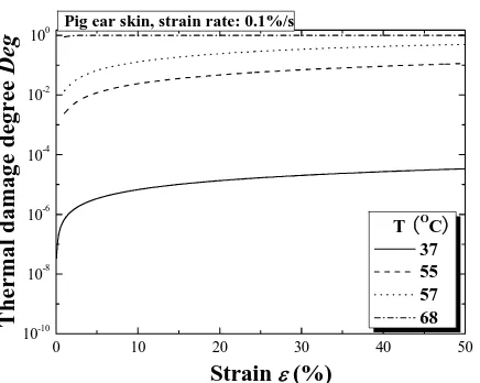
Pig ear skin, strain rate: 0.1%/s
Fig. 8 Stress-thermal damage degree relation of hydrothermal compressive tests under different temperature
B. Compressive behaviour
The hydrothermal compressive tests of back skin under different temperatures have also been performed and results are given in Fig. 6. Form the figure, it is found that, similar to the tensile response, all stress/strain relationships also display two distinct regions: a toe region of low stiffness at low strain, and a region of high stiffness at high strain, with the transition from low to high stiffness occuring around strains of 0.1~0.3. Similar results have also been observed by other researchers for compressive behaviour of skin tissue under room temperature [16]. It is interesting that, contrary to the tensile tests, the compressive stiffness increases with increasing temperature although both the thermal damage and the degree of thermal denaturation also increase, as shown in Fig. 7 and Fig. 8. One key difference is that the compressive tests are performed through the thickness of the skin samples, in a direction normal to the principal orientation of collagen and elastin fibres. Even though there may exist dehydration effects and denaturation of collagen, similar to the tensile tests, the compressive behaviour is governed by the mechanical properties of the gel-like ground substance, inside which the fibres are located [17], [18]. Very little is known about the mechanical properties of the ground substance, and therefore would be subject of future work; we can only speculate that its stiffness increases with increasing temperatures.
IV. CONCLUSION
In summary, this study has examined the tensile and compressible behaviours of skin tissue under different temperatures, and has discussed the effect of temperature and corresponding dermal collagen denaturation on the mechanical properties of skin tissue. The results show that: in tensile tests, the skin stiffness decreases with increasing temperature due to the thermal denaturation of skin collagen, while a contrary trend has been observed in compressive tests, which is maybe due to the hydration change caused by thermal denaturation. More experiments are needed to better understand these phenomena and to quantify the variation of skin properties with temperature and corresponding collagen denaturation, so that these properties can be considered in the future models.
REFERENCES
[1] F. Ebling, R. Eady, and I. Leigh, "Anatomy and organization of human skin," in Textbook of Dermatology, 5th ed, R. H. Champion, J. L. Burrington, and F. J. G. Ebling, Eds. New York: Blackwell Scientific Publications, 1992.
[2] P. J. Flory and R. R. Garrett, "Phase transition in collagen and gelatin systems," J. Am. Chem. Soc., vol. 80, 1958, pp. 4836-4845.
[3] J. M. Davidson, M. Giro, M. Sutcli.e, O. Zoia, Q. D., J. Liu, J. M., E. Perkett, B. Meryck, K. N. Broadley, S. Russell, and G. C. Sephel, "Regulation of elastin synthesis," in Elastin: Chemical and Biological
Aspects, A. M. Tamburro and J. M. Davidson, Eds. Galatina, Italy:
Congedo Editore, 1990.
[image:4.612.63.281.516.690.2][5] K. R. Diller and J. A. Pearce, "Issues in modeling thermal alterations in tissues," Annals New York Academy of Science, vol. 888, 1999, pp. 153-164.
[6] F. C. Henriques and A. R. Moritz, "Studies of thermal injury, 1. The conduction of heat to and through skin and the temperatures attained therein. A theoretical and an experimental investigation," A. J. Pathol.,
vol. 23, 1947, pp. 531-549.
[7] F. Xu, T. Wen, K. A. Seffen, and T. J. Lu, "Characterization of Thermomechanical Behaviour of Skin Tissue II. Viscoelastic behaviour," in World Conference on Engineering London, UK, 2007. [8] M. Le Lous, L. Cohen-Solal, J. C. Allain, J. Bonaventure, and P.
Maroteaux, "Age related evolution of stable collagen reticulation in human skin," Connect Tissue Res., vol. 13, 1985, pp. 145-155. [9] M. Melling, W. Pfeiler, D. Karimian-Teherani, M. Schnallinger, G.
Sobal, C. Zangerle, and E. J. Menzel, "Differential scanning calorimetry, biochemical, and biomechanical analysis of human skin from individuals with diabetes mellitus," The Anatomical Record, vol. 59, 2000, pp. 327-333.
[10] R. Reihsner, M. Melling, W. Pfeiler, and E. J. Menzel, "Alterations of biochemical and two-dimensional biomechanical properties of human skin in diabetes mellitus as compared to effects of in vitro non-enzymatic glycation," Clinical Biomechanics, vol. 15, 2000, pp. 379-386. [11] M. C. Pierce, R. L. Sheridan, B. H. Park, B. Cense, and J. F. De Boer,
"Collagen denaturation can be quantified in burned human skin using polarization-sensitive optical coherence tomography," Burns, vol. 30, 2004, pp. 511-517.
[12] O. A. Shergold, N. A. Fleck, and D. Radford, "The uniaxial stress versus strain response of pig skin and silicone rubber at low and high strain rates," International Journal of Impact Engineering, vol. 32, Sep 2006, pp. 1384-1402.
[13] E. Middelkoop, A. J. Van den Bogaerdt, E. N. Lamme, M. J. Hoekstra, K. Brandsma, and M. M. W. Ulrich, "Porcine wound models for skin substitution and burn treatment," Biomaterials, vol. 25, 2004, pp. 1559-1567.
[14] F. Xu, "Thermomechanical analysis of skin tissue, PhD First Year Report," Cambridge, UK Engineering Department, Cambridge University, 2005.
[15] F. Xu, "Skin biothermomechanics and thermal pain, PhD Second Year Report," Engineering Department, Cambridge University, 2006. [16] J. Z. Wu, R. G. Cutlip, M. E. Andrew, and R. G. Dong, "Simultaneous
determination of the nonlinear-elastic properties of skin and subcutaneous tissue in unconfined compression tests," Skin Research
and Technology, vol. 13, Feb 2007, pp. 34-42.
[17] R. T. Tregear and P. Dirnhuber, "Viscous flow in compressed human and rat skin," J. Invest. Dermatol., vol. 45, 1965, pp. 119-125.