Ann. Rev. Biochem. 1983. 53.’263-300
Copyright © 1983 by Annual Reviews lnc. All rights reserved
DYNAMICS OF PROTEINS:
ELEMENTS AND FUNCTION
M. Karplus and J. ,4. McCammon
Department of Chemistry, Harvard University, Cambridge, Massachusetts 02138
Department of Chemistry, Fleming Building, University of Houston, Houston, Texas 77004
CONTENTS
INTRODUCTION ... 263
OVERVIEW ... 265
DYNAMICS METHODOLOGY ... 268
Molecular Dynamics ... 270
Stochastic Dynamics ... 270
Harmonic Dynamics ... . ... 272
Activated Dynamics ... 272
Simplified Model Dynamics ... 273
ATOMIC FLUCTUATIONS ... 273
Mean-Square Fluctuations and Temperature Factors ... 273
Time-Dependence: Local and Collective Effects ... 277
Biological Function ... 278
SIDECHAIN MOTIONS ... 279
Tyrosines in PT1 ... 279
Ligand-Protein Interaction in Myoglobin ... 283
Exterior Sidechain and Loop Motions ... 287
RIGID BODY MOTIONS ... 288
Hinge Bending ... 288
Quaternary Structural Change ... 290
a-HELIX MOTION: HARMONIC AND SIMPLIFIED MODEL DYNAMICS ... 291
PERSPECTIVE ... 292
INTRODUCTION
The classic view of proteins has been static in character, primarily because
of the dominant role of the information provided by high-resolution X-ray
crystallography for these very complex systems. The intrinsic beauty and 263 0066-4154/83/0701-0263502.00
Annu. Rev. Biochem. 1983.52:263-300. Downloaded from arjournals.annualreviews.org by MASSACHUSETTS INSTITUTE OF TECHNOLOGY on 03/20/08. For personal use only.
remarkable detail of the drawings of protein structures led to an image in wi~ich each protein atom is fixed in place; an article on lysozyme by Phillips (1), the books by Dickerson & Geis (2), and by Perutz & Fermi (3), the review by Richardson (4) give striking examples. Stating clearly the static viewpoint, Tanford (5) suggested that as a result of packing consider- ations "the structure of native proteins must be quite rigid." Phillips (6)
wrote recently "... the period 1965-75 may be described as the decade of the rigid macromolecule. Brass models of double helical DNA and a variety of protein molecules dominated the scene and much of the thinking."
Most attempts to explain enzyme function have been based on the exami- nation of the average structure obtained from crystallography; e.g. the high specificity of enzymes for their substrates has been likened to the com- plementarity of two pieces of a jigsaw puzzle. Cases in which conforma- tional changes were known from X-ray data to be induced by ligand or substrate binding (e.g. the allosteric transition in hemoglobin) were gener- ally treated as abrupt transitions between otherwise static structures.
The static view of protein structure is being replaced by a dynamic picture. The atoms of which the protein is composed are recognized to be in a state of constant motion at ordinary temperatures. From the X-ray structure of a protein, the average atomic positions are obtained, but the atoms exhibit fluidlike motions of sizable amplitudes around these average positions. Crystallographers have acceded to this viewpoint and have come so far as to sometimes emphasize the parts of a protein molecule they do
not see in a crystal structure as evidence of motion or disorder (7).
The new understanding of protein dynamics subsumes the static picture in that use of the average positions still allows discussion of many aspects of protein function in the language of structural chemistry. However, the recognition of the importance of fluctuations opens the way for more sophis- ticated and accurate interpretations of protein function. The dynamic pic- ture incorporates a variety of phenomena known to be involved in the biological activity of proteins, but whose detailed description was not possi- ble under the static view. Transient packing defects due to atomic motions play an essential role in the penetration of oxygen to the berne-binding site in myoglobin and hemoglobin (8, 9). Functional interactions of flexible ligands with their binding sites often require conformational adjustments in both the ligand and the binding protein; the ligands involved include drugs, hormones, and enzyme substrates (I0, 11). The structural changes in the binding proteins regulate the activity of many of these molecules through induced fit and allosteric effects (12-14). The chemical transformations substrates by enzymes typically involve significant atomic displacements in the enzyme-substrate complexes. The mechanisms and rates of such trans- formations are sensitive to the dynamic properties of these systems; for example, the differences in the vibrational modes of the initial and transition
Annu. Rev. Biochem. 1983.52:263-300. Downloaded from arjournals.annualreviews.org by MASSACHUSETTS INSTITUTE OF TECHNOLOGY on 03/20/08. For personal use only.
states affect the free energies of activation and catalytic rates (13-15).
Electron transfer processes may depend strongly on vibronic coupling and fluctuations that alter the distance between the donor and acceptor (16-19).
The relative motion of distinct structural domains is important in the activities of myosin (20-22), other enzymes (23-25), and antibody mole- cules.(26-28), as well as in the assembly of supramolecular structures such as viruses (29).
Any attempt to understand the function of proteins requires an investiga- tion of the dynamics of the structural fluctuations and their relation to activity and conformational change. The review deals primarily with theo- retical approaches to protein dynamics. This rapidly developing field of study is founded on efforts to supplement our understanding of protein structure with concepts and techniques from modern chemical theory, including reaction dynamics and quantum and statistical mechanics. From a knowledge of the potential energy surface, the forces on the component atoms can be calculated and used to determine phase space trajectories for a protein molecule at a given temperature. Such molecular dynamics simu- lations, which have been successfully applied to gases and liquids containing a large number of atoms, provide information concerning the thermody- namic properties and the time-dependence of processes in the system of interest (30). More generally, statistical rnechanical techniques have suc- ceeded very well in characterizing molecular motion and chemical reaction in condensed phases (31-33). The application of these methods to proteins is natural in that proteins contain many atoms, are densely packed, and function typically in liquid environments (34).
In this review we present first a brief overview of the wide range of motions that occur in proteins. We then outline the methods that can be used to study the various motions, and review the results obtained so far.
We emphasize the role of the motions in the biological activity and compare with experiments where data exist. We conclude with an outlook for the future of this new and exciting field.
A number of reviews of the theory of protein dynamics has already appeared (35-39a). Specific aspects of protein dynamics, including the rap- idly growing body of experimental data, have been reviewed (36, 40-50).
The proceedings of a Ciba Foundation meeting (March 2-4, 1982) devoted to Protein Motion andltsRelation toFunction are to be published (51). Two detailed reviews surveyed protein folding recently from the structural (52) and dynamic viewpoints (53).
OVERVIEW
Globular proteins have a wide variety of internal motions. They can be classified for convenience in terms of their amplitude, energy, and time
Annu. Rev. Biochem. 1983.52:263-300. Downloaded from arjournals.annualreviews.org by MASSACHUSETTS INSTITUTE OF TECHNOLOGY on 03/20/08. For personal use only.
scale, or by their structural type. Table 1 lists the ranges involved for these quantities; Careri, Fasella & Gratton (54) give a complementary summary.
One expects an increase in one quantity (e.g. the amplitude of the fluctua- tion) to correspond to an increase in the others (e.g. a larger energy and longer time scale). This is often true, but not always. Some motions are slow because they are complex, involving the correlated displacements of many atoms. An example might be partial-to-total unfolding transitions, in which the correlation of amplitude, energy, and time scale is expected to hold.
However, in much more localized events, often involving small displace- ments of a few atoms, the motion is slow because of a high activation barrier; an example is the aromatic ring flips in certain proteins (55-60).
this case the macroscopic rate constant can be very slow (k ~ 1 sec -1 at 300°K), not because an individual event is slow (a ring flip occurs in ~ 10-12 sec), but because the probability is very small (~ 10-12) that a ring sutticient energy to get over an activation barrier on the order of 16 Kcal.
At any given time, a typical protein exhibits a wide variety of motions;
they range from irregular elastic deformations of the whole protein driven by collisions with solvent molecules to chaotic librations of interior groups driven by random collisions with neighboring atoms in the protein. Consid- ering only typical motions at physiological temperatures, the smallest effec- tive dynamical units in proteins are those that behave nearly as rigid bodies because of their internal covalent bonding. Examples include the phenyl group in the side chain of phenylalanine, the isopropyl group in the side chains of valine or leucine, and the amide groups of the protein backbone.
Except for the methyl rotations in the isopropyl group, these units display only relatively small internal motions owing to the high energy cost asso- ciated with deformations of bond lengths, bond angles, or dihedral angles about multiple bonds. The important motions in proteins involve relative displacements of such groups associated with torsional motions about the
Table 1 Classification of internal motions of globular proteins Scales of motions (300°K)
Amplitude 0.01 to 100 .~
Energy 0.1 to 100 Kcal Time 10-15 to 103 sec Types of motions
Local Atom fluctuations, side chain oscillations, loop and "arm"
displacements
Rigid body Helices, domains, subunits
Large-scale Opening fluctuation, folding and unfolding
Collective Elastic-body modes, coupled atom fluctuations, soliton and other non-linear motional contributions Annu. Rev. Biochem. 1983.52:263-300. Downloaded from arjournals.annualreviews.org by MASSACHUSETTS INSTITUTE OF TECHNOLOGY on 03/20/08. For personal use only.
rotationally permissive single bonds that link the groups together. High frequency vibrations occur within the local group, but these are not of primary importance in the relative displacements.
Most groups in a protein are tightly encaged by atoms of the protein or of the surrounding solvent. At very short times (~< 10- la s), such a group may display a rattling motion in its cage, but such motions are of relatively small amplitude (~0.2 A). More substantial displacements of the group occur over longer time intervals; these displacements involve concomitant dis- placements of the cage atoms. Broadly speaking, such "collective" motions may have a local or rigid-body character. The former involves changes of the cage structure and relative displacements of neighboring groups, while the latter involves relative displacements of different regions of the protein but only small changes on a local scale.
The presence of such motional freedom implies that a native protein at room temperature samples a range of conformations. Most are in the gen- eral neighborhood of the average structure, but at any given moment an individual protein molecule is likely to differ significantly from the average structure. This in no way implies that the X-ray structure, which corre- sponds to the average in the crystal, is not important. Rather, it suggests that fluctuations about that average are likely to play a role in protein function. In a protein, as in any polymeric system in which rigidity is not supplied by covalent cross-links, significant fluctuations cannot be avoided;
they must, therefore, have been taken into account in the evolutionary development.
Although the existence of the fluctuations is now well established, our understanding of their biological role in specific areas is incomplete. Both conformational and energy fluctuations with local to global character are expected to be important. In a protein, as in other nonrigid condensed systems, structural changes arise from correlated fluctuations. Perturba- tions, such as ligand binding, that produce tertiary or quaternary alterations do so by introducing forces that bias the fluctuations in such a way that the protein makes a transition from one structure to another. Alternatively, the fluctuations can be regarded as searching out the path or paths along which the transition takes place.
In considering the internal motions of proteins, one must separate the dynamic from the thermodynamic aspects; in the latter, the presence of flexibility is important (e.g. entropy of binding), while in the former the directionality and time scale play a role. Another way of categorizing the two is that in the second, equilibrium behavior is the sole concern, while in the first, the dynamics is the essential element. In certain cases, some
Annu. Rev. Biochem. 1983.52:263-300. Downloaded from arjournals.annualreviews.org by MASSACHUSETTS INSTITUTE OF TECHNOLOGY on 03/20/08. For personal use only.
aspects of the dynamics may be unimportant because they proceed on a time scale that is much faster than the phenomenon of interest. An example might be the fast local relaxation of atoms involved in a much slower hinge bending motion; here only the time scale of the latter would be expected to be involved in determining a rate process, though the nature of the former would be of considerable interest. In other situations, the detailed aspects of the atomic fluctuations may be the essential factor.
DYNAMICS METHODOLOGY
To study theoretically the dynamics of a macromolecular system, one must have a knowledge of the potential energy surface, thc energy of the system as a function of the atomic coordinates. The potential energy can be used directly to determine the relative stabilities of the different possible struc- tures of the system (30). The forces acting on the atoms of the systems are obtained from the first derivatives of the potential with respect to the atom positions. These forces can be used to calculate dynamical properties of the system, e.g. by solving Newton’s equations of motion to determine how the atomic positions change with time (30, 31, 61). From the second derivatives of the potential surface, the force constants for small displacements can be evaluated and used to find the normal modes (62); this serves as the basis for an alternative approach to the dynamics in the harmonic limit (62, 63).
Although quantum mechanical calculations can provide potential sur- faces for small molecules, empirical energy functions of the molecular mechanics type (64-67) are the only possible source of such information for proteins and their solvent surroundings. Since most of the motions that occur at ordinary temperatures leave the bond lengths and bond angles of the polypeptide chains near their equilibrium values, which appear not to vary significantly throughout the protein (e.g. the standard dimensions of the peptide group first proposed by Pauling et al in 1951; 68), the energy- function representation of the bonding can be hoped to have an accuracy on the order of that achieved in the vibrational analysis of small molecules.
Where globular proteins differ from small molecules is that the contacts among nonbonded atoms play an essential role in the potential energy of the folded or native structure. From the success of the pioneering conforma- tional studies of Ramachandran et al in 1963 (69), which used hardsphere nonbonded radii, it is likely that relatively simple functions (Lennard-Jones nonbonded potentials supplemented by a special hydrogen-bonding term and electrostatic interactions) can adequately describe the interactions in- volved.
The energy function used for proteins are generally composed of terms representing bonds, bond angles, torsional angles, van der Waals interac-
Annu. Rev. Biochem. 1983.52:263-300. Downloaded from arjournals.annualreviews.org by MASSACHUSETTS INSTITUTE OF TECHNOLOGY on 03/20/08. For personal use only.
tions, electrostatic interactions, and hydrogen bonds. The resulting expres- sion has the form (64-67, 70):
1 ~ Kb(b_bo)2 + 1 ~. O ( 0-0o)2 1.
E(R)=~ bonds ~ bond
angles +_1 Z K¢ [1 + cos (nq~-6)]
2 torsional
A C
+ Z +qlq2 + ~
nb pairs r12 r6 Dr H
r < 8 ~ bonds
A
t Ct
r12 rlO
The energy is a function of the Cartesian coordinate set, R, specifying the positions of all the atoms involved, but the calculation is carried out by first evaluating the internal coordinates for bonds (b), bond angles (0), dihedral angles (t~), and interparticle distances (r) for any given geometry, R, using them to evaluate the contributions to Equation 1, which depends on the bonding energy parameters Kb, K8, K~, Lennard-Jones parameters A and C, atomic charges ql, dielectric constant D, hydrogen-bond parameters A’ and C’, and geometrical reference values bo, 0o, n, and ~. For most protein atoms an extended atom representation is used; i.e., one extended atom replaces a nonhydrogen atom and any hydrogens bonded to it. How- ever, although the earliest studies employed the extended atom representa- tion for all hydrogens, present calculations treat hydrogen-bonding hydrogens explicitly and generally use a more accurate function to repre- sent hydrogen bonding interactions (e.g. angular terms are included) than that given in Equation 1 (70).
Given a potential-energy function, one may take any of a variety of approaches to study protein dynamics. The most exact and detailed infor- mation is provided by molecular-dynamics simulations, in which one uses a computer to solve the Newtonian equations of motion for the atoms of the protein and any surrounding solvent (70-73). With currently available computers, it is possible to simulate the dynamics of small proteins for up to a few hundred ps. Such periods are long enough to characterize com- pletely the librations of small groups in the protein and to determine the dominant contributions to the atomic fluctuations. To study slower and more complex processes in proteins, it is generally necessary to use methods other than straightforward molecular dynamics simulation. A variety of dynamical approaches, such as stochastic dynamics (74-78), harmonic dy- namics (63, 79-81), and activated dynamics (59, 82-86), can be introduced to study particular problems.
Annu. Rev. Biochem. 1983.52:263-300. Downloaded from arjournals.annualreviews.org by MASSACHUSETTS INSTITUTE OF TECHNOLOGY on 03/20/08. For personal use only.
Molecular Dynamics
To begin a dynamical simulation, one must have an initial set of atomic coordinates and velocities. These are obtained from the X-ray coordinates of the protein by a preliminary calculation that serves to equilibrate the system (72, 73). The X-ray structure is first refined using an energy-minimi- zation algorithm to relieve local stresses caused by nonbonded atomic over- laps, bond length distortions, etc. The protein atoms are then assigned velocities at random from a Maxwellian distribution corresponding to a low temperature, and a dynamical simulation is performed for a period of a few ps. The equilibration is continued by alternating new velocity assignments, chosen from Maxwellian distributions corresponding to successively in- creased temperatures, with similar intervals of dynamical relaxation. The temperature, T, for this microcanonical ensemble is measured in terms of the mean kinetic energy for the system composed of N atoms as:
N
2i=1 mi <vi 2 > =3-NkBT.
2
In this equation, mi and <vi2> are the mass and average velocity squared of the i th atom, and kB is the Boltzmann constant. Any residual overall translational and rotational motion can be removed to simplify analysis of the subsequent conformational fluctuations. The equilibration period is considered finished when no systematic changes in the temperature are evident over a time of about 10 ps (slow fluctuations could be confused with continued relaxation over shorter intervals). It is necessary also to check that the atomic momenta obey a Maxwellian distribution and that different regions of the protein have the same average temperature. The actual dynamical simulation results (coordinates and velocities for all the atoms as a function of time) for determining the equilibrium properties of the protein are then obtained by continuing to integrate the equations of motion for the desired length of time.
Several different algorithms for integrating the equations of motion in Cartesian coordinates are used in protein molecular dynamics calculations.
Most common are the Gear predictor-corrector algorithm, familiar from small molecule trajectory calculations (72) and the Verlet algorithm, widely used in statistical mechanical simulations (87).
Stochastic Dynamics
In certain cases it is advantageous to simplify the dynamical treatment by separating the system under study into two parts. One part is that whose dynamics are to be examined and the other serves as a heat bath for the first;
Annu. Rev. Biochem. 1983.52:263-300. Downloaded from arjournals.annualreviews.org by MASSACHUSETTS INSTITUTE OF TECHNOLOGY on 03/20/08. For personal use only.
this could be a protein in a solvent or one portion of a protein with the surrounding protein serving as the heat bath. In such an analysis (e.g. of a tyrosine sidechain in a protein) the displacement of the part whose dynam- ics is to be studied relative to its neighbors is presumed to be analogous to molecular diffusion in a liquid or solid. The allowed range of motion can be characterized by an effective potential-energy function termed the "po- tential of mean force" (30, 72); this potential corresponds to the free energy of displacement of the elements being studied in the average field due to surrounding bath atoms. The motion of the group under study is deter- mined largely by the time variation of its nonbonded interactions with the neighboring atoms, These interactions produce randomly varying forces that act to speed or slow the motion of the group in a given direction. In favorable cases, these dynamical effects can be represented by a set of Langevin equations of motion (30, 72, 74). For a particle in one dimension, we can write:
rnd~ xdt2 = F(x ) - f "~tt + R (
Here, m and x are the mass and position of the particle, respectively, and t is the time; thus, the term on the left is simply the acceleration of the particle. The term F(x) represents the systematic force on the particle derived from the potential of mean force. The terms -fdx/dt and R(t) represent the effects of the varying forces caused by the bath acting on the particle; the first term is the average frictional force caused by the motion of the particle relative to its surroundings (f is the friction coefficient), and R(t) represents the remaining randomly fluctuating force, The Langevin equation and its generalized forms are phenomenological in character but they are consistent with more detailed models for the atomic dynamics.
The Langevin equation also provides a useful focal point in the discussion of large-scale motions (88, 89). For displacements of whole sections polypeptide chain away from protein surface (local denaturation), the terms corresponding to the one on the left of Equation 3 are typically negligible
in comparison to the others (78). The motion then has no inertial character and the chain displacements have the particularly erratic character of Brownian motion. For elastic deformations of the overall protein shape, such as those involved in interdomain or hinge-bending motions, the poten- tial of mean force may have a simple Hooke’s law or springlike character (88). Finally, the larger-scale structural changes involved in protein folding (e.g. the coming together of two helices connected by a coil region to form part of the native structure) are also likely to have Brownian character (90, 91).
Annu. Rev. Biochem. 1983.52:263-300. Downloaded from arjournals.annualreviews.org by MASSACHUSETTS INSTITUTE OF TECHNOLOGY on 03/20/08. For personal use only.
Harmonic Dynamics
Harmonic dynamics provides an alternative approach to the dynamics of a protein or one of its constituent elements (e.g. an c~-helix). Early attempts to examine dynamical properties of proteins or their fragments used the harmonic approximation. They were motivated by vibrational spectro- scopic studies (92), in which the calculation of normal mode frequencies from empirical potential functions has long been a standard step in the assignment of infrared spectra (62). One assumes that the vibrational dis- placements of the atoms from their equilibrium positions are small enough that the potential energy can be approximated as a sum of terms that are quadratic in the displacements. The coefficients of these quadratic terms form a matrix of force constants which, together with the atomic masses, can be used ~o set up a matrix equation for the vibrational modes of the molecule (62). For a molecule composed of N atoms, 3N-6 eigenvalues provide the internal vibrational frequencies of the molecule; the associated eigenvectors give the directions and relative amplitudes of the atomic dis- placements in each normal mode.
Although the harmonic model may be incomplete because of the contri- bution of anharmonic terms to the potential energy (Equation 1), it nevertheless of considerable importance because it serves as a first approxi- mation for which the theory is highly developed. Further, the harmonic model is essential for some quantum mechanical treatments of vibrational contributions to the heat capacity and free energy (81, 93) and for certain approaches to unimolecular reactions (94).
Activated Dynamics
Enzyme catalyzed reactions generally involve some processes in which the rate is limited by an energy barrier. In many cases the phenomenological time scale of such activated events is a microsecond or longer. Such pro- cesses that are intrinsically fast but occur rarely (i.e. with an average fre- quency much less than 10~ sec -~) are not observed often enough for adequate characterization in an ordinary molecular dynamics simulation.
To study such processes, alternative dynamical methods can be employed.
It is often possible to identify the particular character of the structural change involved (e.g. the reaction path) and then to approximate the asso- ciated energy changes. In the adiabatic-mapping approach, one calculates the minimized energy of the protein consistent with a given structural change (57, 95, 96). Minimization allows the remainder of the protein relax in response to the assumed structural change, so that the resulting energy provides a rough approximation to the potential of mean force.
Accurate potentials of mean force can be calculated by means of specialized
Annu. Rev. Biochem. 1983.52:263-300. Downloaded from arjournals.annualreviews.org by MASSACHUSETTS INSTITUTE OF TECHNOLOGY on 03/20/08. For personal use only.
molecular-dynamics calculations (59), but the computational requirements are greater. To analyze the time-dependence of the process, the potential of mean force is incorporated into a model for the dynamics such as the familiar transition state theory (57, 83). A more detailed understanding the process can be obtained by analyzing trajectories chosen to sample the barrier region (59, 85, 86). The trajectory analysis displays the space and time correlations of the atomic motions involved and provides experimen- tally accessible quantities such as rate constants and activation energies.
Simplified Model Dynamics
To simulate processes that are intrinsically complicated (i.e. that involve the sampling of many configurations), it is sometimes possible to use simplified models for the structure and energetics of the protein. In one model of this kind, each residue in the protein is represented by a single interaction center and these centers are linked by virtual bonds (97, 98). The energy function for this model is obtained by averaging interresidue interactions over all the local atomic configurations within each residue (78, 98-100). Thus, the model incorporates the assumption of separated time scales for local and overall chain motions. The reduced number of degrees of freedom allows rapid calculation of the energy and forces, so that significantly longer dynamical simulations are possible than with a more detailed model. Such an approach may be particularly useful for studying local unfolding or folding of proteins and their secondary structural elements (78).
ATOMIC FLUCTUATIONS
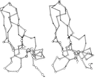
Figure 1 gives a qualitative picture of the fluctuations observed in the molecular dynamics simulation of the basic pancreatic trypsin inhibitor (PTI), a small protein with 58 amino acids and 454 heavy atoms; only the
a-carbon atoms plus the three disulfide bonds are shown. The left-hand drawing represents the X-ray structure and the right-hand drawing an instantaneous picture of the equilibrated structure after 3 ps (71). The two structures are very similar, but there are small differences throughout. The largest displacements appear in the C-terminal end, which interacts with a neighboring molecule in the crystal, and in the loop in the lower left, which has rather weak interactions with the rest of the molecule. Corresponding behavior and deviations from the X-ray structure would be observed in
"snap shots" taken at any other time during the simulation.
Mean-Square Fluctuations and Temperature Factors
A more quantitative measure of the motions is obtained from the mean- square fluctuations of the atoms from their average positions. These can be
Annu. Rev. Biochem. 1983.52:263-300. Downloaded from arjournals.annualreviews.org by MASSACHUSETTS INSTITUTE OF TECHNOLOGY on 03/20/08. For personal use only.
Figure 1 Drawing of c~-carbon skeleton plus S-S bonds of PTI; left-hand drawing is the X-ray structure and right-hand drawing is a typical "snapshot" during the simulation.
related to the atomic temperature or Debye-Waller factors, B, determined in an X-ray diffraction study of a protein crystal (101-105). The mean- square positional fluctuation, < Ar2 > dyn, with the assumption of isotropic and harmonic motion can be written:
_ 3B ~ A r~ ~is" 4.
~Ar2 )’dyn 8rr2
<A r 2> dis is the contribution to B from lattice disorder and other effects that are difficult to evaluate experimentally. For a number of proteins at ambient temperatures (101 - 105), the measured value of (3 B/8 zr 2) averaged over all of the nonsurface atoms of the protein is in the range 0.48-0.58
~2. Comparison of this result with the mean value of < A r 2 > dyn from protein simulations (0.28-0.36 ,~2; 101, 106, 107) suggests that the nonmo- tional contribution to the B-factor <Ar2>’dis, is in the range 0.20-0.25 /~z. The only experimental estimate of <At2> dis is from Mtissbauer data for the heme iron in myoglobin (102); for that one atom a somewhat smaller value (0.14 ~2) was obtained. Thus, in the cases examined, approximately half of the experimental B-factor is associated with thermal fluctuations in
Annu. Rev. Biochem. 1983.52:263-300. Downloaded from arjournals.annualreviews.org by MASSACHUSETTS INSTITUTE OF TECHNOLOGY on 03/20/08. For personal use only.
the atomic positions and half with other sources. However, some protein crystals, particularly those with a high percentage of water, appear to have a larger disorder contribution (109).
There is generally an increase in the magnitude of the experimental and theoretical fluctuations with distance from the center of the molecule. The magnitudes of the rms fluctuations range from "~ 0.4/~ for backbone atoms to ~ 1.5/~ for the ends of long sidechains. The hydrogen-bonded secondary structural elements (a-helices,/~-sheets) tend to have smaller fluctuations than the random coil parts of the protein 006, 108). The magnitude of the fluctuations vary widely throughout the protein interior, suggesting that the system is inhomogeneous and that some regions are considerably more flexible than others.
To examine the importance of bond length and bond angle fluctuations, simulations were performed on PTI in which the bond lengths or both the bond lengths and the bond angles were ~xed at their average values (73).
It was found that use of fixed bond lengths (normal fluctuations +0.03
~) does not significantly alter the dynamical properties on a time scale longer than 0.05 ps, but that constraint of the bond angles (normal fluctua- tions, _+ 5°) reduces the mean amplitude of the atomic motions by a factor of two. This result demonstrates that in a closely packed system, such as a protein in its native configuration, the excluded volume effects of repulsive van der Waals interactions introduce a strong coupling between the dihe- dral angle and bond-angle degrees of freedom.
Figure 2 shows a comparison of the calculated and experimental rms fluctuations on a residue-by-residue basis for reduced cytochrome c (I01).
The experimental values were corrected for an estimated disorder contribu- tion by subtracting from all of them < A r 2 ~ dis = 0.25 ,/]k 2, obtained from
the average calculated results for the protein interior. There is generally good agreement between the experimental and theoretical values. This correlation confirms the reliablity of both the simulation results and the temperature factors as detailed measures of the internal mobility of pro- teins.
Since most of the molecular dynamics simulations have been done for a protein in vacuum, it is expected that, particularly for the exterior residues, the results will be in error owing to the absence of solvent and, with regard to X-ray temperature factors, the absence of the crystal environment. For cytochrome c, the most prominent differences between theoretical and ex- perimental mean displacements (Figure 2) involve the residues calculated to have very large fluctuations; these are all charged sidechains (particularly lysines) that protrude from the protein and so are not correctly treated in the vacuum simulation. This result is confirmed by molecular-dynamics
Annu. Rev. Biochem. 1983.52:263-300. Downloaded from arjournals.annualreviews.org by MASSACHUSETTS INSTITUTE OF TECHNOLOGY on 03/20/08. For personal use only.
simulations of PTI in a Lennard-Jones solvent and in a crystal environment.
The simulations show that the motion of the outside residues is significantly perturbed by the surrounding medium (107, 110); in particular, the interac- tion between charged sidechains of a given protein and its crystal neighbors can produce a reduction in the rms values (66, 110). Such results for the external residues contrast with those for the protein interior, where the environmental effects on the amplitude of fluctuations are found to be small.
The dominant medium effect on the equilibrium properties of the PTI molecule is that the average structure in the solvent or crystal field is significantly closer to the X-ray structure than is the vacuum result (e.g. for the Ca-atoms, the vacuum simulation has an rms deviation from the X-ray structure equal to 2.2/~, while those for the solvent and crystal simulations are 1.35 /~ and 1.52/~, respectively).
Recently, a crystal simulation of PTI, including the water molecules, was completed (111). Although the simulation is too short (12 ps) for definitive conclusions, the magnitude of the fluctuations corresponded to those found in the earlier simulations of PTI, while the dynamic average structure was somewhat closer to the X-ray result (C°-atom rms deviation is 1.06/~). That the integrity of the dynamic average structure is necessary to obtain the
1.5
|,0
¸
0.5-
i 1 ¯ I i 1 ¯ I J !
0.0 I I I I I I I I A i
0 20 40 60 80 100
Residue no.
Figure 2 Calculated and experimental rms fluctuations of ferrocytochrome c; residue aver- ages are shown as a function of residue number: (a) molecular-dynamics simulation; (b) X-ray temperature factor estimation corrected for mean disorder contribution.
Annu. Rev. Biochem. 1983.52:263-300. Downloaded from arjournals.annualreviews.org by MASSACHUSETTS INSTITUTE OF TECHNOLOGY on 03/20/08. For personal use only.
correct rms fluctuations was noted in a simulation of rubredoxin (112);the agreement between calculated and experimental (113) temperature factors was poor, apparently because of a significant perturbation in the structure during the simulation.
Of interest also are the results from the dynamic simulation concerning deviations of the atomic motions from the isotropic, harmonic behavior assumed in most X-ray analyses of proteins. The motions of many of the atoms were found in the simulations to be highly anisotropic and somewhat anharmonic. The rms fluctuation of an atom in its direction of largest displacement is typically twice that in its direction of smallest displacement;
larger ratios are not uncommon (106, 110, 114, 115). It is sometimes possi- ble to rationalize these directional preferences in terms of local bonding, e.g.
torsional oscillation of a small group around a single bond (115). In most cases, however, the directional preferences appear to be determined by larger-scale collective motions involving the atom and its neighbors (115- 118). The atom fluctuations are generally also anharmonic; that is, the potentials of mean force for the atom displacements deviate from the simple parabolic forms that would obtain at sut~ciently low temperature (106, 110, 119). The most markedly anharmonic atoms are those having multiple minima in their potentials of mean force. The shape of the PTI potential
surface in the region of the native structure indicates that the anharmonicity is primarily associated with the softest collective modes of displacement in the protein (120).
Time-Dependence: Local and Collective Effects
Analyses of the time development of the atomic fluctuations were made for PTI (117, 118) and cytochrome c (116). The atomic fluctuations that tribute to the temperature factor (thermal ellipsoid) can be separated into local oscillations superposed on motions with a more collective character.
The former have a subpicosecond time scale; the latter, which can involve only a few neighboring atoms, a residue, or groups of many atoms in a given region of the protein, have time scales ranging from 1-10 ps or longer (v ~ 3 to 30 cm- x). By following the time development of the atomic fluctua- tions of PTI from 0.2-25 ps, it was shown that the high-frequency oscilla- tions, which contribute about 40% of the average rms fluctuations of mainchain atoms, tend to be uniform over the structure. It is the longer- time-scale, more collective motions that introduce the variations in the fluctuation magnitudes that characterize different parts of the protein struc- ture (117, 118). The correlations of anisotropy with local bonding are often destroyed by these large amplitude collective motions (115, 116).
The time dependence of atom and group motions in proteins can be characterized more fully by calculating appropriate time correlation func-
Annu. Rev. Biochem. 1983.52:263-300. Downloaded from arjournals.annualreviews.org by MASSACHUSETTS INSTITUTE OF TECHNOLOGY on 03/20/08. For personal use only.
tion (72, 117, 118). The time correlation function of a fluctuating quantity describes the average manner in which a typical fluctuation decays (30, 31).
For the positional fluctuations of individual atoms in the protein interior, the time correlation function has a partial loss of amplitude within the first 0.2 ps, followed by much slower decay on a time scale of several ps; the slow component has significant oscillations in many cases (72, 116-118). The decay times of the correlation functions are increased by including external solvent in the dynamic simulation; this effect is most pronounced for atoms at the protein surface (110, ll7, 118). An analysis of the relaxation times for the atoms in PTI plus solvent yields a wide range (0.45-10 ps);
vacuum, the times shift to somewhat shorter values (0.2-6 ps).
Although there is no direct experimental measure of the time scale of the atomic fluctuations, it has been shown that NMR-relaxation parameters (T1, T2, and NOE values) are sensitive to picosecond motions. Of particular relevance are IaC-NMR data since for protonated carbons, the C-H bond reorientation provides the dominant relaxation mechanism. Preliminary comparisons of ~3C-relaxation data for PTI suggest that the a-carbon mo- bility has a small effect on the relaxation parameters and that increasing effects are expected as the observed carbon is further out along a sidechain (121, 122). The effect of internal motions on other NMR parameters, such as chemical shifts (123) and vicinal-coupling constants, was also examined by molecular-dynamics simulations.
Biological Function
Although many of the individual atom fluctuations observed in the simula- tions or obtained from temperature factors may in themselves not be impor- tant for protein function, they contain information that is of considerable significance. The calculated fluctuations are such that the conformational space available to a protein at room temperature includes the range of local structural changes observed on substrate or inhibitor binding for many enzymes. There may be a correlated directional character to the active-site fluctuations that play a role in catalysis. Further, the small amplitude fluctuations are essential to all other motions in proteins; they serve as the
"lubricant" which makes possible larger-scale displacements, such as do- main motions (see Table I), on a physiological time scale. It may be possible to extrapolate from the short time fluctuations to larger-scale protein mo- tions. This is suggested by the approximate correspondence between the rms fluctuations of hydrogen bond lengths in a dynamical simulation of PTI (124) and the relative exchange rate of the hydrogens as measured by NMR (42). Changes in the fluctuations induced by perturbations, e.g. ligand binding, are likely to be important as well, e.g. the entropy differences, for the study of which molecular-dynamics techniques were developed (125), may make a significant contribution to the binding free energy (15).
Annu. Rev. Biochem. 1983.52:263-300. Downloaded from arjournals.annualreviews.org by MASSACHUSETTS INSTITUTE OF TECHNOLOGY on 03/20/08. For personal use only.
The collective modes are likely to be of particular significance in the biological function; they may be involved in the displacements of side- chains, loops or other structural units required for the transition from an inactive to the active configuration of a globular protein and in the corre- lated fluctuations that play a direct role in enzyme catalysis. Further, the extended nature of these motions makes them more sensible to the environ- ment, e.g., differences in the simulation results between vacuum and solu- tion results for PTI (117, 118). Because they involve sizable portions of the protein surface, the collective motions may be involved in transmitting external solvent effects to the protein interior (45). They might also expected to be quenched at low temperature by freezing of the solvent. Their contribution to the mean-square fluctuations could explain the transition observed near 200°K in the temperature dependence of the fluctuations in proteins like in myoglobin (126).
SIDECHAIN MOTIONS
The motions of aromatic sidechains serve as a convenient probe of protein dynamics. The sidechain motions span a time range from picoseconds, during which local oscillations occur, to milliseconds or longer required for
180° rotations. To cover this range of motions requires use of a variety of approaches that complement each other in t~e analysis of protein dynamics.
Further, the results obtained are typical of a class of motional phenomena that play a significant role.
Tyrosines -in PTI
The torsional librations of buried tyrosines in PTI were studied in some detail (72). W~ focus on a particular aromatic sidechain, Tyr 21, whose ring is surrounded by and has a significant nonbonded interaction with atoms
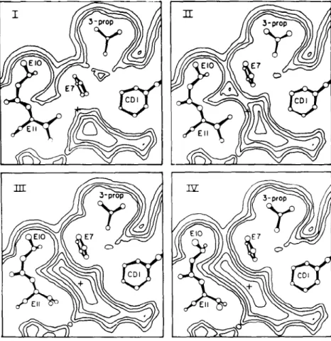
of its own backbone and of surrounding residues that are more distant along the polypeptide chain. Figure 3 shows a potential energy contour map for the sidechain dihedral angles X~ and Xz of Tyr 21 in the free dipeptide (top) and inthe protein (bottom) (57). The minimum energy conformations are very similar in the two cases~ this appears to be true for most interior residues of proteins. Where the plots differ is that the sidechain is much more rigidly fixed in position by its nonbonded neighbors in the protein than it is by interactions with the backbone of the chain in the dipeptide.
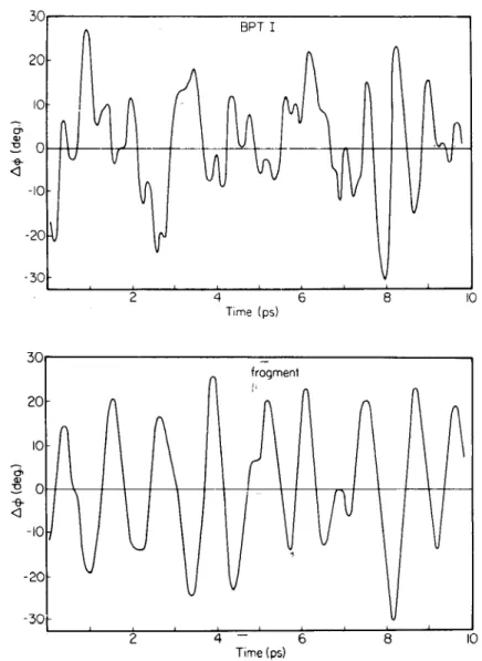
Figure 4 (top) shows the torsional fluctuations of Tyr 21 observed during a PTI simulation (72); the quantity plotted is A~b = ~- "<~b> where
<~b> is the time average of the ring torsional angle. Figure 4 (bottom) shows corresponding torsional fluctuation history for the ring in an isolated tyrosine fragment simulation.
Annu. Rev. Biochem. 1983.52:263-300. Downloaded from arjournals.annualreviews.org by MASSACHUSETTS INSTITUTE OF TECHNOLOGY on 03/20/08. For personal use only.
~2
360
°
~80
°
go° ~
0 o
360
90
° 180° 270° 360°
X~
270-
180"
9O
o 9b 1~0 2~0 360
Figure 3 (K1,K/)) maps for Tyr-21 in PTI: (top) free peptide; (bottom) peptide in protein; the black dot corresponds to the X-ray value for (K~,x2) in the protein; energy contours in kcal/- tool.
Annu. Rev. Biochem. 1983.52:263-300. Downloaded from arjournals.annualreviews.org by MASSACHUSETTS INSTITUTE OF TECHNOLOGY on 03/20/08. For personal use only.
The torsional motion of the ring is less regular when it is surrounded by the protein matrix than in the separated fragment. In PTI, the rms fluctua- tion of the Tyr 21 torsion ang!e is 12°, while that for the tyrosine fragment is 15°. This relatively, small difference in amplitudes as compared with the forms of the rigid rotation potentials (Figure 3) indicates that protein relaxation involving correlated fluctuations must play an important role in
3O 2O
-I0
-20 -5O
BPT I
’ i ’ i ’ ’ ’
Time (ps)
50,
I0
-2O 2O
frogmen1
2 4 -- 6 8 I0
Time
Figure 4 Evolution of the Tyr-21 ring torsional angle during 9.8 ps of dynamical simulation:
(top) in the protein; (bottom) in the isolated tyrosine fragment.
Annu. Rev. Biochem. 1983.52:263-300. Downloaded from arjournals.annualreviews.org by MASSACHUSETTS INSTITUTE OF TECHNOLOGY on 03/20/08. For personal use only.
the ring oscillations. The short time, local motion in the protein is consistent with a torsional Langevin equation that contains a harmonic restoring force (see Equation 3). The frictional random force terms are similar to those expected for ring rotation in an organic solvent; this is consistent with the hydrophobic environments of the rings in the protein. The time correlation functions for the torsional fluctuations decay to small values in a short time (~0.2 ps). However, the quantities involved in the relaxation times (110,
127) measured in fluorescence depolarization (trigonometric functions the angles) decay much more slowly. For the tyrosine rings in PTI there
is rapid partial decay in less than a picosecond to a plateau value equal to about 75% of the initial value; this behavior was recently confirmed by fluorescent depolarization measurements (128). Corresponding calculations
(130) for the fluorescent depolarization of the tryptophan residues in lyso- zyme based on a molecular dynamics simulation (106) indicate a wide range of variation in the depolarization behavior. Since there are six trypto- phans in a variety of environments, their behavior is expected to correspon d
to that which occurs more generally in proteins (129). Certain interior tryptophans have almost no decay over the time scale of the simulation while one in the active site (Trp 62) has its anisotropy reduced to 0.6 after
5 ps.
Tyrosine and phenylalanine ring rotations by 180° were studied by NMR in proteins (40, 43, 44, 56). Such ring "flips" occur very infrequently be- cause of the large energy barrier due to steric hindrance (57-59). The long time intervals separating flips preclude systematic study by conventional molecular-dynamics methods. A modified molecular-dynamics method was recently developed to handle such local activated processes (59). This method is similar to adiabatic mapping in that one starts with an assumed
"reaction coordinate" that defines the fundamental structural, changes in- volved. It differs from the adiabatic method in that it involves consideration of all thermally accessible configurations and not just the minimum energy one for each value of the reaction coordinate. Also it provides a detailed description of the structural and dynamical features of the process. In this method, one calculates separately the factors in the rate constant expression (82, 131):
k = ~ < 1 Ill > [p(¢t)/f.ip(~)d~]. 5.
Here, ~ is the reaction coordinate, ~ = d;~/dt, and Ct is the value of ~ in the transition state region for the process. The factor in square brackets is the probability that the system will be in the transition state region, relative to the probability that it is in the initial stable state. This quantity corresponds roughly to the term exp(-AGt/RT) in more familiar expres-
Annu. Rev. Biochem. 1983.52:263-300. Downloaded from arjournals.annualreviews.org by MASSACHUSETTS INSTITUTE OF TECHNOLOGY on 03/20/08. For personal use only.
sions for rate constants; it can be calculated by carrying out a sequence of simulations in which the system is constrained to stay near particular values of ~:. The remaining factors can be evaluated by analysis of trajectories initiated in the transition state region (59, 85, 86). The transmission coeffi- cient K is equal to one in ideal transition state theory (equilibrium popula- tions maintained in the stable states and uninterrupted crossings through the transition state region); for real systems K is less than one.
Application of this modified molecular dynamics method to the flipping of a tyrosine ring in PTI shows that the rotations themselves required only 0.5-1.0 ps (85, 86). At the microscopic level, the processes responsible for flipping are the same as those responsible for the smaller amplitude libra- tions. The ring goes over the barrier not as the result of a particularly energetic collision with some cage atom, but as the result of a transient decrease in frequency and intensity of collisions that would drive the ring away from the barrier. These alterations of the collision frequency are caused by small, transient packing defects (86). The packing defects help to initiate ring rotation, but they are much too small to allow free rotation of the ring by a simple vacancy or free-volume mechanism (86, 132). The ring tends to be tightly encaged even in the transition state orientation.
Collisions with cage atoms in the transition state produce frictional forces similar to those that occur in the stable state librations; these frictional effects reduce the transition rate to about 20% of the ideal transition-state theory value (59). As to the free energy of activation, the calculations suggest that the activation enthalpy contribution is similar to that found by adiabatic mapping techniques (57, 58) and that the activation entropy small.
Although no enzyme has yet been studied by the techniques applied to the tyrosine ring flips, the methodology is applicable to the activated pro- cesses central to most enzymatic reactions. Further, many of the qualitative features found for the tyrosines (e.g. lowering of the potential of mean force by cage relaxation, alteration of the rate by frictional effects) should be present in general.
Ligand-Protein Interaction in Myoglobin
A biological problem where sidechain fluctuations are important concerns the manner in which ligands like carbon monoxide and oxygen are able to get from the solution through the protein matrix to the heme group in myoglobin and hemoglobin and then out again. The high-resolution X-ray structure of myoglobin (8, 9, 133, 134) does not reveal any path by which ligands such as 02 or CO can move between the heme-binding site and the outside of the protein. Since this holds true both for the unliganded and
Annu. Rev. Biochem. 1983.52:263-300. Downloaded from arjournals.annualreviews.org by MASSACHUSETTS INSTITUTE OF TECHNOLOGY on 03/20/08. For personal use only.