warwick.ac.uk/lib-publications
Manuscript version: Author’s Accepted ManuscriptThe version presented in WRAP is the author’s accepted manuscript and may differ from the published version or Version of Record.
Persistent WRAP URL:
http://wrap.warwick.ac.uk/111369
How to cite:
Please refer to published version for the most recent bibliographic citation information. If a published version is known of, the repository item page linked to above, will contain details on accessing it.
Copyright and reuse:
The Warwick Research Archive Portal (WRAP) makes this work by researchers of the University of Warwick available open access under the following conditions.
© 2018 Elsevier. Licensed under the Creative Commons Attribution-NonCommercial-NoDerivatives 4.0 International http://creativecommons.org/licenses/by-nc-nd/4.0/.
Publisher’s statement:
Please refer to the repository item page, publisher’s statement section, for further information.
1
Connection between circadian rhythms and neurodegenerative diseases
Yue Leng, PhD1 2*, Erik S. Musiek, MD, PhD3, Kun Hu, PhD4 5, Francesco P. Cappuccio, MD, FRCP6 Kristine Yaffe, MD7
1. Department of Psychiatry, University of California, San Francisco, 4150 Clement Street, San Francisco VA Medical Center, CA 94121, USA
2. Shanghai Jiao Tong University School of Medicine, 280 South Chongqing Road, Shanghai, China 3. Department of Neurology, Hope Center for Neurological Disorders, and Knight Alzheimer Disease
Research Center, Washington University School of Medicine, St. Louis, MO 63110, USA 4. Medical Biodynamcis Program, Division of Sleep and Circadian Disorders, Departments of
Medicine and Neurology, Brigham and Women’s Hospital, Boston, MA 02115, USA
5. Division of Sleep Medicine, Department of Medicine, Harvard Medical School, Boston, MA 02115, USA
6. Division of Health Sciences (Mental Health and Wellbeing), Warwick Medical School, University of Warwick, Coventry CV4 7AL, UK.
7. Departments of Psychiatry, Neurology, and Epidemiology, University of California, San Francisco, San Francisco VA Medical Center, San Francisco, CA 94121, USA.
2 I. Abstract
Increasingly circadian rhythms have been linked to age-related diseases. We provide an
up-to-date review (2013-2018) of the association between circadian rhythm disruptions (CRD) and the
most common types of neurodegenerative diseases, including Alzheimer’s disease and related
dementias (ADRD) and Parkinson’s disease (PD). CRD differ according to type and severity of
neurodegenerative diseases, and importantly, could occur early during the course of
neurodegeneration as a prodrome. Evolving evidence also supports CRD as a potential risk factor
for developing ADRD and PD, although confirmatory studies are needed. Proposed mechanistic
pathways underlying this association include alterations in sleep, protein homeostasis, and
immune and inflammatory function. While promising, more studies of CRD and treatment trials
are needed to determine whether circadian interventions may prevent or delay the onset of
neurodegenerative diseases. The study of circadian rhythms holds great promise for a role in the
3 Panel 1.
Glossary Panel
Amplitude: Magnitude of a cycle, or the difference between peak and trough values. In relation to a hormonal cycle, for example, it would be the difference in the levels of the hormone from the trough to the peak within a time period (i.e. 24 hours).
Circadian rhythm: An approximately 24-hour cycle in the physiological processes of most organisms, endogenously generated, and modulated by external cues.
Circadian rhythm sleep-wake disorders: Disorders related to the timing of sleep and wakefulness that are characterized by the inability to fall asleep, remain asleep, and/or wake at the desired time.
Circadian misalignment: A loss of synchrony between internal circadian rhythms, behaviors and the external environment, as a result of a variety of circumstances (e.g. jet lag, shift work).
Entrainment: The resetting of circadian cycle to maintain synchrony with daily environmental and behavioral cycle.
Irregular sleep-wake rhythm disorder: A lack of clear 24-hour sleep-wake pattern, usually with long periods of wakefulness during the night and irregular bouts of sleep throughout the day; most common in patients with neurodegenerative diseases.
Mesor: The mean value around which the rhythm oscillates.
Period: Time interval between two reference points within a rhythm or recurring wave.
Phase: Timing of a reference point in the cycle relative to a fixed event. In relation to a sleep-wake cycle, for example, a phase advance (delay) would mean that sleep timing moves earlier (later).
Sundowning: Increasing behavioral and neuropsychiatric symptoms around the time of sunset, commonly seen in dementia patients.
4 II. Introduction
The award of the 2017 Nobel Prize in Physiology or Medicine to Hall, Rosbash, and Young for
their work on the molecular mechanisms of the circadian clock highlights the importance of
circadian rhythms and their relation to health and wellbeing (1). In humans, circadian rhythm
activities change markedly as people age, and these changes might further accelerate the aging
process (2). While it is recognized that circadian dysfunction in older adults can be partly
attributed to the degeneration of the suprachiasmatic nucleus (SCN), known as the “master
circadian clock” in mammals, the link between circadian rhythms and neurodegeneration is not
fully understood. Patients with neurodegenerative diseases frequently experience circadian
rhythm disruptions (CRD) (3-5). For example, they become more active during the night, less
active during the day, and sometimes have complete reversal or loss of the 24-h rest-activity
pattern (6, 7). Importantly, growing evidence suggests that disruptions of circadian functions
could be early manifestations of neurodegeneration, and might even be a risk factor to the
development of neurodegenerative diseases in healthy older adults (8-10). Greater understanding
of the relationship between circadian rhythms and neurodegeneration could be key to the early
identification and management of neurodegenerative diseases.
This review discusses the association between circadian rhythms and neurodegenerative diseases
by summarizing evidence from both human and animal studies. In order to provide a most
up-to-date review of the evidence, we mainly discuss the empirical findings published within the past 5
years, except for certain landmark studies. We referred to a few previous reviews (11-13) for
detailed discussion of earlier studies. This review focuses on Alzheimer’s disease and related
dementias (ADRD) and Parkinson’s disease (PD), as these are the most common
5
introduce key concepts related to circadian rhythms (See Glossary Panel), present both
behavioral and biological circadian features in patients with ADRD and PD, summarize findings
from clinical and longitudinal epidemiologic studies regarding the effects of CRD on the
development of ADRD and PD, and discuss underlying mechanisms. Finally, we describe the
results of different circadian interventions, especially in those with ADRD and PD, and discuss
future directions.
III. Circadian rhythms
A circadian rhythm (from the Latin ‘circa’ = about and ‘diem’ = a day) is an approximately
24-hour cycle in the physiological processes of most organisms that is endogenously generated and
can be modulated by external cues (14). A circadian cycle is characterized by several features. It
is self-sustained, as the rhythm persists in the absence of any exogenous time signals
(Zeitgebers), including dark-light cycles. This characteristic indicates the presence of an intrinsic
time-keeping mechanism (i.e. biological clock). It shows rhythmicity, as they persist with a cycle
of approximately 24 hours. It also shows the ability to be synchronized by external cues, such as
the dark-light cycle or other social and environmental modulators, like activity and temperature.
The circadian rhythmicity is typically measured by three parameters: amplitude, phase and
period. Amplitude is defined as the magnitude of a cycle, or the difference between crest and
trough values. In relation to a hormonal cycle, for example, it would be the difference in the
levels of the hormone from the trough to the peak within a time period (i.e. 24 hours). Phase
(advanced or delayed) is defined as the timing of a reference point in the cycle relative to a fixed
event. In relation to a sleep-wake cycle, for example, a phase advance (delay) would mean that
sleep timing moves earlier (later). Period is the time interval between two reference points within
6
Circadian rhythms are generated in highly specialized cells of specific structures of the brain that
control a complex network of coupled self-sustained clocks in the brain and in the peripheral
organs. In mammals, the central or master clock of the circadian network is located in two
groups of neurons called the SCN, in the anterior hypothalamus. The SCN consists of
approximately 20,000 specialized neurons, which receive direct synaptic input from the retina,
synchronizing activity to the external light- dark cycle (15). Light input serves to synchronize the
core cellular clock machinery in SCN neurons, which keeps 24-hour time and in turn
synchronize cellular clocks throughout the body via neurohormonal modulation. At the
molecular level, the properties of circadian clocks are based on changes in the expression of
certain genes and consist of proteins which form a transcriptional-translation feedback loop that
is tuned to a 24-hour period (16). The clock proteins BMAL1 and Clock interact to drive
transcription of clock-controlled genes, including their own negative feedback repressors, which
include PERIOD, CRYTOCHROME, and REV-ERB proteins (17). This transcriptional
feedback loop maintains 24-hour rhythms in gene expression which are required for behavioral
and physiologic rhythmicity at the organismal level. While light is the primary circadian cue,
resetting the circadian cycle in synchrony with the daily environmental and behavioral cycle
(entrainment) is achieved through the 24-h cycle of light input (photic synchronizer) to the SCN
and neurohormonal modulations (non-photic synchronizers) (e.g. temperature, food availability,
social interactions) for the peripheral ones. Importantly, in the absence of external cues, such as
in constant darkness, the circadian system retains a near 24-hour rhythm, while light cues that are
7
The pattern of one’s circadian rhythm can be measured with both biological and behavioral
markers. Landmark experiments by Czeisler et al. (18) identified core body temperature (CBT),
as well as melatonin and cortisol secretions, as ‘circadian’ biomarkers, oscillations of which are
controlled by the SCN. In normally entrained individuals, CBT has a rhythm that falls during the
night and rises in the early hours of the morning; cortisol peaks in blood and saliva early in the
morning, then regularly decreases throughout late morning and afternoon, to reach low values
during evening and night, thereby availing sleep; melatonin is generated by the pineal gland,
with its onset near sunset, peak during the nighttime hours and offset after sun rise, thereby
stimulating wakefulness. The circadian rhythm of melatonin in saliva or plasma is one of the
most commonly used circadian phase biomarkers in human beings (19). The onset time of
melatonin secretion under dim light conditions, known as the dim light melatonin onset
(DLMO), has been suggested as the single most accurate circadian phase marker in humans (20).
Behavioral markers of circadian rhythm mainly include sleep-wake cycles and rest-activity
rhythms. The circadian system has powerful influence over the sleep-wake cycle, such that it is
often difficult to distinguish the relative contributions of the two sleep regulatory systems (the
sleep-wake homeostasis and the circadian timing system) to behavior. The circadian clock
regulates the timing of sleep, as mutations in core circadian clock genes in mice and humans
manifest as sleep phenotypes, including short sleep time, early or late sleep phase, or fragmented
sleep-wake rhythms (21, 22). Moreover, clock gene expression can be influenced by sleep
deprivation, emphasizing that these systems are interrelated. While specific circadian analyses
can be used to parse out aspects of CRD from behavioral data, activity must be monitored around
the clock for several days. Some circadian biomarkers, such as the timing of melatonin secretion
8
face of sleep deprivation (23, 24). Therefore, it is important for studies to include both
behavioral and biological markers of circadian rhythms to more robustly identify CRD. Given
the scope of this review, we include studies if they present information on biological markers or
behavioral markers related to sleep timing, daytime sleep/sleepiness and rest-activity rhythm;
studies that only present nocturnal sleep disruptions are excluded.
Age-related changes in any of the structures or processes involved in generating or entraining
circadian rhythms may modify circadian rhythmicity with advancing age. In particular, circadian
phase has been shown to move earlier, or advance, with age (25), while the amplitude of the
rhythms tend to decrease (26). For example, older adults have decreased peak melatonin,
elevated nadir level of CBT, and a phase advance (earlier onset) in the peak of these rhythms (2)
(27) (28). Age-related changes in sleep-wake cycles may be related to circadian dysfunction and
include earlier bedtimes and rise times, increased sleep fragmentation, and increased daytime
sleepiness that has been frequently suggested as an early indicator of declining health in the
elderly (29, 30). Duffy et al. reported that older adults are more prone to several circadian
rhythm sleep-wake disorders (CRSWDs), including advanced sleep wake phase disorders
(ASWPD), jet lag disorder and shift-work disorder, due to an inability to fall asleep or remain
asleep, conflicting with desired sleep timing (31, 32). The circadian system is paramount for
optimal biological functioning, maintaining synchrony between internal physiology, behavior,
and the cues deriving from the external environment. When this synchrony is lost, e.g. due to jet
lag, shift work, or chronic sleep deprivation, a “circadian misalignment” occurs, leading to
significant health consequences affecting cardiovascular, metabolic, cognitive, immunological
9 IV. Disruption in neurodegeneration
a. Alzheimer disease and related dementias
The prevalence of circadian disruption in patients with moderate-to-severe AD has been
recognized for more than two decades (6). AD patients were often considered to have much more
severe circadian disruptions compared to healthy older adults, including higher fragmentations,
dampened amplitude and phase delay, as opposed to more typical advanced circadian phase
associated with normal aging (6). It was suggested that “sundowning”, known as the increasing
behavioral and neuropsychiatric symptoms in AD patients around the time of sunset, could also
partly be attributed to the phase delay of temperature and hormone rhythms in AD (36)(37). The
most common CRSWD seen in AD patients is irregular sleep-wake rhythm disorder (ISWRD),
as opposed to ASWRD in healthy older adults. ISWRD is defined as a lack of clear 24-hour
sleep-wake pattern, usually with long periods of wakefulness during the night and irregular bouts
of sleep throughout the day which might get worse in severe AD (38, 39).
Over the past five years, a growing number of studies focused on patients of various levels of
cognitive impairment and found their circadian patterns differed from those reported in previous
studies (6). These studies (Table 1) included patients with pre-clinical AD (8), mild cognitive
impairment (MCI) (40) (41), mild AD (3) (42) (43), moderate to severe AD (43), global AD(44),
as well as early onset dementia (EOD) (4). All of these studies have reported on behavioral
markers of CRD, including disruptions of rest-activity rhythms and sleep timing. Two studies (3,
40) examined melatonin rhythms using saliva melatonin assay, one study (41) assessed
temperature rhythms using a wrist temperature sensor, and one study (3) also examined
peripheral clock gene expression. Overall, studies have found high rest-activity rhythm
10
melatonin rhythms (3, 4, 8, 42, 44).One recent US study in 189 cognitive normal older adults
(50 with preclinical AD pathology) showed decreased rhythm amplitude associated with aging,
but not with AD pathology(8). Another study in 16 mild-moderate AD patients from Italy found
large variability among individual actigraphic profiles, which could have also contributed to the
overall minor changes in the amplitude of rhythms in these patients (42). There are mixed
findings with regard to changes in circadian phases. The Rush Memory and Aging Project
suggested a significant phase delay in rest-activity rhythm among 7 AD patients compared to 10
controls (44), whereas a study of 48 AD patients from Italy showed an advanced bedtime in AD,
especially for moderate to severe cases of AD (43). Meanwhile, two recent studies of MCI
patients both found a phase advance, one in melatonin and sleep onset (40), and another in CBT
and activity rhythm (41).
In general, studies that focused on more severe AD found more circadian disruptions, while
studies in MCI, preclinical AD and mild AD suggested moderate circadian changes (3) (8) (40).
Weissova et al. found no correlation between circadian features and severity measures of AD in
16 mild to moderate AD patients (42). No study to date has examined change in circadian
rhythms with the progression of AD symptoms. Few studies have examined molecular
perturbations in circadian clock oscillations in ADRD, though alteration in clock gene
methylation and expression have been described in patient fibroblasts (45), and altered clock
gene expression noted in post-mortem tissue (46). Further, evidence specifically pertaining to
circadian disruptions among patients with non-AD dementia is sparse. Larger and longitudinal
studies are needed to determine the correlation between both behavioral and biological markers
11
markers and features specific to each type of dementia might help with the differential diagnosis
of the disease.
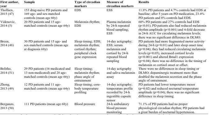
b. Parkinson’s disease
Both motor and non-motor manifestations of PD show disruptions in their typical 24-h
oscillations. Unlike patients with ADRD, CRD among PD patients is featured by a reduction in
the amplitude of the circadian rhythm but no significant shift in circadian phases (5) (47) (48)
(49) (50) (51). Sleep-wake disturbances as a whole are the most common non-motor symptom of
PD patients, affecting up to 80% of PD patients (52). Indeed, five of the six studies (Table 2) that
examined circadian features in PD patients reported on either excessive daytime sleepiness
(EDS) (5) (47) (48) or sleep timing (48) (51) (49). It was reported that PD patients were at least
twice as likely to experience EDS compared to healthy older adults (5, 47). Only one study
reported slightly later sleep onset time in 30 PD patients compared to 15 healthy controls from
England (53), while the others did not find significant differences in sleep timing (49, 51). One
Australian study found among 12 PD patients a significant reduction in the mesor (mean value
around which the rhythm oscillates) and amplitude of their CBT rhythm, compared to 11 healthy
controls(49). Three studies examined rhythms of melatonin secretion, using plasma (5), serum
(48) and saliva melatonin (51), respectively. While none of these studies found a difference in
the timing of melatonin onset, most found significantly reduced circulating melatonin levels
among PD patients (5) (48). Importantly, the usual circadian dip in blood pressure during the
night may be lost in PD, putting patients at significantly higher risk for cardiovascular
complications including nocturnal hypertension (50). For example, a study of 111 PD patients
from Spain reported that 71.1% of patients did not have the usual dip in blood pressure as
12
Despite the consistently reported CRD among PD patients, it remains unclear whether these
circadian changes result from dopaminergic treatment or PD disease progression itself. Earlier
studies reported that the dopaminergic treatment might lead to phase advance of the melatonin
rhythm(54, 55), while a more recent study among 29 PD patients (16 medicated and 13
non-medicated) and 28 healthy controls from Australia found more than double the melatonin
secretion and uncoupling of circadian and sleep-wake regulations in the treatment group (51).
EDS is another potential consequence of dopaminergic treatment (47). One study in Norway
suggested a doubled frequency of EDS among 153 drug-naive patients with early PD, compared
to 169 age- and sex-matched controls at baseline, and a tripled frequency of EDS among these
patients after 5 years of dopaminergic treatment compared to the controls (47). Larger studies
with other circadian markers are needed to help clarify the effects of dopaminergic treatment on
circadian rhythms, relative to neurodegeneration per se.
V. Disruption and risk of neurodegeneration
A critical question is whether CRD is a cause or consequence of neurodegeneration, or both. If
CRD were contributing to neurodegeneration, it would be expected to occur early in disease
course (or precede disease), and would increase disease risk or rate of progression. While this
question is still unanswered, growing evidence suggests that CRD might precede the
development of clinical symptoms of neurodegenerative diseases. One recent study of 189
cognitively normal older adults (50 with preclinical AD) reported that circadian rest-activity
rhythm fragmentation appeared very early on in the preclinical phase of AD and correlated with
AD-related pathology as assessed with PET imaging and cerebrospinal fluid (CSF)
phosphorylated tau to amyloid β (Aβ)42 ratio (8). Several studies found a correlation between
13
change in cognitively normal older adults, though other biological markers of CRD were not
specifically examined (10, 56, 57). Alterations in circadian melatonin rhythm were also found in
healthy middle-aged men with worse cognitive trajectories in midlife (58). These cross-sectional
findings suggested that CRD could be a result of preclinical AD pathology and may be a
prodromal symptom or sign.
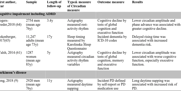
Several longitudinal studies with long follow-up periods also reported greater cognitive decline,
increased risk of all-cause dementia and increased risk of PD among those with circadian
disturbances, including shift work (59-63). Table 3 shows longitudinal studies on CRD and risk
of developing ADRD or PD published over the past five years. These studies all examined
behavioral indicators of CRD, including actigraphy-measured rest-activity rhythm and daytime
napping (9, 61, 64) and self-reported sleep timing(65). Two studies both found an association
between lower circadian amplitude and greater cognitive decline over the next 3-5 years, in
cognitively normal older men (64) and women (61) from the US. Bokenberger et al. reported in
11,247 individuals from the Swedish Twin Registry that delayed rising time predicted dementia
incidence over 17 years (65). Another recent study of 2920 older men from the US suggested
that those who napped for at least 1h per day were twice as likely to develop PD in 11 years (9).
While all together these studies suggest that reduced circadian amplitude and circadian phase
shifts precede the risk of ADRD, and that daytime inactivity precedes the risk of PD in healthy
older adults, the number of published studies is small especially for PD. Additional confirmatory
studies with long follow-up period are needed to determine whether CRD is a risk factor for
ADRD and PD. Comprehensive and repeated measures of CRD with simultaneous assessment of
preclinical disease biomarkers (such as amyloid and tau pathology) will also help understand the
14 VI. Underlying mechanisms
a. Effects of neurodegenerative disease pathology on circadian clock function
The mechanisms by which neurodegenerative pathology affects circadian function likely vary by
specific disease. In AD, human post-mortem neuropathological studies have demonstrated loss
of critical neuronal populations in the SCN, including those expressing arginine vasopressin
(AVP) or vasoactive intestinal peptide (VIP) (44, 66). Both age- and AD-associated loss of
VIP-expressing neurons in the SCN were correlated with pre-mortem circadian dysfunction (Fig.1).
However, the mechanisms driving SCN neuronal loss are unclear, as it is not a major site of
amyloid plaque or neurofibrillary pathology. Circadian abnormalities are observed in transgenic
mouse models of AD, including those expressing human mutant amyloid precursor protein
(APP), tau, or both. However, there is great heterogeneity across mouse models, and little
correlation with pathology, obscuring any definitive mechanistic conclusions (67-69). Aβ
peptide has been implicated as a mediator of circadian dysfunction, and in cultured cells it can
induce degradation of the master clock protein BMAL1 (70, 71). However, this direct interaction
between Aβ and the circadian clock has not been demonstrated in vivo in animals, or in humans.
Altered methylation of the BMAL1 promoter, leading to altered BMAL1 expression and
disrupted circadian rhythms, was described in fibroblasts from AD patients and in post-mortem
AD brain samples, suggesting an underlying epigenetic mechanism of circadian disruption in AD
(Fig.1) (45).
b. Effects of circadian disruption on neurodegeneration
There are several proposed mechanisms by which the circadian clock influences
neurodegenerative disease (Fig. 1). Circadian dysfunction could promote neurodegeneration by
15
napping. Sleep deprivation causes altered Aβ dynamics in humans and increased Aβ and tau
pathology in mouse models, and can increase inflammatory and neuronal injury markers in
human cerebrospinal fluid (72-75).Sleep deprivation can also impact other aspects of
neurodegeneration including protein clearance from the brain, inflammation, and synaptic
homeostasis (76, 77). In this case, intervention to promote sleep should overcome any effect of
circadian disruption. However, in mouse models, clock gene deletion in the brain can cause
neuropathology without altering sleep, suggesting that sleep alone may not explain the brain
effects of circadian disruption (78).
Circadian regulation of immune responses may also contribute to the effects of circadian
dysfunction on neurodegeneration. The circadian system strongly modulates the peripheral
immune response to inflammogens, as the degree of inflammation is highly dependent on
time-of-day of exposure (79, 80). In a mouse experimental autoimmune encephalitis model of
neuroinflammation, the time of day of immunization has a striking impact on disease severity
weeks later, while deletion of Bmal1 in myeloid cells exacerbates pathology (81, 82). In the
brain, microglia and astrocytes represent the primary innate immune cells, and both cell types
possess functional circadian clocks which regulate inflammatory activation (83, 84). Deletion of
Bmal1 in the brain, which disrupts all circadian clock function, causes widespread astrocyte
activation and synaptic degeneration, emphasizing the importance of core clock function in
maintaining innate immune homeostasis in the brain (78). In mouse models of Amyotrophic
Lateral Sclerosis and PD, circadian disruption using non-24 hour light dark cycles led to
increased glial activation and neuroinflammation and exacerbated neuropathology (85, 86).
Thus, circadian dysfunction appears to promote aspects of neuorinflammation, which could
16
The circadian clock could directly regulate protein homeostasis and quality control, thereby
influencing protein aggregation in neurodegenerative diseases (87). In AD, levels of interstitial
fluid Aβ peptide in the hippocampus show clear diurnal oscillation, which require an intact
circadian system (88, 89). Similar diurnal oscillations in Aβ are observed in human
cerebrospinal fluid (90). Moreover, disruption of the circadian clock in a mouse β-amyloidosis
model of AD leads to accelerated amyloid plaque deposition (89). Circadian regulation in
protein quality control systems, such as autophagy, may contribute to the circadian influence on
protein aggregation in general (91, 92) . Bulk removal of aggregated proteins from the brain by
the glymphatic system, a glia-mediated perivascular fluid flow, has been associated with sleep,
but its relation to the circadian system and the role of glial clocks in the process are still
unclear(76). Recent studies demonstrating circadian clock control of blood-brain barrier
permeability may also have implications for protein aggregates clearance from the brain (93, 94).
Finally, numerous studies reveal a complex, bidirectional relationship between the circadian
clock and oxidative stress, a key pathogenic process in neurodegeneration (78, 95-98). Thus, a
number of potential identified mechanisms, as well as those which are not yet known, could link
the circadian clock to neurodegenerative diseases.
VII. Circadian Interventions
If circadian dysfunction is a risk factor contributing to the development of neurodegenerative
diseases, one of the appealing testable hypotheses is that enhancing circadian rhythms might
prevent or halt the progress of these diseases as well as mitigating their related symptoms.
Limited earlier studies have tested this hypothesis using timed light and/or melatonin treatments
but provided inconsistent results (see Review by Forbes, et al.(99)). For instance, in a
17
in the Netherlands (87% had dementia), Riemersma-van der Lek et al. examined the effects of
daily treatment with whole-day bright of 1000 lux (as compared to dim light of 300 lux) and
daily evening melatonin treatment (as compared to placebo) and found that the long-term light
treatment (up to 3.5 years) attenuated cognitive decline with aging and improved depressive
symptoms (100). However, Burn et al. did not find similar cognitive benefit of bright light in
their randomized controlled trial of 48 patients in two nursing homes in the UK with diagnosed
dementia, sleep disruption, and agitated behavior (101). The discrepancy may be attributed to
uncontrolled treatment dose such as exposure duration and intensity of light that are especially
important for the elderly with reduced response of the circadian system to light exposure (102);
future studies should examine these possibilities.
In the last five years, only two published circadian intervention studies examined patients with
ADRD or PD. In a multicenter (one in the UK and four in the USA), double-blinded,
parallel-group study (103), sixty patients diagnosed with mild to moderate AD dementia (13 of them had
insomnia) were randomized to receiving daily treatment of a prolonged-release melatonin
formulation for 24 weeks or placebo. The study showed a positive effect of melatonin treatment
on cognitive performance, especially for those with insomnia. The other study was performed in
PD centers at Northwestern University and Rush University, where 31 patients with PD and
coexistent excessive daytime sleepiness who received stable dopaminergic therapy underwent a
14-day light intervention with twice 1-h exposure to bright or dim light each day (104). The light
intervention improved daily activity rhythms and reduced daytime sleepiness, and the effects
were stronger with bright light.
The application of circadian interventions in neurodegenerative diseases is a promising but
18
also be entrained or shifted by many other non-photic time cues or zeitgebers (105), including
food (106), caffeine consumption (107) and exercise (108). These zeitgebers affect circadian
rhythms likely through direct influences on the peripheral clocks and their feedback to the central
circadian clock (109). How to appropriately implement these time cues in circadian interventions
requires better understanding of the interactions between the central and peripheral clocks. (ii)
The intrinsic properties such as the period of the central circadian clock can be different between
individuals, leading to different chronotypes (i.e., evening- and morning-types) and different
circadian timings (relative to time of day) of behavior and physiological functions including
melatonin secretion. Thus, individuals of different chronotypes have different responses even
when light exposure and melatonin are scheduled at the same time of day (110). However, no
clinical trials have incorporated chronotype into personalized circadian interventions. (iii)
Though circadian control and sleep regulation are tightly coupled, they have different underlying
mechanisms. Understanding these specific mechanistic pathways in addition to distinguishing
whether the observed beneficial effects of interventions are through the influences on the
circadian clocks or directly on the neural circuitry of sleep homeostasis may improve strategies
for future drug and therapeutic design. (iv) Despite the association between circadian
disturbances and cognitive impairment, more evidence for the impacts of circadian interventions
on cognitive decline and the progression of neurodegenerations over a long term (e.g., >5 years),
especially after the intervention period, is required. (v) No circadian intervention study has
considered neuropathological biomarkers. Using structural MRI or PET scans of the brain and
examining longitudinal changes in CSF Aβ and tau levels will help clarify the contributions of
circadian disturbances to neuropathological and anatomical changes in the brain, which may
19
on the stages of neurodegenerative diseases after the clinical onset of the diseases. It will be
important to test the benefits of circadian therapies for the prevention of the diseases and related
symptoms at preclinical stages.
VIII. Conclusions and future directions
People with ADRD or PD frequently experience disruptions in both behavioral and biological
markers of CRD, including disrupted sleep-wake cycles, impaired hormonal and body
temperature rhythms, dysregulated autonomic system as well as fluctuated neuropsychiatric
symptoms. CRD in neurodegeneration is often presented in a much more severe form than
typical age-related CRD and also has distinct features. Unlike healthy older adults who usually
have reduced circadian amplitude and advanced circadian phase, patients with ADRD tend to
have high fragmentation and slightly reduced amplitude of circadian rhythms. There are mixed
findings regarding phase shift among these patients, and they are likely to have irregular
sleep-wake patterns. PD patients tend to have reduced circadian amplitude but no change in circadian
phases. In general, behavioral CRD markers have been examined more than biological markers.
Recent evidence has also suggested that the stage and severity of the disease, as well as the
treatment, increase variation in markers of CRD. Larger longitudinal clinical studies are needed
to examine the change in circadian rhythms with the progression of neurodegeneration, including
non-AD dementias, and to disentangle the effects of PD progression and dopaminergic treatment
on circadian rhythms. The integration of non-behavioral circadian biomarkers into these studies
would help disentangle CRD from sleep/behavioral confounds (see Directions for future
research). This will help identify circadian features that are important for differentiating various
types and stages of neurodegenerative diseases, and is important for the management of circadian
20
Several epidemiologic studies suggested the presence of circadian disturbances at the preclinical
stage of ADRD. CRD might be considered as a useful preclinical marker or prodromal for
neurodegenerative diseases and help with the early detection of the disease. Emerging evidence
from longitudinal studies also showed that CRD precedes the development of ADRD or PD.
Additional confirmatory studies with longer follow-up are needed to examine the relationship
between different circadian markers and subsequent risk of developing neurodegenerative
diseases, and should consider the use of biomarkers to help understand potential mechanisms.
For example, using structural MRI or PET scans of the brain and examining longitudinal changes
in CSF Aβ and tau levels will help clarify if circadian disturbances might contribute to AD
pathology or structural change in the brain. Studies of biological mechanisms and intervention
trials are required to determine if CRD is a cause of neurodegenerative diseases.
Finally, personalized multicomponent circadian intervention should be developed and tested for
its benefits on circadian synchronization as well as symptom management of ADRD or PD. In
addition, larger longitudinal clinical trials with longer follow-up are also needed to examine the
long-term benefits of these interventions, and especially to determine whether these interventions
might help prevent or delay the onset of neurodegenerative diseases among healthy older adults,
or delay symptoms in those at the preclinical stage. In this way, CRD may be a promising
therapeutic target for the prevention and management of neurodegenerative diseases.
VIIII. Search strategy and selection criteria
We identified references for this Review by searches of PubMed between Jan 2013 and Oct
2018, and by hand searches of reference lists from relevant articles. We used the search terms:
21
“Parkinson disease”, “neurodegeneration” and "circadian rhythm", “circadian clock”,
“twenty-four-hour rhythm”, “sleep-wake”, “melatonin” or “chronotherapy”. There were no language
restrictions. We included only references published within the past 5 years, except for key or
landmark studies in the field. The final reference list was made based on relevance to the theme
22 Panel 2.
Directions for future research
Studies of CRD in neurodegeneration should incorporate the assessment of both biological and
behavioral markers of CRD.
Larger, longitudinal studies are needed to determine circadian features for different types and
severities of ADRD, and clarify the link between the progression of ADRD and change in
circadian rhythm disruptions.
The interaction between PD disease progression, dopaminergic treatment and circadian changes
should be clarified.
Additional studies with long follow-up period are needed to confirm the effects of CRD on
subsequent cognitive decline and risk of developing ADRD or PD.
Establishment of underlying mechanisms for the bi-directional relationship between circadian
rhythms and neurodegeneration are needed to help draw causal inference and inform
therapeutic targets.
The use of circadian interventions in patients with neurodegenerative diseases should be further
explored, and personalized circadian treatment should be considered given the large
between-individual differences.
Randomized controlled trials of individuals at preclinical stages are needed to test the benefits
23 Declaration of interests
E.S.M receives personal fees from Eisai Pharmaceuticals and GLG Consulting, outside the
submitted work.
F.P.C. receives book royalties from Oxford University Press.
K.Y. serves on DSMBs for Takeda, Eli Lilly, and an NIH sponsored study and is a member of
the Beeson Scholars in Aging Scientific Advisory Board and a Senate member of the German
Center for Neurodegenerative Diseases.
Acknowledgement (Funding Source)
Y.L. is supported by National Institute on Aging (NIA) 1K99AG056598-01. E.S.M is supported
by NIA R01AG054517 and the Coins for Alzheimer's Research Trust (CART) Fund. K.H. is
supported by NIA R01AG048108 and NIA R01AG059867. K.Y. is supported in part by NIA
K24AG031155 and NIA R01AG026720. These organizations had no direct input on any aspect
of the paper.
Author contributions
Y.L. and K.Y. conceived the review and developed the outline of this review. All authors
24
Table 1. Case-control studies of circadian rhythm disruptions among patients with dementia (2013-2018)
First author, year
Sample Type of circadian markers Measure of circadian markers Results Musiek, 2018(8)
189 cognitively normal older adults (mean age 67y, 50 with preclinical amyloid pathology and 139 amyloid negative)
Rest-activity rhythm
7 to 14-day actigraphy
Those with preclinical AD had increased rest-activity rhythm fragmentation (p=0.008) but no significant difference in the amplitude or phase, after adjusting for age and sex.
Weissova, 2016(3)
13 mild AD patients and 13 age-matched controls (mean age 78y)
Rest-activity rhythm; melatonin rhythm; peripheral clock gene expression 21-day actigraphy; sleep diary; saliva melatonin assay; real-time polymerase chain reaction
There was a significantly higher number of daytime naps among AD patients (1.76 ± 0.61) than among controls (0.17 ± 0.05); AD patients had dampened melatonin profiles and slightly reduced amplitude of melatonin rhythm; There was no significant difference in PER1 and BMAL1 expression.
La Morgia, 2016(42)
16 mild-moderate AD patients and 10 age-matched controls (mean age 68y)
Rest-activity rhythm
7-day actigraphy AD patients had a slightly reduced rhythm amplitude (p=0.04), were less active during the wake period but more active during the night; there were large individual variabilities among AD patients. Wang,
2015(44)
7 AD patients and 10 age-matched controls (mean age 90y at death)
Rest-activity rhythm
≥7-day actigraphy within the 18 months prior to death
AD patients had a significant phase delay (activity nadirs and acrophases occurred 2.9 hours later than in controls); there was no significant difference in rhythm amplitude.
Hooghiemstra, 2015(4)
61 patients with EOD and 67 controls (mean age 62y)
Rest-activity rhythm
7-day actigraphy Patients with EOD had increased rest-activity rhythm fragmentation (p=0.03) but no significant difference in the amplitude or regularity.
Liguori, 2014(43)
48 drug-naïve AD patients (21 mild and 27 moderate to severe) and 29 controls (mean age 71y)
Sleep timing Polysomnography Those who had AD, especially moderate to severe AD had earlier bedtimes (10:15pm and 9:45pm,
respectively), compared to controls (11:30pm) and those with mild AD (10:45); there was no difference in rise times.
Naismith, 2014(40)
26 MCI patients and 26 age-matched controls (mean age 68y)
Sleep timing; melatonin rhythm
14-day actigraphy; saliva melatonin assay
MCI patients had earlier melatonin and sleep onset compared to controls; there was no significant difference in melatonin levels.
Ortiz-Tudela, 2014(41)
21 MCI patients and 19 age-matched controls (mean age 73y)
Rest-activity rhythm;
temperature rhythm
7-day actimeter; wrist temperature sensor
MCI patients showed a significant phase advance in both temperature and activity rhythm.
25
Table 2. Case-control studies of circadian rhythm disruptions among patients with Parkinson’s disease (2013-2018)
First author, year
Sample Type of circadian markers Measure of circadian markers Results Tholfsen, 2015 (47)
153 drug-naive PD patients and 169 age- and sex-matched controls (mean age 66y)
EDS ESS 11.8% PD patients and 4.7% controls had EDS at
baseline; after 5 years on PD medication, 23.4% PD patients and 8% controls had EDS.
Videnovic, 2014 (5)
20 PD patients and 15 age-matched controls (mean age 64y)
Melatonin rhythm; EDS
Plasma melatonin by 24-h repeated blood sampling; ESS
60% PD patients and 27% controls had EDS (p<0.01); PD patients also had reduced melatonin rhythm amplitude (p<0.001) and 4-fold decrease in 24-h AUC for circulating melatonin levels; there was no significant difference in DLMO. Breen,
2014 (48)
30 PD patients and 15 age- and sex-matched controls (mean age at diagnosis 68y)
Sleep timing; EDS; melatonin rhythm; cortisol rhythm; peripheral clock gene expression 14-day actigraphy; ESS; serum melatonin and cortisol by 24-h repeated blood sampling
PD patients had more fragmented motor activity during 24-h (p=0.01) and later sleep onset time (p=0.04); they had reduced circulating melatonin levels (p=0.05), increased cortisol levels
(p<0.001) and altered Bmal1 expression
(p=0.04); there was no difference in the timing of melatonin or cortisol onset or offset.
Bolitho, 2014 (51)
29 PD patients (16 medicated and 13 non-medicated) and 28 age-matched controls (mean age 66y)
Sleep timing; melatonin rhythm; phase angle of entrainment
14-day actigraphy and saliva melatonin assay
There were no differences in sleep timing or DLMO; dopaminergic treatment more than doubled the melatonin secretion and the phase angle of entrainment.
Zhong, 2013 (49)
12 PD patients and 11 age-matched controls (mean age 64y)
Sleep timing; core-body temperature profiling
14-day actigraphy; temperature profile recorded by 24-h ingestible capsule sensor
PD patients had lower temperature mesor (p=0.02) and reduced nocturnal temperature amplitude (p=0.04); there was no significant difference in sleep timing.
Berganzo, 2013 (50)
111 PD patients (mean age 68y) Blood pressure 24-h ambulatory blood pressure monitoring
71.1% of PD patients had no proper
26
Table 3. Longitudinal studies on circadian disturbances and subsequent risk of neurodegenerative diseases (2013-2018)
First author, year
Sample Length of
follow-up
Type& measure of Circadian measure
Outcome measure Results
Cognitive impairment including ADRD
Rogers-Soeder,2018 (64)
2754 men (mean age 76y)
3.4y Actigraphy
measured rest-activity rhythm
Cognitive decline by tests of global cognition and executive function
Lower circadian amplitude and phase advance was associated with greater cognitive decline.
Bokenberger, 2017(65)
11,247 adults (mean age 73y)
17y Sleep timing
measured by Karolinska Sleep Questionnaire
Incident dementia by ICD-10 codes
Delayed rising time was associated with increased dementia risk.
Walsh, 2014 (61) 1287
women (mean age 83y)
5y Actigraphy
measured circadian activity rhythm variables
Cognitive decline by tests of global cognition, memory and executive function
Lower circadian amplitude was associated with worse cognitive function, especially executive function.
Parkinson’s disease
Leng, 2018 (9) 2920 men
(mean age 76y)
11y Actigraphy
measured daytime napping
Incident PD defined by self-report or PD medication use
Long daytime napping was associated with increased risk of PD.
27
Figure 1. Hypothesized bi-directional relationship between CRD and neurodegeneration. The central circadian clock influences
29
References
1. M F. Want to fall asleep? Read this story. National Geographic. 2018:40-77.
2. Hood S, Amir S. The aging clock: circadian rhythms and later life. J Clin Invest. 2017;127(2):437-46.
3. Weissova K, Bartos A, Sladek M, Novakova M, Sumova A. Moderate Changes in the Circadian System of Alzheimer's Disease Patients Detected in Their Home Environment. PLoS One.
2016;11(1):e0146200.
4. Hooghiemstra AM, Eggermont LH, Scheltens P, van der Flier WM, Scherder EJ. The rest-activity rhythm and physical activity in early-onset dementia. Alzheimer Dis Assoc Disord. 2015;29(1):45-9. 5. Videnovic A, Noble C, Reid KJ, Peng J, Turek FW, Marconi A, et al. Circadian melatonin rhythm and excessive daytime sleepiness in Parkinson disease. JAMA Neurol. 2014;71(4):463-9.
6. Videnovic A, Lazar AS, Barker RA, Overeem S. 'The clocks that time us'--circadian rhythms in neurodegenerative disorders. Nat Rev Neurol. 2014;10(12):683-93.
7. Witting W, Kwa IH, Eikelenboom P, Mirmiran M, Swaab DF. Alterations in the circadian rest-activity rhythm in aging and Alzheimer's disease. Biol Psychiatry. 1990;27(6):563-72.
8. Musiek ES, Bhimasani M, Zangrilli MA, Morris JC, Holtzman DM, Ju YS. Circadian Rest-Activity Pattern Changes in Aging and Preclinical Alzheimer Disease. JAMA Neurol. 2018;75(5):582-90. 9. Leng Y, Goldman SM, Cawthon PM, Stone KL, Ancoli-Israel S, Yaffe K. Excessive daytime
sleepiness, objective napping and 11-year risk of Parkinson's disease in older men. Int J Epidemiol. 2018. 10. Ju YS, Ooms SJ, Sutphen C, Macauley SL, Zangrilli MA, Jerome G, et al. Slow wave sleep
disruption increases cerebrospinal fluid amyloid-beta levels. Brain. 2017;140(8):2104-11.
11. Musiek ES, Holtzman DM. Mechanisms linking circadian clocks, sleep, and neurodegeneration. Science (New York, NY. 2016;354(6315):1004-8.
12. Videnovic A, Zee PC. Consequences of Circadian Disruption on Neurologic Health. Sleep Med Clin. 2015;10(4):469-80.
13. Mattis J, Sehgal A. Circadian Rhythms, Sleep, and Disorders of Aging. Trends Endocrinol Metab. 2016;27(4):192-203.
14. Vitaterna MH, Takahashi JS, Turek FW. Overview of circadian rhythms. Alcohol Res Health. 2001;25(2):85-93.
15. Herzog ED. Neurons and networks in daily rhythms. Nat Rev Neurosci. 2007;8(10):790-802. 16. Menet JS, Pescatore S, Rosbash M. CLOCK:BMAL1 is a pioneer-like transcription factor. Genes Dev. 2014;28(1):8-13.
17. Buhr ED, Takahashi JS. Molecular components of the Mammalian circadian clock. Handb Exp Pharmacol. 2013(217):3-27.
18. Czeisler CA, Duffy JF, Shanahan TL, Brown EN, Mitchell JF, Rimmer DW, et al. Stability, precision, and near-24-hour period of the human circadian pacemaker. Science (New York, NY.
1999;284(5423):2177-81.
19. Klerman EB, Gershengorn HB, Duffy JF, Kronauer RE. Comparisons of the variability of three markers of the human circadian pacemaker. J Biol Rhythms. 2002;17(2):181-93.
20. Pandi-Perumal SR, Smits M, Spence W, Srinivasan V, Cardinali DP, Lowe AD, et al. Dim light melatonin onset (DLMO): a tool for the analysis of circadian phase in human sleep and chronobiological disorders. Prog Neuropsychopharmacol Biol Psychiatry. 2007;31(1):1-11.
30
23. Davies SK, Ang JE, Revell VL, Holmes B, Mann A, Robertson FP, et al. Effect of sleep deprivation on the human metabolome. Proc Natl Acad Sci U S A. 2014;111(29):10761-6.
24. Lech K, Ackermann K, Revell VL, Lao O, Skene DJ, Kayser M. Dissecting Daily and Circadian Expression Rhythms of Clock-Controlled Genes in Human Blood. J Biol Rhythms. 2016;31(1):68-81. 25. Monk TH, Buysse DJ, Reynolds CF, 3rd, Kupfer DJ, Houck PR. Circadian temperature rhythms of older people. Exp Gerontol. 1995;30(5):455-74.
26. Duffy JF, Zitting KM, Chinoy ED. Aging and Circadian Rhythms. Sleep Med Clin. 2015;10(4):423-34.
27. Carrier J, Paquet J, Morettini J, Touchette E. Phase advance of sleep and temperature circadian rhythms in the middle years of life in humans. Neurosci Lett. 2002;320(1-2):1-4.
28. Duffy JF, Zeitzer JM, Rimmer DW, Klerman EB, Dijk DJ, Czeisler CA. Peak of circadian melatonin rhythm occurs later within the sleep of older subjects. Am J Physiol Endocrinol Metab.
2002;282(2):E297-303.
29. Mander BA, Winer JR, Walker MP. Sleep and Human Aging. Neuron. 2017;94(1):19-36. 30. Leng Y, Stone K, Ancoli-Israel S, Covinsky K, Yaffe K. Who Take Naps? Self-Reported and Objectively Measured Napping in Very Old Women. J Gerontol A Biol Sci Med Sci. 2018;73(3):374-9. 31. Kim JH, Duffy JF. Circadian Rhythm Sleep-Wake Disorders in Older Adults. Sleep Med Clin. 2018;13(1):39-50.
32. Sack RL, Auckley D, Auger RR, Carskadon MA, Wright KP, Jr., Vitiello MV, et al. Circadian rhythm sleep disorders: part II, advanced sleep phase disorder, delayed sleep phase disorder, free-running disorder, and irregular sleep-wake rhythm. An American Academy of Sleep Medicine review. Sleep. 2007;30(11):1484-501.
33. Morris CJ, Purvis TE, Hu K, Scheer FA. Circadian misalignment increases cardiovascular disease risk factors in humans. Proc Natl Acad Sci U S A. 2016;113(10):E1402-11.
34. Morris CJ, Purvis TE, Mistretta J, Scheer FA. Effects of the Internal Circadian System and Circadian Misalignment on Glucose Tolerance in Chronic Shift Workers. J Clin Endocrinol Metab. 2016;101(3):1066-74.
35. Kecklund G, Axelsson J. Health consequences of shift work and insufficient sleep. BMJ. 2016;355:i5210.
36. Volicer L, Harper DG, Manning BC, Goldstein R, Satlin A. Sundowning and circadian rhythms in Alzheimer's disease. Am J Psychiatry. 2001;158(5):704-11.
37. Canevelli M, Valletta M, Trebbastoni A, Sarli G, D'Antonio F, Tariciotti L, et al. Sundowning in Dementia: Clinical Relevance, Pathophysiological Determinants, and Therapeutic Approaches. Front Med (Lausanne). 2016;3:73.
38. Auger RR, Burgess HJ, Emens JS, Deriy LV, Thomas SM, Sharkey KM. Clinical Practice Guideline for the Treatment of Intrinsic Circadian Rhythm Sleep-Wake Disorders: Advanced Sleep-Wake Phase Disorder (ASWPD), Delayed Sleep-Wake Phase Disorder (DSWPD), Non-24-Hour Sleep-Wake Rhythm Disorder (N24SWD), and Irregular Sleep-Wake Rhythm Disorder (ISWRD). An Update for 2015: An American Academy of Sleep Medicine Clinical Practice Guideline. J Clin Sleep Med. 2015;11(10):1199-236.
39. Abbott SM, Zee PC. Irregular Sleep-Wake Rhythm Disorder. Sleep Med Clin. 2015;10(4):517-22. 40. Naismith SL, Hickie IB, Terpening Z, Rajaratnam SM, Hodges JR, Bolitho S, et al. Circadian misalignment and sleep disruption in mild cognitive impairment. J Alzheimers Dis. 2014;38(4):857-66. 41. Ortiz-Tudela E, Martinez-Nicolas A, Diaz-Mardomingo C, Garcia-Herranz S, Pereda-Perez I, Valencia A, et al. The characterization of biological rhythms in mild cognitive impairment. Biomed Res Int. 2014;2014:524971.
31
43. Liguori C, Romigi A, Nuccetelli M, Zannino S, Sancesario G, Martorana A, et al. Orexinergic system dysregulation, sleep impairment, and cognitive decline in Alzheimer disease. JAMA Neurol. 2014;71(12):1498-505.
44. Wang JL, Lim AS, Chiang WY, Hsieh WH, Lo MT, Schneider JA, et al. Suprachiasmatic neuron numbers and rest-activity circadian rhythms in older humans. Ann Neurol. 2015;78(2):317-22. 45. Cronin P, McCarthy MJ, Lim ASP, Salmon DP, Galasko D, Masliah E, et al. Circadian alterations during early stages of Alzheimer's disease are associated with aberrant cycles of DNA methylation in BMAL1. Alzheimers Dement. 2017;13(6):689-700.
46. Cermakian N, Lamont EW, Boudreau P, Boivin DB. Circadian clock gene expression in brain regions of Alzheimer 's disease patients and control subjects. J Biol Rhythms. 2011;26(2):160-70. 47. Tholfsen LK, Larsen JP, Schulz J, Tysnes OB, Gjerstad MD. Development of excessive daytime sleepiness in early Parkinson disease. Neurology. 2015;85(2):162-8.
48. Breen DP, Vuono R, Nawarathna U, Fisher K, Shneerson JM, Reddy AB, et al. Sleep and circadian rhythm regulation in early Parkinson disease. JAMA Neurol. 2014;71(5):589-95.
49. Zhong G, Bolitho S, Grunstein R, Naismith SL, Lewis SJ. The relationship between thermoregulation and REM sleep behaviour disorder in Parkinson's disease. PLoS One. 2013;8(8):e72661.
50. Berganzo K, Diez-Arrola B, Tijero B, Somme J, Lezcano E, Llorens V, et al. Nocturnal hypertension and dysautonomia in patients with Parkinson's disease: are they related? J Neurol. 2013;260(7):1752-6. 51. Bolitho SJ, Naismith SL, Rajaratnam SM, Grunstein RR, Hodges JR, Terpening Z, et al.
Disturbances in melatonin secretion and circadian sleep-wake regulation in Parkinson disease. Sleep Med. 2014;15(3):342-7.
52. Chahine LM, Amara AW, Videnovic A. A systematic review of the literature on disorders of sleep and wakefulness in Parkinson's disease from 2005 to 2015. Sleep Med Rev. 2017;35:33-50.
53. Breen DP, Vuono R, Nawarathna U, Fisher K, Shneerson JM, Reddy AB, et al. Sleep and circadian rhythm regulation in early Parkinson disease. JAMA Neurol. 2014;71(5):589-95.
54. Bordet R, Devos D, Brique S, Touitou Y, Guieu JD, Libersa C, et al. Study of circadian melatonin secretion pattern at different stages of Parkinson's disease. Clin Neuropharmacol. 2003;26(2):65-72. 55. Fertl E, Auff E, Doppelbauer A, Waldhauser F. Circadian secretion pattern of melatonin in de novo parkinsonian patients: evidence for phase-shifting properties of l-dopa. J Neural Transm Park Dis Dement Sect. 1993;5(3):227-34.
56. Sprecher KE, Koscik RL, Carlsson CM, Zetterberg H, Blennow K, Okonkwo OC, et al. Poor sleep is associated with CSF biomarkers of amyloid pathology in cognitively normal adults. Neurology.
2017;89(5):445-53.
57. Van Someren EJW, Oosterman JM, Van Harten B, Vogels RL, Gouw AA, Weinstein HC, et al. Medial temporal lobe atrophy relates more strongly to sleep-wake rhythm fragmentation than to age or any other known risk. Neurobiol Learn Mem. 2018.
58. Waller KL, Mortensen EL, Avlund K, Fagerlund B, Lauritzen M, Gammeltoft S, et al. Melatonin and cortisol profiles in late midlife and their association with age-related changes in cognition. Nat Sci Sleep. 2016;8:47-53.
59. Tranah GJ, Blackwell T, Stone KL, Ancoli-Israel S, Paudel ML, Ensrud KE, et al. Circadian activity rhythms and risk of incident dementia and mild cognitive impairment in older women. Ann Neurol. 2011;70(5):722-32.
32
61. Walsh CM, Blackwell T, Tranah GJ, Stone KL, Ancoli-Israel S, Redline S, et al. Weaker circadian activity rhythms are associated with poorer executive function in older women. Sleep.
2014;37(12):2009-16.
62. Gao J, Huang X, Park Y, Hollenbeck A, Blair A, Schatzkin A, et al. Daytime napping, nighttime sleeping, and Parkinson disease. Am J Epidemiol. 2011;173(9):1032-8.
63. Bokenberger K, Sjolander A, Dahl Aslan AK, Karlsson IK, Akerstedt T, Pedersen NL. Shift work and risk of incident dementia: a study of two population-based cohorts. Eur J Epidemiol. 2018;33(10):977-87. 64. Rogers-Soeder TS, Blackwell T, Yaffe K, Ancoli-Israel S, Redline S, Cauley JA, et al. Rest-Activity Rhythms and Cognitive Decline in Older Men: The Osteoporotic Fractures in Men Sleep Study. J Am Geriatr Soc. 2018.
65. Bokenberger K, Strom P, Dahl Aslan AK, Johansson AL, Gatz M, Pedersen NL, et al. Association Between Sleep Characteristics and Incident Dementia Accounting for Baseline Cognitive Status: A Prospective Population-Based Study. J Gerontol A Biol Sci Med Sci. 2017;72(1):134-9.
66. Zhou JN, Hofman MA, Swaab DF. VIP neurons in the human SCN in relation to sex, age, and Alzheimer's disease. Neurobiol Aging. 1995;16(4):571-6.
67. Oyegbami O, Collins HM, Pardon MC, Ebling FJ, Heery DM, Moran PM. Abnormal clock gene expression and locomotor activity rhythms in two month-old female APPSwe/PS1dE9 mice. Curr Alzheimer Res. 2017.
68. Stevanovic K, Yunus A, Joly-Amado A, Gordon M, Morgan D, Gulick D, et al. Disruption of normal circadian clock function in a mouse model of tauopathy. Exp Neurol. 2017;294:58-67.
69. Duncan MJ, Smith JT, Franklin KM, Beckett TL, Murphy MP, St Clair DK, et al. Effects of aging and genotype on circadian rhythms, sleep, and clock gene expression in APPxPS1 knock-in mice, a model for Alzheimer's disease. Exp Neurol. 2012;236(2):249-58.
70. Schmitt K, Grimm A, Eckert A. Amyloid-beta-Induced Changes in Molecular Clock Properties and Cellular Bioenergetics. Front Neurosci. 2017;11:124.
71. Song H, Moon M, Choe HK, Han DH, Jang C, Kim A, et al. Abeta-induced degradation of BMAL1 and CBP leads to circadian rhythm disruption in Alzheimer's disease. Mol Neurodegener. 2015;10:13. 72. Lucey BP, Hicks TJ, McLeland JS, Toedebusch CD, Boyd J, Elbert DL, et al. Effect of sleep on overnight CSF amyloid-beta kinetics. Ann Neurol. 2017.
73. Benedict C, Cedernaes J, Giedraitis V, Nilsson EK, Hogenkamp PS, Vagesjo E, et al. Acute sleep deprivation increases serum levels of neuron-specific enolase (NSE) and S100 calcium binding protein B (S-100B) in healthy young men. Sleep. 2014;37(1):195-8.
74. Di Meco A, Joshi YB, Pratico D. Sleep deprivation impairs memory, tau metabolism, and synaptic integrity of a mouse model of Alzheimer's disease with plaques and tangles. Neurobiol Aging.
2014;35(8):1813-20.
75. Rothman SM, Herdener N, Frankola KA, Mughal MR, Mattson MP. Chronic mild sleep restriction accentuates contextual memory impairments, and accumulations of cortical Abeta and pTau in a mouse model of Alzheimer's disease. Brain Res. 2013;1529:200-8.
76. Xie L, Kang H, Xu Q, Chen MJ, Liao Y, Thiyagarajan M, et al. Sleep drives metabolite clearance from the adult brain. Science (New York, NY. 2013;342(6156):373-7.
77. de Vivo L, Bellesi M, Marshall W, Bushong EA, Ellisman MH, Tononi G, et al. Ultrastructural evidence for synaptic scaling across the wake/sleep cycle. Science (New York, NY. 2017;355(6324):507-10.
33
80. Curtis AM, Fagundes CT, Yang G, Palsson-McDermott EM, Wochal P, McGettrick AF, et al. Circadian control of innate immunity in macrophages by miR-155 targeting Bmal1. Proc Natl Acad Sci U S A. 2015;112(23):7231-6.
81. Druzd D, Matveeva O, Ince L, Harrison U, He W, Schmal C, et al. Lymphocyte Circadian Clocks Control Lymph Node Trafficking and Adaptive Immune Responses. Immunity. 2017;46(1):120-32.
82. Sutton CE, Finlay CM, Raverdeau M, Early JO, DeCourcey J, Zaslona Z, et al. Loss of the molecular clock in myeloid cells exacerbates T cell-mediated CNS autoimmune disease. Nat Commun.
2017;8(1):1923.
83. Hayashi Y, Koyanagi S, Kusunose N, Okada R, Wu Z, Tozaki-Saitoh H, et al. The intrinsic microglial molecular clock controls synaptic strength via the circadian expression of cathepsin S. Sci Rep.
2013;3:2744.
84. Fonken LK, Frank MG, Kitt MM, Barrientos RM, Watkins LR, Maier SF. Microglia inflammatory responses are controlled by an intrinsic circadian clock. Brain Behav Immun. 2015;45:171-9.
85. Huang Z, Liu Q, Peng Y, Dai J, Xie Y, Chen W, et al. Circadian Rhythm Dysfunction Accelerates Disease Progression in a Mouse Model With Amyotrophic Lateral Sclerosis. Front Neurol. 2018;9:218. 86. Lauretti E, Di Meco A, Merali S, Pratico D. Circadian rhythm dysfunction: a novel environmental risk factor for Parkinson's disease. Mol Psychiatry. 2017;22(2):280-6.
87. Hastings MH, Goedert M. Circadian clocks and neurodegenerative diseases: time to aggregate? Curr Opin Neurobiol. 2013;23(5):880-7.
88. Kang JE, Cirrito JR, Dong H, Csernansky JG, Holtzman DM. Acute stress increases interstitial fluid amyloid-beta via corticotropin-releasing factor and neuronal activity. Proc Natl Acad Sci U S A.
2007;104(25):10673-8.
89. Kress GJ, Liao F, Dimitry J, Cedeno MR, FitzGerald GA, Holtzman DM, et al. Regulation of amyloid-beta dynamics and pathology by the circadian clock. J Exp Med. 2018.
90. Huang Y, Potter R, Sigurdson W, Santacruz A, Shih S, Ju YE, et al. Effects of age and amyloid deposition on Abeta dynamics in the human central nervous system. Arch Neurol. 2012;69(1):51-8. 91. Woldt E, Sebti Y, Solt LA, Duhem C, Lancel S, Eeckhoute J, et al. Rev-erb-alpha modulates skeletal muscle oxidative capacity by regulating mitochondrial biogenesis and autophagy. Nat Med.
2013;19(8):1039-46.
92. Huang G, Zhang F, Ye Q, Wang H. The circadian clock regulates autophagy directly through the nuclear hormone receptor Nr1d1/Rev-erbalpha and indirectly via Cebpb/(C/ebpbeta) in zebrafish. Autophagy. 2016;12(8):1292-309.
93. Zhang SL, Yue Z, Arnold DM, Artiushin G, Sehgal A. A Circadian Clock in the Blood-Brain Barrier Regulates Xenobiotic Efflux. Cell. 2018;173(1):130-9.
94. Nakazato R, Kawabe K, Yamada D, Ikeno S, Mieda M, Shimba S, et al. Disruption of Bmal1 Impairs Blood-Brain Barrier Integrity via Pericyte Dysfunction. J Neurosci. 2017;37(42):10052-62. 95. Stangherlin A, Reddy AB. Regulation of circadian clocks by redox homeostasis. J Biol Chem. 2013;288(37):26505-11.
96. Kuintzle RC, Chow ES, Westby TN, Gvakharia BO, Giebultowicz JM, Hendrix DA. Circadian deep sequencing reveals stress-response genes that adopt robust rhythmic expression during aging. Nat Commun. 2017;8:14529.
97. Pekovic-Vaughan V, Gibbs J, Yoshitane H, Yang N, Pathiranage D, Guo B, et al. The circadian clock regulates rhythmic activation of the NRF2/glutathione-mediated antioxidant defense pathway to
modulate pulmonary fibrosis. Genes Dev. 2014;28(6):548-60.
34
99. Forbes D, Blake CM, Thiessen EJ, Peacock S, Hawranik P. Light therapy for improving cognition, activities of daily living, sleep, challenging behaviour, and psychiatric disturbances in dementia.
Cochrane Database Syst Rev. 2014(2):CD003946.
100. Riemersma-van der Lek RF, Swaab DF, Twisk J, Hol EM, Hoogendijk WJ, Van Someren EJ. Effect of bright light and melatonin on cognitive and noncognitive function in elderly residents of group care facilities: a randomized controlled trial. JAMA. 2008;299(22):2642-55.
101. Burns A, Allen H, Tomenson B, Duignan D, Byrne J. Bright light therapy for agitation in dementia: a randomized controlled trial. Int Psychogeriatr. 2009;21(4):711-21.
102. Figueiro MG. Light, sleep and circadian rhythms in older adults with Alzheimer's disease and related dementias. Neurodegener Dis Manag. 2017;7(2):119-45.
103. Wade AG, Farmer M, Harari G, Fund N, Laudon M, Nir T, et al. Add-on prolonged-release melatonin for cognitive function and sleep in mild to moderate Alzheimer's disease: a 6-month, randomized, placebo-controlled, multicenter trial. Clin Interv Aging. 2014;9:947-61.
104. Videnovic A, Klerman EB, Wang W, Marconi A, Kuhta T, Zee PC. Timed Light Therapy for Sleep and Daytime Sleepiness Associated With Parkinson Disease: A Randomized Clinical Trial. JAMA Neurol. 2017;74(4):411-8.
105. Mrosovsky N. Locomotor activity and non-photic influences on circadian clocks. Biol Rev Camb Philos Soc. 1996;71(3):343-72.
106. Yoshizaki T, Tada Y, Hida A, Sunami A, Yokoyama Y, Yasuda J, et al. Effects of feeding schedule changes on the circadian phase of the cardiac autonomic nervous system and serum lipid levels. Eur J Appl Physiol. 2013;113(10):2603-11.
107. Burke TM, Markwald RR, McHill AW, Chinoy ED, Snider JA, Bessman SC, et al. Effects of caffeine on the human circadian clock in vivo and in vitro. Sci Transl Med. 2015;7(305):305ra146.
108. Youngstedt SD, Kline CE, Elliott JA, Zielinski MR, Devlin TM, Moore TA. Circadian Phase-Shifting Effects of Bright Light, Exercise, and Bright Light + Exercise. J Circadian Rhythms. 2016;14:2.
109. Schibler U, Gotic I, Saini C, Gos P, Curie T, Emmenegger Y, et al. Clock-Talk: Interactions between Central and Peripheral Circadian Oscillators in Mammals. Cold Spring Harb Symp Quant Biol.
2015;80:223-32.