Contents lists available atScienceDirect
Environment International
journal homepage:www.elsevier.com/locate/envint
A novel sulfonamide resistance mechanism by two-component
fl
avin-dependent monooxygenase system in sulfonamide-degrading actinobacteria
Dae-Wi Kim
a,1, Cung Nawl Thawng
a,1, Kihyun Lee
a, Elizabeth M.H. Wellington
b,
Chang-Jun Cha
a,⁎aDepartment of Systems Biotechnology and Center for Antibiotic Resistome, Chung-Ang University, Anseong 17456, Republic of Korea bSchool of Life Sciences, University of Warwick, Coventry CV4 7AL, United Kingdom
A R T I C L E I N F O
Handling Editor: Yong-Guan Zhu
Keywords:
Sulfonamide resistance Sulfonamide degradation Genomic island Composite transposon Horizontal gene transfer
A B S T R A C T
Sulfonamide-degrading bacteria have been discovered in various environments, suggesting the presence of novel resistance mechanisms via drug inactivation. In this study,Microbacteriumsp. CJ77 capable of utilizing various sulfonamides as a sole carbon source was isolated from a composting facility. Genome and proteome analyses revealed that a gene cluster containing aflavin-dependent monooxygenase and aflavin reductase was highly up-regulated in response to sulfonamides. Biochemical analysis showed that the two-component monooxygenase system was key enzymes for the initial cleavage of sulfonamides. Co-expression of the two-component system in Escherichia coliconferred decreased susceptibility to sulfamethoxazole, indicating that the genes encoding drug-inactivating enzymes are potential resistance determinants. Comparative genomic analysis revealed that the gene cluster containing sulfonamide monooxygenase (renamed assulX) andflavin reductase (sulR) was highly conserved in a genomic island shared among sulfonamide-degrading actinobacteria, all of which also contained sul1-carrying class 1 integrons. These results suggest that the sulfonamide metabolism may have evolved in sulfonamide-resistant bacteria which had already acquired the class 1 integron under sulfonamide selection pressures. Furthermore, the presence of multiple insertion sequence elements and putative composite transposon structures containing thesulXgene cluster indicated potential mobilization. This is thefirst study to report that sulXresponsible for both sulfonamide degradation and resistance is prevalent in sulfonamide-degrading acti-nobacteria and its genetic signatures indicate horizontal gene transfer of the novel resistance gene.
1. Introduction
Antibiotic resistance has become one of the most serious global health issues because of the dissemination of pre-existing resistance from many known pathogens (Surette and Wright, 2017), limited drug discoveries (Adu-Oppong et al., 2017) and emergence of novel re-sistance mechanisms (Liu et al., 2016). Sulfonamides are synthetic an-timicrobial agents that have been widely used in human and veterinary medicines (Huovinen, 2001). Extensive use of sulfonamides worldwide not only causes environmental pollution but also threatens public health because of the potential development and dissemination of an-tibiotic resistance (Larcher and Yargeau, 2012).
Bacterial resistance to sulfonamides mainly occurs because of mu-tations in folP gene encoding dihydropteroate synthase (DHPS) in-volved in nucleotide biosynthesis or through acquisition of alternative DHPS genes (sul1, sul2, and sul3), the products of which have low
affinity to sulfonamides (Perreten and Boerlin, 2003;Skold, 2000;Yun et al., 2012). Recently, the fourth mobile sulfonamide resistance gene sul4was found to be widespread across Asia and Europe (Razavi et al., 2017). Thus, sulgenes commonly located in plasmids are the most common mechanism of sulfonamide resistance and have been detected in a wide range of bacterial species from many different environments, including agricultural soils and wastewaters (Byrne-Bailey et al., 2009; Phuong Hoa et al., 2008).
Bacterial catabolism of sulfonamides is important not only for an-tibiotic degradation to clean up pollutants in the environment, but also for antibiotic resistance, considering that enzymes involved in de-gradation can be regarded as a potential resistance mechanism (Yang et al., 2004). Sulfonamides were regarded as recalcitrant chemicals (Ingerslev and Halling-Sørensen, 2000) until several sulfonamide-de-grading bacteria were isolated from various environmental sites such as acclimated membrane reactors, agricultural soil, seawater, and
https://doi.org/10.1016/j.envint.2019.03.046
Received 23 January 2019; Received in revised form 18 March 2019; Accepted 19 March 2019 ⁎Corresponding author.
E-mail address:cjcha@cau.ac.kr(C.-J. Cha). 1These authors contributed equally to this work.
0160-4120/ © 2019 Published by Elsevier Ltd. This is an open access article under the CC BY-NC-ND license (http://creativecommons.org/licenses/BY-NC-ND/4.0/).
activated sludge in recent years. Many of these bacteria were found to utilize these drugs as a sole carbon and energy source (Deng et al., 2016; Jiang et al., 2014; Mao et al., 2018; Reis et al., 2018;Ricken et al., 2013; Tappe et al., 2013;Topp et al., 2013; Wang and Wang, 2018), but there was limited information available on the genes re-sponsible for sulfonamide degradation and exact mechanism involved in degradation. Recently, an FMNH2-dependent monooxygenase was found to initiate the catabolism of sulfonamides inMicrobacteriumsp. strain BR1 (Ricken et al., 2017). Sulfonamide monooxygenase (SadA) and flavin reductase (SadC) were responsible for the initial ipso -hy-droxylation and the subsequent cleavage of sulfonamides and proposed to be related to sulfonamide resistance (Ricken et al., 2013; Ricken et al., 2017), but their roles in the resistance have never been eluci-dated.
Here we report the isolation ofMicrobacteriumsp. CJ77 from a se-diment sample near a composting facility which was capable of de-grading sulfonamides as a sole carbon source. Using genomic and proteomic approaches, we identified the conserved gene cluster and analyzed expression profiles of the gene cluster in response to sulfo-namide treatment. The reaction mechanism and biochemical properties were elucidated using purified enzymes. We also demonstrated that the acquisition of the genes encoding these enzymes conferred resistance to sulfonamides and their genetic signatures were associated with mobile genetic elements.
2. Materials and methods
2.1. Chemicals and culture media
Sulfamethoxazole, sulfamethazine, sulfadiazine, and sulfanilamide were purchased from Sigma. Sulfathiazole was purchased from TCI (Tokyo, Japan). Minimal medium contained the following components per liter: 7 g of Na2HPO4∙12H2O, 1 g of KH2PO4, 10 mg of CaCl2∙2H2O, 2 mg of ferric citrate, 20 mg of MgSO4∙7H2O, and 53 mg of NH4Cl. Sulfonamides (0.5 to 5 mM) were added as a sole carbon source for growth. Culture media were supplemented with 1% (v/v) BME vitamins solution 100× (Sigma Aldrich, St. Louis, MO, USA), 0.001 gp ‑amino-benzoic acid and 0.05% yeast extract (Difco Laboratories, Detroit, MI, USA) to enhance growth if necessary.
2.2. Isolation and identification of sulfonamide-degrading bacterium
A degrading bacterium was isolated from sulfonamide-contaminated sediment samples near a swine manure composting fa-cility in Gangwon province, South Korea. Isolation was conducted by enrichment culture using sulfathiazole (100μg/mL) as a sole carbon source in the above minimal medium at 30 °C for four weeks. After subculture once in a week, colonies were isolated by spreading on agar plates of the same medium as enrichment culture. Identification of the isolate was performed by PCR amplification of the 16S rRNA gene and sequencing at Macrogen (Seoul, Korea). The 16S rRNA gene was aligned with the nearest sequences obtained from the database of the EzBioCloud server (http://www.ezbiocloud.net) (Yoon et al., 2017a).
2.3. Sulfonamide degradation assay
Microbacterium sp. CJ77 was grown in 50 mL of the minimal medium described above at 30 °C. Cultures without cells were used as controls to examine abiotic degradation. Heat-killed cells were used to monitor adsorption of sulfonamides. Culture supernatants were subject to HPLC analysis after centrifugation at 13,000 ×gfor 20 min. For the crude extract assay, sulfonamide-grown cells were disrupted by soni-cation in 50 mM Tris-HCl buffer (pH 7.5). Cell debris was removed by centrifugation at 13,000 ×g at 4 °C for 1 h and filtered (0.2μm) to obtain cell-free protein extracts. The reaction mixture contained 250μg of protein, 200μM sulfonamide, 1 mM NADH, and 5μM FMN in 1 mL of
50 mM Tris-HCl buffer (pH 7.5) and incubated at 30 °C. The reaction was stopped by adding 12% phosphoric acid. Samples taken from the reaction mixture were analyzed by HPLC. To detect metabolites in the reaction, the reaction mixture was extracted with an equal volume of ethyl acetate three times. The ethyl acetate extract was evaporated to dryness under nitrogen gas. The residue was dissolved in methanol for HPLC analysis. For the activity assay for recombinant strains,E. coli BL21(DE3) harboring appropriate plasmid constructs were grown at 37 °C overnight in 5 mL of LB medium supplemented with 100 mg/L of ampicillin. The overnight culture was transferred into 50 mL of fresh LB medium containing ampicillin and incubated at 30 °C. Isopropyl-β-D -thiogalactopyranoside (IPTG) was added at a final concentration of 0.1 mM when the cells reached an OD600of 0.4–0.6 and the culture was induced for 3 h. Sulfamethoxazole was added to the culture at afinal concentration of 0.5 mM and the culture was further incubated for 16 h. The culture supernatant was taken at intervals for HPLC analysis.
2.4. HPLC and LC-MS/MS analyses
Degradation of sulfonamides and detection of metabolites were analyzed by HPLC using an Atlantis dC-18 column (4.6 × 250 mm; Waters) and Varian ProStar HPLC (Varian Inc., Walnut Creek, CA, USA) system with a diode-array detector. The mobile phase consisted of water (solvent A) and acetonitrile (solvent B) both of which contained 0.1% (v/v) formic acid. The following gradient was applied at aflow rate of 1 mL/min; 5% solvent B for 1 min, solvent B from 5% to 95% for 11 min, 95% solvent B for 1 min, and 5% solvent B for 2 min. An LTQ Velos mass spectrometer (Thermo Scientific, Waltham, MA, USA) with Accela PDA detector (Thermo Scientific) was used for liquid chroma-tographic/electrospray ionization mass spectrometric (LC/ESI-MS) and tandem MS (LC/ESI-MS/MS) analyses. The column, mobile phase and gradient conditions were same as used for HPLC analysis. Survey full-scan MS spectra (m/z50 to 500) were acquired to determine the pre-cursor ions and charge states, and MS/MS spectra from the survey scan were acquired with options of normalized collision energy of 35% and dynamic exclusion duration for 20 s. Mass spectral data were analyzed with Xcalibur software v. 2.1 (Thermo Scientific). Chemical structures were confirmed by comparison with those of authentic compounds.
2.5. Genome sequencing and annotation
Genomic DNA of the isolate was extracted using the DNEasy Blood and Tissue Kit (Qiagen, Valencia, CA, USA) according to the manufac-turer's instructions. Genome sequencing was performed at Chunlab (Seoul, Korea). The draft genome sequence of strain CJ77 was de-termined by a combination of Illumina MiSeq (250-bp paired end) and Roche 454 (8-kb insert paired end) sequencing platforms. Generated paired-end sequencing reads were assembled using CLC genomics workbench 6.5 (CLC bio). The contigs were assembled using CodonCode Aligner 3.7.1 (CodonCode Corp., Centerville, MA, USA). Coding sequences (CDS) were predicted by Glimmer 3.02 (Delcher et al., 2007). For functional annotation, the predicted CDS were com-pared to those from catalytic families (catFam), the COG database, NCBI reference sequences (RefSeq), and SEED subsystem (Overbeek et al., 2005;Pruitt et al., 2009;Tatusov et al., 2000;Yu et al., 2009). The genome sequence has been deposited in the NCBI GenBank data-base under the accession number NZ_PQBR00000000.1.
2.6. Proteome analysis
(Kim et al., 2017). MS/MS data were acquired and deconvoluted using Xcalibur 2.1 (Thermo Scientific), and the whole dataset was searched by the SEQUEST algorithm implemented in Proteome Discoverer 1.3 software (Thermo Scientific). The genome sequence ofMicrobacterium sp. CJ77 was used as database for protein identification. Filter para-meters for peptide identification (high peptide confidence ofΔCn > 0.1 and false discovery rate of < 5%) and protein identification (more than two peptides per protein) were applied to the spectra searched by SEQUEST. The shared proteome of biological duplicate samples was used for further analysis, and protein expression level was determined by normalized spectral counts.
2.7. Cloning, expression, and purification of monooxygenase andflavin reductases
Cloning and expression of genes were conducted using pET28-(a) vector for single gene expression and pETDuet-1 vector for co-expres-sion of two genes in E. coliBL21 (DE3). Monooxygenase and flavin reductase genes were amplified by PCR using appropriate primers (Table S1).E.coliBL21(DE3) harboring appropriate plasmid constructs were cultivated at 37 °C. At an OD600of 0.5, cultures were induced with IPTG at afinal concentration of 0.1 mM, and then further incubated at 37 °C for 3 h or at 20 °C for 12 h as required. Cells were harvested and re-suspended in 20 mM Tris-HCl buffer (pH 7.5). Cell-free protein ex-tracts were obtained as described previously. The recombinant His-tagged proteins were purified using a His GraviTrap column (GE Healthcare, Chicago, IL, USA) according to the manufacturer's in-structions.
2.8. Enzyme assay for kinetic studies
Sulfonamide degradation activity by purified enzymes was de-termined by HPLC analysis as described above. The reaction mixture contained 50μM sulfamethazine, 0.75μM sulfonamide mono-oxygenase, 0.5μM,flavin reductase, 1.0μM FMN and 400μM NADH in 50 mM Tris-HCl buffer (pH 7.5) and was incubated at 25 °C. The reac-tion was stopped by adding 12% phosphoric acid and sulfamethazine and its metabolites were quantified at every 1 min for 5 min. Steady state kinetic parameters were obtained byfitting initial velocity data to the standard Michaelis-Menten equation. The initial velocities for var-ious concentrations of sulfonamides were obtained with sulfonamide monooxygenase (0.5, 5.0 and 2.5μM), the equivalent amounts offlavin reductase and FMN, and 200μM NADH at 25 °C for 1 min.
2.9. Antibiotic susceptibility test
The minimum inhibitory concentration (MIC) was determined by the broth microdilution method according to CLSI recommendations (Wiegand et al., 2008). LB broth medium containing 0.1 mM IPTG was used for susceptibility testing. The susceptibility ofE.coliBL21(DE3) harboring appropriate plasmid constructs was tested against sulfa-methoxazole. The test was performed in duplicate. For disk diffusion assay,E.coliBL21(DE3) harboring appropriate plasmid constructs was grown at 37 °C overnight in 5 mL of LB medium supplemented with 100 mg/L of ampicillin. The overnight culture was transferred into fresh medium containing ampicillin and incubated at 37 °C up to an OD600= 0.4. The bacterial suspension was spread on LB agar supple-mented with ampicillin and IPTG (0.2 mMfinal concentration). Filter paper disks with sulfamethoxazole (20μg) were overlaid onto theE.coli lawn and plates incubated at 30 °C overnight.
2.10. Phylogenomic analysis
Genome assembly data of 173Microbacteriumstrains,Micrococcus luteus NCTC 2665, Arthrobacter sp. D2 and Arthrobactersp. D4 were downloaded from GenBank and compared with the genome of strain
CJ77. Average nucleotide identity (ANI) between the genomes was calculated by OrthoANIu tool (Yoon et al., 2017b). Phylogenetic tree of all available genomes of the genus Microbacteriumwas reconstructed using 697 orthologous CDS shared by 95% of the strains. Substitution model selection and maximum likelihood phylogenetic analysis were performed by concatenated alignment using IQ-TREE (Nguyen et al., 2015). For the selectedMicrobacteriumgenomes and twoArthrobacter genomes, 453 orthologous CDS shared by the selected strains were used for phylogenetic analysis.
2.11. Comparative genomic analysis
Blastn was performed against the genomes of 38 representative Microbacterium spp. andfive sulfonamide-degrading strains for every 500-bp fragment of the contigs of strain CJ77 genome to detect homologous genomic sequences. The resulting identity values were visualized as circular heat maps using Circos software (Krzywinski et al., 2009). Orthologs of the CDS of strain CJ77 were determined by searching for bi-directional blastp best hits (Wolf and Koonin, 2012).
2.12. Codon usage analysis
Codon usage was analyzed for CDS in the strain CJ77 genome. Relative synonymous codon usage (RSCU) of each CDS was calculated, and correspondence analysis was performed based on 59-dimensional vectors of RSCU values. A distance matrix of the genes based on their RSCU values was used for permutated multivariate analysis of variance (Adonis) with 999 permutations. Calculation of RSCU values, their distance matrix and corresponding analysis were performed using the GCUA program (McInerney, 1998). The Adonis test was performed using vegan R package (vegan: Community Ecology Package. R package version 2.4-4.2017.https://CRAN.R-project.org/package=vegan).
3. Results
3.1. Sulfonamide-dependent expression of a gene cluster in sulfonamide-degrading Microbacterium sp. CJ77
A bacterial strain capable of degrading sulfonamides was isolated from a sulfonamide-contaminated site (Ok et al., 2011) by enrichment culture using sulfathiazole as a sole carbon source. The isolate desig-nated Microbacterium sp. CJ77 was Gram-positive, rod-shaped and yellow-pigmented bacterium. It was able to utilize various types of sulfonamides as a carbon source for its growth (Fig. S1). Abiotic de-gradation and adsorption of sulfonamides were not observed. De-gradation of sulfamethazine, sulfamethoxazole, sulfathiazole, and sul-fadiazine was followed by accumulation of the corresponding dead-end metabolites, 2‑amino‑4,6‑dimethylpyrimidine, 3‑amino‑5‑ methylisox-azole, 2‑aminothimethylisox-azole, and 2‑aminopyrimidine, respectively (Figs. S1 and S2), while the benzyl ring parts of sulfonamides were not detected in the culture supernatants. When the expression levels of proteins from cells grown on glucose (control), sulfanilamide, sulfamethoxazole and sulfamethazine were compared by proteome analysis, several genes in a gene cluster were highly up-regulated in cultures containing each sul-fonamide as a carbon source (Fig. 1and Table S2). The gene cluster was found to contain homologs of sulfonamide monooxygenase (SadA) and flavin reductase (SadC) which were previously identified to be re-sponsible for the initial cleavage of sulfonamides inMicrobacteriumsp. BR1 (Ricken et al., 2013, 2017).
3.2. Reaction mechanism of sulfonamide degradation
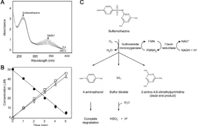
products and 4-aminophenol in stoichiometric manners (Fig. 2A and B). Our results are consistent with the reaction mechanism previously proposed inMicrobacteriumsp. BR1 (Ricken et al., 2013), where SadA and SadC were reported to initiate the catabolism of sulfonamides in Microbacteriumsp. BR1, but the reaction was conducted using partially purified enzymes (Ricken et al., 2017). In the present study, the me-chanism of the initial cleavage reaction of sulfonamide drugs was
[image:4.595.49.556.61.264.2]demonstrated using purified enzymes (Fig. 2C); Flavin reductase re-duces the oxidized form offlavin cofactor (FMN) through the oxidation of NADH. The reducedflavin cofactor (FMNH2) functions as electron donor for theipso-hydroxylation of sulfonamide substrates by sulfona-mide monooxygenase. Subsequently, the hydroxylation of sulfonasulfona-mides results in the cleavage of the drugs, releasing 4‑aminophenol, sulfite and the corresponding dead-end metabolites. Purified monooxygenase
Fig. 1.Genetic organization and expression profiles of a gene cluster for sulfonamide degradation. The locations of transposases and integrases are shown as red bars in the genome ofMicrobacteriumsp. CJ77. Expression levels are displayed by normalized spectral counts below the genetic map of the cluster. GLU, SNM, SMX, and SMZ indicate glucose, sulfanilamide, sulfamethoxazole and sulfamethazine, respectively, used as a sole carbon source. (For interpretation of the references to colour in thisfigure legend, the reader is referred to the web version of this article.)
[image:4.595.98.496.323.577.2]andflavin reductase showed the degradation activities towards several sulfonamides with different substrate specificities (Table 1). Kinetic studies indicated that the highestVmaxwas observed with
sulfametha-zine, while the substrate affinity was highest (lowestKm value) for
sulfathiazole (Table 1). The order of catalytic efficiency (Vmax/Km) for
these substrates is as follows: sulfamethazine, sulfathiazole, sulfa-methoxazole and sulfadiazine (Table 1). In addition toflavin reductase (MCJ23810) in the gene cluster, four other paralogousflavin reductases present in the genome of strain CJ77 displayed sulfonamide degrada-tion activities when combined with sulfonamide monooxygenase (Table S3), indicating thatflavin reductase is not specific for the reaction.
3.3. Sulfonamide monooxygenase as a novel class Dflavin-dependent monooxygenase
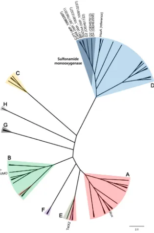
Sulfonamide monooxygenase was considered to belong to the two-component flavin-dependent monooxygenase (FDM) family (Huijbers et al., 2014; Ricken et al., 2017). Interestingly, sulfonamide mono-oxygenase from strain CJ77 exhibited relatively low sequence simila-rities (< 50%) with other known monooxygenases available in the GenBank database, except for homologs found in the genomes of pre-viously reported sulfonamide-degrading actinobacteriaMicrobacterium spp. BR1, SDZm4, and C448, andArthrobacterspp. D2 and D4, many of which were initially annotated as hypothetical proteins. Phylogenetic analysis based on amino acid sequences from all classes of FDMs from the RCSB protein data bank (PDB) and class D FDMs from the Unitprot database (Huijbers et al., 2014; Mascotti et al., 2016) revealed that sulfonamide monooxygenases from these sulfonamide-degrading acti-nobacteria formed a distinct lineage of class D FDM within other known FDMs (Fig. 3). Antibiotic-inactivating monooxygenases such as TetX, Rox, and Baeyer-Villiger monooxygenase, which conferred resistance to tetracycline, rifamycin, and imipenem (Hoshino et al., 2010; Koteva et al., 2018;Minerdi et al., 2015;Yang et al., 2004), respectively, were previously characterized to be single-component FDMs belonging to class A or B (Fig. 3). Sulfonamide monooxygenases identified in this study are distinguished in that they are two-component FDMs in class D. Structural homology modeling with the closest characterized protein (HsaA fromMycobacterium tuberculosis) revealed that several residues at the flavin-binding site were well-conserved in sulfonamide mono-oxygenase of strain CJ77, while residues at the substrate-binding site varied (Fig. S4).
3.4. Two-component monooxygenase system as a novel sulfonamide resistance determinant
Like other known antibiotic-inactivating monooxygenases (Forsberg et al., 2015;Hoshino et al., 2010;Koteva et al., 2018;Minerdi et al., 2015;Yang et al., 2004), the decomposition of sulfonamides indicate a potential resistance mechanism via inactivation of the drugs. To clarify their roles in resistance, genes encoding sulfonamide monooxygenase andflavin reductase were introduced into a sulfamethoxazole-suscep-tibleE.colistrain. Both genes were successfully expressed inE.colicells,
which exhibited sulfonamide degradation activity (Fig. 4). When anti-biotic susceptibility was tested, E. coli cells harboring both of two component genes showed a significant increase in resistance compared to the control E. coli cells (Fig. 4). E. coli cells harboring only the monooxygenase gene also displayed a lower level of resistance (Fig. 4), suggesting that indigenousflavin reductases present inE.colicontribute to the slight increase in resistance, as also shown in the degradation activity, and both genes were required for the acquisition of resistance to the drugs. In conclusion, our results demonstrate that the two-com-ponent system consisting of sulfonamide monooxygenase and flavin reductase is key enzymes for both sulfonamide degradation activity and novel resistance mechanism via drug inactivation. Therefore, we pro-pose that the heretofore unrecognized monooxygenase responsible for sulfonamide resistance should be renamed as SulX in analogy to TetX, which is distinguished from previously known sulfonamide resistance genes (sul1234) (Perreten and Boerlin, 2003;Razavi et al., 2017;Skold, 2000). Flavin reductase as a two-component system is renamed as SulR.
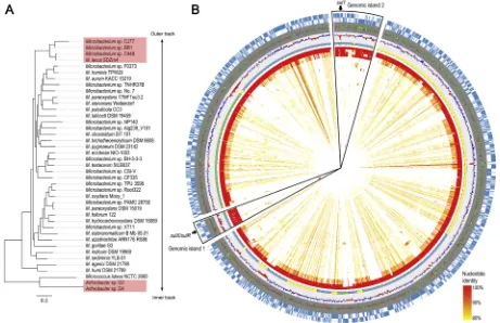
3.5. Comparative genomic analysis of sulfonamide-degrading actinobacteria
To date, genes homologous tosulX have been found only in the genomes of sulfonamide-degrading actinobacteria including Microbacteriumspp. BR1, SDZm4 and C448, andArthrobacterspp. D2 and D4. Phylogenetic analysis of the genomes ofMicrobacteriumspp. placed the four sulfonamide-degrading strains in a distinct lineage (Fig. 5A and Fig. S5). Based on average nucleotide identity (ANI) values (Richter and Rossello-Mora, 2009), strains CJ77 and BR1 (99.2%), and strains C448 and SDZm4 (97.8%) belong to the same species respec-tively. In addition, genomic comparison showed that the four sulfona-mide-degrading Microbacterium strains had higher similarities com-pared to other non-sulfonamide-degrading strains (Fig. 5B). Particularly, two genomic island regions were highly conserved among the four sulfonamide-degradingMicrobacteriumstrains in the genome comparison map (Fig. 5B). Interestingly, the two regions contained the sulX/sulR-containing gene cluster (genomic island 1) andsul1-carrying class 1 integron (genomic island 2), respectively. Codon usage and G+C content of the protein-coding sequences (CDSs) in genomic island 1 were significantly different from those ofMicrobacteriumcore genes (Fig. S6), suggesting that these sequences were acquired from different origins. The occurrence of horizontal gene transfer was also demon-strated by the presence of tRNA genes at the 5′and 3′ends of genomic island 1 (Boyd et al., 2009) (Fig. S7).
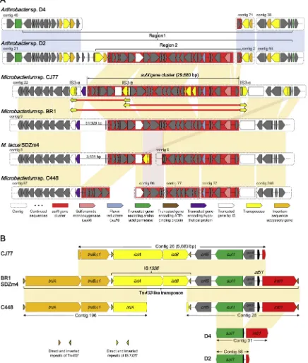
3.6. Comparative analysis of sulX gene clusters and sul1-carrying class 1 integrons associated with mobile genetic elements
ThesulXgene cluster (29,680 bp) containing genes encoding sul-fonamide monooxygenase (SulX) and flavin reductase (SulR) in the strain CJ77 genome was highly conserved in sulfonamide-degrading actinobacteria (shown in red;Fig. 6A). It remains ambiguous whether this gene cluster was present inArthrobactersp. D4 or missing during genome sequencing. In the genomes ofArthrobacter spp. D2 and D4, another gene homologous tosulX(73.4%) was detected in other regions of the genomes. ThesulXgene cluster of strain CJ77 was unique com-pared to those of other sulfonamide-degrading strains in that the cluster was located in a transposase-rich region (Figs. 1 and 6A). Three intact insertion sequences (IS) of the IS3family whose sequences were iden-tical except for direct repeat sequences were detected around the gene cluster (Fig. 6A and Fig. S8). These features suggest that three insertion events occurred independently after strain CJ77 had acquired thesulX gene cluster. Similar IS elements were found to be prevalent in the genomes of various actinobacteria (Table S4). Interestingly, repeated insertion of the identical IS resulted in the formation of three possible genetic structures of composite transposon, two of which contained the sulX gene cluster (Fig. 6A). The presence of composite transposons harboring the intact sulX gene cluster indicates the possibility of
Table 1
Steady state kinetic parameters for the initial cleavage reaction by sulfonamide monooxygenase.
Substrate⁎ Km(μM)† Vmax(U/mg protein)† Vmax/Km
Sulfamethazine 16.83 ± 2.27 0.5809 ± 0.0530 0.03452 Sulfamethoxazole 23.44 ± 0.95 0.0634 ± 0.0025 0.00270 Sulfathiazole 9.76 ± 1.15 0.0321 ± 0.0033 0.00329 Sulfadiazine 24.35 ± 1.81 0.0111 ± 0.0007 0.00046
⁎ 200μM NADH, 50 mM Tris- HCl (pH 7.5) and an equal amount of FMN to
sulfonamide monooxygenase concentration were used in the reactions.
† Kinetic values are shown with standard deviations offit of the data to the
transposition events and subsequent emergence of sulfonamide re-sistance in the clinical settings.
Mobilization of thesulXgene cluster was indicated by two insertion events that may have occurred inArthrobacterspp. strains D2 and D4 (Fig. 6A). First, compared withArthrobactersp. ATCC 21022 as a re-ference genome (Deng et al., 2016), a larger transposon structure (re-gion 1) with intact direct repeats and imperfect inverted repeats was identified to be inserted into a gene encoding amino acid permease (shown in green) of strains D2 and D4 (Figs. 6A and S9A). A part of structure and sequence of the transposon were highly similar to those of the previously reported Tn552transposon of megaplasmid pAO1 from A. nicotinovoransATCC 49919 (Fig. S9A) (Igloi and Brandsch, 2003). Another insertion event (region 2) was detected inside region 1 in strain D2 (Fig. 6A). ThesulXgene cluster and an intact IS element belonging to the IS21 family were inserted into a gene encoding ATP-binding protein (Fig. S9B). The presence of transposon-associated sulX gene cluster in bothMicrobacterium andArthrobacterstrains indicated that horizontal gene transfer occurred among these groups of bacteria.
In the genomes of sulfonamide-degrading Microbacterium strains,
another sulfonamide resistance gene,sul1, was found to be located in a typical structure of the clinical class 1 integron (Gillings, 2014), in-cludingqacEΔ1,sul1,orf5andtnimodule (Fig. 6B). Class 1 integrons have been regarded to play an important role in disseminating anti-biotic resistance genes (Gillings, 2014) and proposed as a proxy for anthropogenic pollution (Gillings et al., 2015). In these bacterial gen-omes, the class 1 integron and IS element IS1326were carried in a Tn402-like transposon (Fig. 6B). IS1326-inserted class 1 integrons have been reported to be prevalent in proteobacteria (Jones-Dias et al., 2016) but not in actinobacteria. The prevalence ofsul1associated with the class 1 integron has been reported in many bacterial isolates from manured agricultural soils (Byrne-Bailey et al., 2009; Wang et al., 2014), suggesting that sulfonamide resistance evolved under sulfona-mide selective pressures through horizontal gene transfer ofsul1 -car-rying class 1 integron among disparate taxa.
4. Discussion
[image:6.595.43.358.55.532.2]As the environmental resistome has been regarded as a reservoir of
novel antibiotic resistance genes (Perry et al., 2014), a more extensive understanding of the resistome gained in the past few decades has
enabled studies of the evolution and dissemination of antibiotic re-sistance. Among the various antibiotic resistance mechanisms (Crofts et al., 2017), the enzymatic inactivation mechanism remains relatively unexplored and should be rigorously examined to identify undiscovered resistance determinants in the environment (Morar and Wright, 2010), considering the enormous bacterial diversity and their functional ver-satility (Morar and Wright, 2010; Wright, 2007). Several novel re-sistance mechanisms by antibiotic-inactivating enzymes have been discovered in environmental bacteria (Pawlowski et al., 2016; Spanogiannopoulos et al., 2012). Furthermore, recent advances in metagenomics revealed previously unrecognized sequences that were functionally demonstrated to confer novel resistance (Forsberg et al., 2015;Kim et al., 2018).
[image:7.595.39.287.56.244.2]Because sulfonamides are synthetic antibiotics, naturally occurring enzymes that degrade or modify these drugs may not be readily de-veloped compared to antimicrobials of natural origin (Morar and Wright, 2010). Sulfonamide-degrading bacteria were relatively recently discovered mainly in sulfonamide-contaminated sites and all of those strains whose genome sequences are available contained bothsulXand sul1, suggesting that sulfonamide degradation is associated with sulfo-namide resistance. Notably, two genomic islands shared only among the genomes of sulfonamide-degrading Microbacterium strains con-tained thesulX gene clusters andsul1-carrying class 1 integrons re-spectively (Fig. 5): two independent sulfonamide resistance determi-nants co-existed and were distantly located in the genomes of sulfonamide-degrading actinobacteria. Among the sulfonamide-de-grading bacteria reported, the genome sequences of three
Fig. 4.Sulfonamide-cleavage activity associated with resistance ofE.colicells where sulfonamide monooxygenase andflavin reductase were heterologously expressed. Activity was assayed using cells of E.coli strains harboring the plasmid pET-Duet derivatives. Susceptibility of E. coli cells against sulfa-methoxazole was tested by broth dilution assay and disk-diffusion assay.
[image:7.595.67.528.353.651.2]proteobacterial species Pseudomonas psychrophila HA-4 (Jiang et al., 2014),Shewanella oneidensisMR-1 andShewanellasp. strain MR-4 (Mao et al., 2018) are available in addition to the six actinobacteria analyzed in this study. These proteobacteria were not found to possesssulXgene, suggesting that different mechanisms may be involved in the sulfona-mide degradation. The co-existence ofsul1and sulfonamide degrada-tion genes in sulfonamide-degrading actinobacteria is consistent with the prevailing idea of resistance to antibiotics as a condition for de-gradation (Islas-Espinoza et al., 2012). To evaluate the contribution of these two genes to sulfonamide resistance, gene deletion studies were performed using strain CJ77 but knock-out mutants for sulX orsul1
have yet to be isolated. However, we observed that mutant strains which lost sulfonamide degradation activity were still highly resistant to sulfonamides, suggesting thatsul1plays a major role in sulfonamide resistance in strain CJ77.
[image:8.595.81.517.63.576.2]Codon usage and GC content of thesulXgene cluster distinguished from those ofMicrobacteriumcore genes suggest that the gene clusters were acquired at later stages of species evolution. Clearly defined in-sertion events observed inArthrobacterspp. D2 and D4 provide strong evidence of mobilization. Particularly, in strain CJ77, the presence of multiple IS elements and putative composite transposon structures containing thesulXgene cluster also indicate potential mobilization of
sulfonamide resistance. Many studies have reported composite trans-posons carrying metabolic gene clusters that may have been acquired under certain selection pressures (Clark et al., 2013;Mei et al., 2014). Considering that the sulfonamide-dependent expression of sulX gene cluster can provide a selective advantage for the use of sulfonamides as carbon sources, sulfonamide metabolism may have evolved in sulfo-namide-resistant bacteria that had already acquired the sul1-carrying class 1 integron under sulfonamide selection pressures. Currently,sulX has been found in only a few sulfonamide-degrading actinobacteria. This may be because of the low number of sulfonamide-degrading bacteria reported or relatively recent evolution of this gene. The pre-sence of thesulX gene cluster at geographically distant locations in-cluding Europe, North America and Asia suggests that evolution of the gene cluster occurred independently (Bouju et al., 2012; Deng et al., 2016; Tappe et al., 2013; Topp et al., 2013) and it was much more globally widespread than discovered so far, as acquisition of the gene cluster confers selective advantages in sulfonamide-contaminated en-vironments. Furthermore, the emergence of sulfonamide-degrading bacteria in a particular ecological niche may lead to elimination of the selective pressure which can allow sulfonamide-susceptible bacteria to survive, influencing the microbial community structure in the niche (Deng et al., 2018).
Since the tetracycline-degrading monooxygenase (TetX) conferring resistance was first identified in the transposons of commensal Bacteroidesspp. (Speer et al., 1991;Whittle et al., 2001),tetXgene has been discovered in environmental Sphingobacteriumsp. (Ghosh et al., 2009), the duck pathogenRiemerella anatipestifer(Chen et al., 2010), Myroides sp. from a meat processing plant (Li et al., 2016), clinical isolates ofEnterobacteriaceaeandPseudomonadaceae(Leski et al., 2013), and in sequence data of uncultured bacteria. Remarkably, transposon structures (Tn4351 and CTnDOT) harboring tetX gene were sig-nificantly conserved in commensal, environmental and clinical isolates (Ghosh et al., 2009;Leski et al., 2013;Speer et al., 1991;Whittle et al., 2001), indicating widespread horizontal gene transfer between dis-parate taxa (Ghosh et al., 2015). As sulfonamides have been extensively used worldwide,sulXassociated with mobile genetic elements as well as sul1-carrying class 1 integron may be now under mobilization and subsequently emergent in animal and clinical isolates as shown fortetX.
5. Conclusions
Although sulfonamide monooxygenase wasfirst identified to cata-lyze the initial cleavage of sulfonamides inMicrobacteriumsp. BR1, the role of this protein in the resistance was never demonstrated. Furthermore, the association of sulfonamide-degrading genes with mobile genetic elements was not elucidated in detail. In the present study, through a combination of proteomics, heterologous protein ex-pression, and in vitro enzyme assays, we successfully identified the flavin-dependent monooxygenase SulX in non-pathogenic environ-mental actinobacteria, which not only catalyzed the degradation of sulfonamides but also conferred resistance to these antibiotics. Comparative genomic analysis revealed that sulXorthologs were pre-valent in sulfonamide-degrading actinobacteria and contained genetic contexts for mobilization. Our study suggests that much wider diversity of resistome might be present in the environment than previously thought, which may be associated with the bacterial metabolism of antimicrobials. Indeed, numerous antibiotic-resistant bacterial strains subsisting on antibiotic chemicals were isolated from the natural en-vironment (Dantas et al., 2008). Therefore, exploring microbial meta-bolic versatility related to the degradation of antimicrobials is im-portant for expanding our knowledge of antibiotic resistance mechanisms, recollecting the concept “microbial infallibility” which states that most organic chemicals including antimicrobials have been degraded and recycled on the planet throughout history (Alexander, 1965).
Acknowledgments
This work was supported by a grant from the National Institute of Biological Resources (NIBR) (NIBR201618201) and funded by the Korea Ministry of Environment (MOE) as“the Environmental Health Action Program (2016001350004)”.
Declarations of interest
None.
Appendix A. Supplementary data
Supplementary data to this article can be found online athttps:// doi.org/10.1016/j.envint.2019.03.046.
References
Adu-Oppong, B., Gasparrini, A.J., Dantas, G., 2017. Genomic and functional techniques to mine the microbiome for novel antimicrobials and antimicrobial resistance genes. Ann. N. Y. Acad. Sci. 1388, 42–58.
Alexander, M., 1965. Biodegradation: problems of molecular recalcitrance and microbial fallibility. Adv. Appl. Microbiol. 7, 35–80.
Bouju, H., Ricken, B., Beffa, T., Corvini, P.F., Kolvenbach, B.A., 2012. Isolation of bac-terial strains capable of sulfamethoxazole mineralization from an acclimated mem-brane bioreactor. Appl. Environ. Microbiol. 78, 277–279.
Boyd, E.F., Almagro-Moreno, S., Parent, M.A., 2009. Genomic islands are dynamic, an-cient integrative elements in bacterial evolution. Trends Microbiol. 17, 47–53. Byrne-Bailey, K.G., Gaze, W.H., Kay, P., Boxall, A.B., Hawkey, P.M., Wellington, E.M.,
2009. Prevalence of sulfonamide resistance genes in bacterial isolates from manured agricultural soils and pig slurry in the United Kingdom. Antimicrob. Agents Chemother. 53, 696–702.
Chen, Y.P., Tsao, M.Y., Lee, S.H., Chou, C.H., Tsai, H.J., 2010. Prevalence and molecular characterization of chloramphenicol resistance inRiemerella anatipestiferisolated from ducks and geese in Taiwan. Avian Pathol 39, 333–338.
Clark, I.C., Melnyk, R.A., Engelbrektson, A., Coates, J.D., 2013. Structure and evolution of chlorate reduction composite transposons. MBio 4, e00379-13.
Crofts, T.S., Gasparrini, A.J., Dantas, G., 2017. Next-generation approaches to understand and combat the antibiotic resistome. Nat. Rev. Microbiol. 15, 422–434.
Dantas, G., Sommer, M.O., Oluwasegun, R.D., Church, G.M., 2008. Bacteria subsisting on antibiotics. Science 320, 100–103.
Delcher, A.L., Bratke, K.A., Powers, E.C., Salzberg, S.L., 2007. Identifying bacterial genes and endosymbiont DNA with Glimmer. Bioinformatics 23, 673–679.
Deng, Y., Mao, Y., Li, B., Yang, C., Zhang, T., 2016. Aerobic degradation of sulfadiazine by
Arthrobacterspp.: kinetics, pathways, and genomic characterization. Environ. Sci. Technol. 50, 9566–9575.
Deng, Y., Li, B., Zhang, T., 2018. Bacteria that make a meal of sulfonamide antibiotics: blind spots and emerging opportunities. Environ. Sci. Technol. 52, 3854–3868. Forsberg, K.J., Patel, S., Wencewicz, T.A., Dantas, G., 2015. The tetracycline destructases:
a novel family of tetracycline-inactivating enzymes. Chem. Biol. 22, 888–897. Ghosh, S., Sadowsky, M.J., Roberts, M.C., Gralnick, J.A., LaPara, T.M., 2009.
Sphingobacteriumsp. strain PM2-P1-29 harbours a functionaltet(X) gene encoding for the degradation of tetracycline. J. Appl. Microbiol. 106, 1336–1342.
Ghosh, S., LaPara, T.M., Sadowsky, M.J., 2015. Transformation of tetracycline by TetX and its subsequent degradation in a heterologous host. FEMS Microbiol. Ecol. 91, fiv059.
Gillings, M.R., 2014. Integrons: past, present, and future. Microbiol. Mol. Biol. Rev. 78, 257–277.
Gillings, M.R., Gaze, W.H., Pruden, A., Smalla, K., Tiedje, J.M., Zhu, Y.G., 2015. Using the class 1 integron-integrase gene as a proxy for anthropogenic pollution. ISME J 9, 1269–1279.
Hoshino, Y., Fujii, S., Shinonaga, H., Arai, K., Saito, F., Fukai, T., Satoh, H., Miyazaki, Y., Ishikawa, J., 2010. Monooxygenation of rifampicin catalyzed by theroxgene product ofNocardia farcinica: structure elucidation, gene identification and role in drug re-sistance. J. Antibiot. (Tokyo) 63, 23–28.
Huijbers, M.M., Montersino, S., Westphal, A.H., Tischler, D., van Berkel, W.J., 2014. Flavin dependent monooxygenases. Arch. Biochem. Biophys. 544, 2–17. Huovinen, P., 2001. Resistance to trimethoprim-sulfamethoxazole. Clin. Infect. Dis. 32,
1608–1614.
Igloi, G.L., Brandsch, R., 2003. Sequence of the 165-kilobase catabolic plasmid pAO1 fromArthrobacter nicotinovoransand identification of a pAO1-dependent nicotine uptake system. J. Bacteriol. 185, 1976–1986.
Ingerslev, F., Halling-Sørensen, B., 2000. Biodegradability properties of sulfonamides in activated sludge. Environ. Toxicol. Chem. 19, 2467–2473.
Islas-Espinoza, M., Reid, B.J., Wexler, M., Bond, P.L., 2012. Soil bacterial consortia and previous exposure enhance the biodegradation of sulfonamides from pig manure. Microb. Ecol. 64, 140–151.
98, 4671–4681.
Jones-Dias, D., Manageiro, V., Ferreira, E., Barreiro, P., Vieira, L., Moura, I.B., Canica, M., 2016. Architecture of class 1, 2, and 3 integrons from Gram negative bacteria re-covered among fruits and vegetables. Front. Microbiol. 7, 1400.
Kim, D.W., Thawng, C.N., Lee, S.H., Cha, C.J., 2017. Unique features ofAeromonas
plasmid pAC3 and expression of the plasmid-mediated quinolone resistance genes. mSphere 2, e00203–17.
Kim, D.W., Thawng, C.N., Choi, J.H., Lee, K., Cha, C.J., 2018. Polymorphism of antibiotic-inactivating enzyme driven by ecology expands the environmental resistome. ISME J 12, 267–276.
Koteva, K., Cox, G., Kelso, J.K., Surette, M.D., Zubyk, H.L., Ejim, L., Stogios, P., Savchenko, A., Sorensen, D., Wright, G.D., 2018. Rox, a rifamycin resistance enzyme with an unprecedented mechanism of action. Cell Chem. Biol. 25, 403–412. Krzywinski, M., Schein, J., Birol, I., Connors, J., Gascoyne, R., Horsman, D., Jones, S.J.,
Marra, M.A., 2009. Circos: an information aesthetic for comparative genomics. Genome Res. 19, 1639–1645.
Larcher, S., Yargeau, V., 2012. Biodegradation of sulfamethoxazole: current knowledge and perspectives. Appl.Mcrobiol. Biotechnol. 96, 309–318.
Leski, T.A., Bangura, U., Jimmy, D.H., Ansumana, R., Lizewski, S.E., Stenger, D.A., Taitt, C.R., Vora, G.J., 2013. Multidrug-resistanttet(X)-containing hospital isolates in Sierra Leone. Int. J. Antimicrob. Agents 42, 83–86.
Li, L., Ye, L., Zhang, S., Meng, H., 2016. Isolation and identification of aerobic bacteria carrying tetracycline and sulfonamide resistance genes obtained from a meat pro-cessing plant. J. Food Sci. 81, M1480–M1484.
Liu, Y.Y., Wang, Y., Walsh, T.R., Yi, L.X., Zhang, R., Spencer, J., Doi, Y., Tian, G., Dong, B., Huang, X.,et al. 2016. Emergence of plasmid-mediated colistin resistance mechanism MCR-1 in animals and human beings in China: a microbiological and molecular biological study. Lancet Infect. Dis. 16, 161–168.
Mao, F., Liu, X., Wu, K., Zhou, C., Si, Y., 2018. Biodegradation of sulfonamides by
Shewanella oneidensisMR-1 andShewanellasp. strain MR-4. Biodegradation 29, 129–140.
Mascotti, M.L., Juri Ayub, M., Furnham, N., Thornton, J.M., Laskowski, R.A., 2016. Chopping and changing: the evolution of theflavin-dependent monooxygenases. J. Mol. Biol. 428, 3131–3146.
McInerney, J.O., 1998. GCUA: general codon usage analysis. Bioinformatics 14, 372–373. Mei, X., Xu, K., Yang, L., Yuan, Z., Mahillon, J., Hu, X., 2014. The genetic diversity of
cereulide biosynthesis gene cluster indicates a composite transposon Tnces in emetic
Bacillus weihenstephanensis. BMC Microbiol. 14, 149.
Minerdi, D., Zgrablic, I., Castrignano, S., Catucci, G., Medana, C., Terlizzi, M.E., Gribaudo, G., Gilardi, G., Sadeghi, S.J., 2015.Escherichia colioverexpressing a Baeyer-Villiger monooxygenase fromAcinetobacter radioresistensbecomes resistant to imipenem. Antimicrob. Agents Chemother. 60, 64–74.
Morar, M., Wright, G.D., 2010. The genomic enzymology of antibiotic resistance. Annu. Rev. Genet. 44, 25–51.
Nguyen, L.T., Schmidt, H.A., von Haeseler, A., Minh, B.Q., 2015. IQ-TREE: a fast and effective stochastic algorithm for estimating maximum-likelihood phylogenies. Mol. Biol. Evol. 32, 268–274.
Ok, Y.S., Kim, S.C., Kim, K.R., Lee, S.S., Moon, D.H., Lim, K.J., Sung, J.K., Hur, S.O., Yang, J.E., 2011. Monitoring of selected veterinary antibiotics in environmental compart-ments near a composting facility in Gangwon Province, Korea. Environ. Monit. Assess. 174, 693–701.
Overbeek, R., Begley, T., Butler, R.M., Choudhuri, J.V., Chuang, H.Y., Cohoon, M., de Crecy-Lagard, V., Diaz, N., Disz, T., Edwards, R., et al., 2005. The subsystems ap-proach to genome annotation and its use in the project to annotate 1000 genomes. Nucleic Acids Res. 33, 5691–5702.
Pawlowski, A.C., Wang, W., Koteva, K., Barton, H.A., McArthur, A.G., Wright, G.D., 2016. A diverse intrinsic antibiotic resistome from a cave bacterium. Nat. Commun. 7, 13803.
Perreten, V., Boerlin, P., 2003. A new sulfonamide resistance gene (sul3) inEscherichia coli
is widespread in the pig population of Switzerland. Antimicrob. Agents Chemother. 47, 1169–1172.
Perry, J.A., Westman, E.L., Wright, G.D., 2014. The antibiotic resistome: what's new? Curr. Opin. Microbiol. 21, 45–50.
Phuong Hoa, P.T., Nonaka, L., Hung Viet, P., Suzuki, S., 2008. Detection of thesul1,sul2, andsul3genes in sulfonamide-resistant bacteria from wastewater and shrimp ponds of North Vietnam. Sci. Total Environ. 405, 377–384.
Pruitt, K.D., Tatusova, T., Klimke, W., Maglott, D.R., 2009. NCBI Reference Sequences: current status, policy and new initiatives. Nucleic Acids Res. 37, D32–D36.
Razavi, M., Marathe, N.P., Gillings, M.R., Flach, C.F., Kristiansson, E., Joakim Larsson, D.G., 2017. Discovery of the fourth mobile sulfonamide resistance gene. Microbiome 5, 160.
Reis, A.C., Cvancarova, M., Liu, Y., Lenz, M., Hettich, T., Kolvenbach, B.A., Corvini, P.F., Nunes, O.C., 2018. Biodegradation of sulfamethoxazole by a bacterial consortium of
Achromobacter denitrificansPR1 andLeucobactersp. GP. Appl. Microbiol. Biotechnol. 102, 10299–10314.
Richter, M., Rossello-Mora, R., 2009. Shifting the genomic gold standard for the pro-karyotic species definition. Proc. Natl. Acad. Sci. U. S. A. 106, 19126–19131. Ricken, B., Corvini, P.F., Cichocka, D., Parisi, M., Lenz, M., Wyss, D., Martinez-Lavanchy,
P.M., Muller, J.A., Shahgaldian, P., Tulli, L.G., et al., 2013.ipso-Hydroxylation and subsequent fragmentation: a novel microbial strategy to eliminate sulfonamide an-tibiotics. Appl. Environ. Microbiol. 79, 5550–5558.
Ricken, B., Kolvenbach, B.A., Bergesch, C., Benndorf, D., Kroll, K., Strnad, H., Vlcek, C., Adaixo, R., Hammes, F., Shahgaldian, P., et al., 2017. FMNH2-dependent
mono-oxygenases initiate catabolism of sulfonamides inMicrobacteriumsp. strain BR1 subsisting on sulfonamide antibiotics. Sci. Rep. 7, 15783.
Skold, O., 2000. Sulfonamide resistance: mechanisms and trends. Drug Resist. Updat. 3, 155–160.
Spanogiannopoulos, P., Thaker, M., Koteva, K., Waglechner, N., Wright, G.D., 2012. Characterization of a rifampin-inactivating glycosyltransferase from a screen of en-vironmental actinomycetes. Antimicrob. Agents Chemother. 56, 5061–5069. Speer, B.S., Bedzyk, L., Salyers, A.A., 1991. Evidence that a novel tetracycline resistance
gene found on twoBacteroidestransposons encodes an NADP-requiring oxidor-eductase. J. Bacteriol. 173, 176–183.
Surette, M.D., Wright, G.D., 2017. Lessons from the environmental antibiotic resistome. Annu. Rev. Microbiol. 71, 309–329.
Tappe, W., Herbst, M., Hofmann, D., Koeppchen, S., Kummer, S., Thiele, B., Groeneweg, J., 2013. Degradation of sulfadiazine byMicrobacterium lacusstrain SDZm4, isolated from lysimeters previously manured with slurry from sulfadiazine-medicated pigs. Appl. Environ. Microbiol. 79, 2572–2577.
Tatusov, R.L., Galperin, M.Y., Natale, D.A., Koonin, E.V., 2000. The COG database: a tool for genome-scale analysis of protein functions and evolution. Nucleic Acids Res. 28, 33–36.
Topp, E., Chapman, R., Devers-Lamrani, M., Hartmann, A., Marti, R., Martin-Laurent, F., Sabourin, L., Scott, A., Sumarah, M., 2013. Accelerated biodegradation of veterinary antibiotics in agricultural soil following long-term exposure, and isolation of a sul-famethazine-degradingMicrobacteriumsp. J. Environ. Qual. 42, 173–178. Wang, S., Wang, J., 2018. Biodegradation and metabolic pathway of sulfamethoxazole by
a novel strainAcinetobactersp. Appl. Microbiol. Biotechnol. 102, 425–432. Wang, N., Yang, X., Jiao, S., Zhang, J., Ye, B., Gao, S., 2014. Sulfonamide-resistant
bacteria and their resistance genes in soils fertilized with manures from Jiangsu Province, Southeastern China. PLoS One 9, e112626.
Whittle, G., Hund, B.D., Shoemaker, N.B., Salyers, A.A., 2001. Characterization of the 13-kilobaseermFregion of theBacteroidesconjugative transposon CTnDOT. Appl. Environ. Microbiol. 67, 3488–3495.
Wiegand, I., Hilpert, K., Hancock, R.E., 2008. Agar and broth dilution methods to de-termine the minimal inhibitory concentration (MIC) of antimicrobial substances. Nat. Protoc. 3, 163–175.
Wolf, Y.I., Koonin, E.V., 2012. A tight link between orthologs and bidirectional best hits in bacterial and archaeal genomes. Genome Biol. Evol. 4, 1286–1294.
Wright, G.D., 2007. The antibiotic resistome: the nexus of chemical and genetic diversity. Nat. Rev. Microbiol. 5, 175–186.
Yang, W., Moore, I.F., Koteva, K.P., Bareich, D.C., Hughes, D.W., Wright, G.D., 2004. TetX is aflavin-dependent monooxygenase conferring resistance to tetracycline anti-biotics. J. Biol. Chem. 279, 52346–52352.
Yoon, S.H., Ha, S.M., Kwon, S., Lim, J., Kim, Y., Seo, H., Chun, J., 2017a. Introducing EzBioCloud: a taxonomically united database of 16S rRNA gene sequences and whole-genome assemblies. Int. J. Syst. Evol. Microbiol. 67, 1613–1617. Yoon, S.H., Ha, S.M., Lim, J., Kwon, S., Chun, J., 2017b. A large-scale evaluation of
algorithms to calculate average nucleotide identity. Antonie Van Leeuwenhoek 110, 1281–1286.