Original citation:
Boniecka, Justyna, Prusińska, Justyna, Dąbrowska, Grażyna B. and Goc, Anna. (2017) Within and beyond the stringent response-RSH and (p)ppGpp in plants. Planta, 246 (5). pp. 817-842.
Permanent WRAP URL:
http://wrap.warwick.ac.uk/93070
Copyright and reuse:
The Warwick Research Archive Portal (WRAP) makes this work of researchers of the University of Warwick available open access under the following conditions.
This article is made available under the Creative Commons Attribution 4.0 International license (CC BY 4.0) and may be reused according to the conditions of the license. For more details see: http://creativecommons.org/licenses/by/4.0/
A note on versions:
The version presented in WRAP is the published version, or, version of record, and may be cited as it appears here.
REVIEW
Within and beyond the stringent response‑RSH and (p)ppGpp
in plants
Justyna Boniecka1 · Justyna Prusińska2 · Grażyna B. Dąbrowska1 · Anna Goc1
Received: 25 May 2017 / Accepted: 17 September 2017 / Published online: 25 September 2017 © The Author(s) 2017. This article is an open access publication
chloroplasts, in plants, unlike in bacteria, (p)ppGpp promote chloroplast DNA replication and division. Next, (p)ppGpp may also perform their functions in cytoplasm, where they would promote plant growth inhibition. Furthermore, (p) ppGpp accumulation also affects nuclear gene expression, i.a., decreases the level of Arabidopsis defense gene tran-scripts, and promotes plants susceptibility towards Turnip mosaic virus. In this review, we summarize recent findings that show the importance of RSH and (p)ppGpp in plant growth and development, and open an area of research aim-ing to understand the function of plant RSH in response to stress.
Keywords RelA/SpoT homologs · Alarmones · Plant growth and development · Stress response · Photosynthesis · Senescence
Abbreviations
CTD C-terminal domain
(p)ppGpp Guanosine tetra- and pentaphosphates RelA Escherichia coli (p)ppGpp synthetase RNAP RNA polymerase
RSH RelA/SpoT homolog SAH Small alarmone hydrolase
SpoT Escherichia coli (p)ppGpp synthetase and hydrolase
TuMV Turnip mosaic virus WT Wild type
Introduction
Prokaryotes have developed different types of stress responses, including chemotaxis, the SOS response and the stringent response (Dabrowska et al. 2006b). The latter was
Abstract
Main conclusion Plant RSH proteins are able to synthetize and/or hydrolyze unusual nucleotides called (p)ppGpp or alarmones. These molecules regulate nuclear and chloroplast transcription, chloroplast translation and plant development and stress response.
Homologs of bacterial RelA/SpoT proteins, designated RSH, and products of their activity, (p)ppGpp—guanosine tetra— and pentaphosphates, have been found in algae and higher plants. (p)ppGpp were first identified in bacteria as the effec-tors of the stringent response, a mechanism that orchestrates pleiotropic adaptations to nutritional deprivation and vari-ous stress conditions. (p)ppGpp accumulation in bacteria decreases transcription—with exception to genes that help to withstand or overcome current stressful situations, which are upregulated—and translation as well as DNA replication and eventually reduces metabolism and growth but promotes adaptive responses. In plants, RSH are nuclei-encoded and function in chloroplasts, where alarmones are produced and decrease transcription, translation, hormone, lipid and metabolites accumulation and affect photosynthetic effi-ciency and eventually plant growth and development. During senescence, alarmones coordinate nutrient remobilization and relocation from vegetative tissues into seeds. Despite the high conservancy of RSH protein domains among bacteria and plants as well as the bacterial origin of plant
* Grażyna B. Dąbrowska [email protected]
1 Department of Genetics, Nicolaus Copernicus University in Toruń, Lwowska 1, 87-100 Toruń, Poland
first identified in Escherichia coli as a reaction to amino acid deprivation (Cashel and Gallant 1969). Further experi-ments showed that it also takes place under limitation of other nutrients, e.g., carbon (Flardh et al. 1994; Gentry and Cashel 1996), iron (Vinella et al. 2005), fatty acid (Sey-fzadeh et al. 1993; Battesti and Bouveret 2006), phosphate (Spira et al. 1995) as well as during various environmental stresses such as temperature change (Gallant et al. 1977; English et al. 2011).
Under amino acid limitation, in E. coli, attachment of uncharged tRNA in the ribosome acceptor site activates the ribosome-associated RelA protein to synthesize pppGpp and ppGpp nucleotides [hereafter referred to as (p)ppGpp] via the transfer of pyrophosphate from ATP to the 3′ site of GTP or GDP, respectively (reviewed in Hauryliuk et al.
2015). According to other model, RelA is rather recruited to ribosomes when uncharged tRNA is already bound in the ribosome acceptor site, then activated to produce (p)ppGpp (Wendrich et al. 2002; Brown et al. 2016), and afterwards dissociates to find another ribosome-uncharged tRNA com-plex (Wendrich et al. 2002). According to some research-ers, RelA dissociates from the ribosome to perform several rounds of (p)ppGpp synthesis in the ribosome-unattached state (English et al. 2011). On the other hand, other scientists stated that RelA is actively displaced from ribosome during translation but not during its working times. RelA conforma-tion required for (p)ppGpp synthesis is stabilized when the protein is bound to uncharged tRNA on ribosome (Loveland et al. 2016), undermining the aforementioned RelA “hop on-hop off” model (English et al. 2011).
(p)ppGpp act swiftly and robustly to regulate molecular targets, such as transcription (Traxler et al. 2011), transla-tion (Milon et al. 2006; Mitkevich et al. 2010), chromosomal and various plasmid DNA replication (Levine et al. 1991; Wegrzyn 1999; Wang et al. 2007b; Maciag et al. 2010), and eventually affect growth (Potrykus and Cashel 2008) as well as bacterial virulence (Dalebroux et al. 2010). The changes invoked by alarmones are hallmarks of the stringent response, whose aim is to prevent excessive energy usage during unfavorable conditions. However, basal (p)ppGpp levels are also effective and modulate bacterial growth, per-form housekeeping functions that regulate general metabo-lism and are even responsible for bacterial antibiotic survival (Potrykus et al. 2011; Kriel et al. 2012; Gaca et al. 2013).
In E. coli, (p)ppGpp are hydrolyzed by SpoT, which removes 3′ site pyrophosphate from pppGpp or ppGpp and generates GTP or GDP, respectively. SpoT is actually a bifunctional protein that is capable of (p)ppGpp degrada-tion and synthesis (Xiao et al. 1991). Other studies show the existence of non-RelA/SpoT enzymes metabolizing (p)ppGpp (reviewed in Hauryliuk et al. 2015; Steinchen and Bange 2016) as well as of other alarmones (i.e., pGp, ppGp, pGpp). Each of them, along with pppGpp and ppGpp,
probably act in a specific manner (Ooga et al. 2009; Ito et al.
2012; Mechold et al. 2013; Gaca et al. 2015).
The accumulation of (p)ppGpp results in decreased levels of ribosomal RNA (rRNA) and transfer RNA (tRNA), due to inhibition of transcription of corresponding genes, and increased expression of stress responsive genes to ensure proper cell adaptation and survival. Alarmones are required for production and function of alternative sigma factors, i.a., sigma factor S, which is responsible for bacterial entrance into a stationary phase and survival in stress conditions (Gentry et al. 1993; Kvint et al. 2000; Dabrowska et al.
2006b). The regulation of transcription occurs either via (p) ppGpp-dependent inhibition of RNA polymerase (RNAP) (Ross et al. 2013; Zuo et al. 2013) or indirectly via the regulation of GTP pool via GDP/GTP consumption during (p)ppGpp synthesis and inhibition of enzymes involved in guanosine nucleotide biosynthesis, such as guanylate kinase (Krasny and Gourse 2004; Kriel et al. 2012; Liu et al. 2015a,
b).
In bacteria, (p)ppGpp also regulate translation, DNA rep-lication, nucleotide metabolism and other targets. The indi-rect way to regulate translation is via the already mentioned transcriptional inhibition of rRNA and tRNA genes. Another way to inhibit translation is by competition between GTP and (p)ppGpp binding to translational guanosine triphosphatase initiation factor If2 and elongation factor Ef-G (Milon et al.
2006; Mitkevich et al. 2010). (p)ppGpp also bind to DNA primase and thus downregulate bacterial DNA replication (Wang et al. 2007b). (p)ppGpp modify other targets as well, such as guanosine triphosphatases involved in the assembly of ribosomal small and large subunits (i.e., Obg, BipA) or lysine decarboxylase LdcI to counteract low pH. All of these mechanisms are described in recent reviews (Hauryliuk et al.
2015; Steinchen and Bange 2016).
activity in Streptococcus equisimilis was shown to be regu-lated by the CTD of the protein (Mechold et al. 2002).
An SMG medium that contains only one carbon amino acid invokes metabolic imbalance and results in isoleucine starvation that in wild-type (WT) strains promotes (p)ppGpp production what helps to overcome the amino acid shortage. Since in E. coli, RelA is the major (p)ppGpp synthase, its mutation (relA−) prevents alarmones production and
sub-sequently cell growth on SMG. spoT− mutant is not able to
survive on SMG because the unabated accumulation of (p) ppGpp is detrimental for cells. relA−spoT− double mutant
behaves similarly to relA−, but an introduction of a RSH
transgene with only (p)ppGpp synthetic activity results in a SpoT− phenotype (Uzan and Danchin 1976; Xiao et al. 1991; Cashel et al. 1996). These strains are commonly used in complementation studies to assess whether a studied RSH protein has (p)ppGpp synthetic and/or hydrolytic activity.
RelA and SpoT belong to the so-called “long” multid-omain group of RSH proteins, which contain the SYNTH and the HD domains in their N-terminal part, and optionally also TGS, helical, conserved cysteine and ACT domains in their CTD part. The CTD of RSH was proposed to regulate the catalytic activity of RSH proteins and to mediate both inter- and intra-molecular interactions. However, no exact residues that accounts for these interactions were identi-fied (Atkinson et al. 2011). Originally, the TGS sequence was found in threonyl-tRNA synthetase, guanosine triphos-phatase as well as in SpoT proteins, and, based on that, the domain was proposed to have a regulatory role in ligand (most likely nucleotide) binding (Wolf et al. 1999). The ACT domain (aspartate kinase chorismate mutase TyrA) is a ligand-binding domain that is present in the proteins involved in amino acid and purine biosynthesis (Chipman and Shaanan 2001), suggesting its role in amino acid bind-ing. Thus, both TGS and ACT domains seem to play a role in ligand binding. Recently, ACT domain was described to be actually an RNA recognition motif (RRM) domain that along with TGS domain play a role in the E. coli RelA inter-action with the uncharged tRNA in the ribosome acceptor site (Brown et al. 2016; Loveland et al. 2016). In silico RelA and SpoT analysis showed that the TGS and ACT domains are the hotspots for rate variation, what suggests their involvement in organism adaptation towards different kinds of stresses (Atkinson et al. 2011). Indeed, the TGS domain of E. coli SpoT protein interacts with acyl carrier protein, the central cofactor in fatty acid synthesis, which signals fatty acids starvation to SpoT (Battesti and Bouveret 2006).
Bacteria and animals also have a single domain RSH called “short RSH” that contain either the SYNTH domain, e.g., firmicute bacteria Streptococcus mutans (Lemos et al.
2007), Bacillus subtilis (Nanamiya et al. 2008), Vibrio chol-era (Das et al. 2009; Pal et al. 2011), or the HD domain (i.e., bacteria, metazoan) and are called Small Alarmone
Synthetases (SAS) or Small Alarmone Hydrolases (SAH), respectively (Atkinson et al. 2011). One of such SAH ani-mal examples is Drosophila melanogaster, Caenorhabditis elegans and human metazoan SpoT homologue 1 (MESH1). In bacteria, SAS and SAH accompany the long RSH and were proposed to fine-tune bacterial sensitivity and to bolster responses to the stringent response-inducing stimuli (Sun et al. 2010; Atkinson et al. 2011).
Genes encoding for (p)ppGpp synthetases and hydro-lases are widespread and highly conserved in green algae (Kasai et al. 2002), a moss Physcomitrella patens (Sato et al.
2015), a monocotyledon plant Oryza sativa (Xiong et al.
2001; Tozawa et al. 2007) and dicotyledon plants, i.e., Arabi-dopsis thaliana (van der Biezen et al. 2000; Tozawa et al.
2007; Masuda et al. 2008a), Nicotiana tabacum (Givens et al. 2004), Suaeda japonica (Yamada et al. 2003), Capsi-cum annnum (Kim et al. 2009) and Pharbitis nil (Dabrowska et al. 2006a). The origin and evolution of the stringent response genes in plants are described in the recent review (Ito et al. 2017), according to which RSH genes were intro-duced into proto-plant cell by lateral gene transfer events from different bacterial phyla.
The aim of the review is to summarize the current state of our knowledge of RSH and (p)ppGpp in plants. We focus on RelA/SpoT Homologs (RSH) across multiple plant species, their domain structure and function, including (p)ppGpp synthetic and hydrolytic activity, gene expression, func-tion in chloroplasts and cytosol as well as their importance for plant growth, development and abiotic and biotic stress responses.
Distribution, structure and enzymatic activity of plant RSH proteins
Plant RSH were divided into four subgroups (RSH1–4), where RSH2 and RSH3 (due to their high similarity) were eventually put into one subgroup. It seems that the three plant RSH families (RSH1, RSH2/3 and RSH4) diverged after the separation of algae and mosses but before the sepa-ration of mosses and seed plants. The green algae Chla-mydomonas reinhardtii has a single RSH protein, with both (p)ppGpp synthetase and hydrolase activities (Kasai et al.
2002), that does not cluster within any other plant RSH (Masuda 2012). P. patens, a moss which belongs to the bryophyte group, intermediates between algae and vascu-lar plants, encodes putatively for nine RSH proteins, which according to in silico studies cluster within RSH1–4 sub-groups (except for PpRSH4) (Sato et al. 2015). Thus, per-haps the three RSH subgroups were established when plant species adapted to terrestrial growth (Masuda 2012).
in some subgroups and gain of the plant specific calcium-binding EF-hand motifs in the RSH4 subgroup (Atkinson et al. 2011).
It seems that bacteria and plants have developed differ-ent ways of achieving the accuracy and magnitude of RSH-mediated responses. Plants, contrary to bacteria, carry nei-ther SAS nor SAH but only the long RSH. However, plant RSH display more complex domain combination in compari-son to non-plant long RSH. Thus, in plants, the complexity of domain composition is probably the source of superior resilience and adaptation to various adversities, whereas bacteria instead equip themselves with SAS and SAH, which accompany the classical long RSH (i.a., RelA and SpoT). Nevertheless, from the evolutionary point of view, it is very intriguing that the domain structure of RSH proteins is so highly conserved across kingdoms. Both bacteria and plants long RSH carry highly similar SYNTH and HD domains. Moreover, the subfunctionalizations observed in bacteria such as loss of a functional HD domain in the RelA pro-tein and the weak synthetic activity of SpoT also take place among plant RSH (Atkinson et al. 2011).
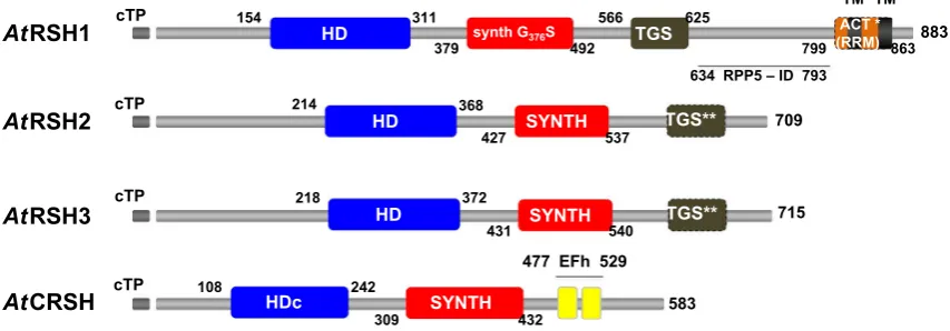
Among the different types of plant RSH, RSH1 has the widest distribution, as it is already present in archaeplastida and chromalveolates (Atkinson et al. 2011). In recent stud-ies with five plant phyla, RSH1 was found among Embryo-phyta, CharoEmbryo-phyta, Chlorophyta and RhodoEmbryo-phyta, but not in Glaucophyta (Ito et al. 2017). In most plants, RSH1 pro-tein carries all four major domains (HD, SYNTH, TGS and ACT; Fig. 1) present in the bacterial long RSH (Atkinson et al. 2011). However, the ACT domain, described recently
as an RNA recognition motif (RRM) domain (Brown et al.
2016), was shown as not conserved in Arabidopsis or Phy-scomitrella (asterisk in Fig. 1; Ito et al. 2017). Plant RSH1 TGS domain was proposed to play a role in RSH1–ribosome interaction in chloroplasts, similarly to its function in bacte-ria (Brown et al. 2016; Loveland et al. 2016; Ito et al. 2017). Even though RSH1 has the SYNTH domain, it rather func-tions as a (p)ppGpp hydrolase since the conserved glycine residue necessary for (p)ppGpp synthetase activity is sub-stituted with serine (Wendrich and Marahiel 1997; Masuda et al. 2008a; Fig. 1). Although van der Biezen et al. (2000) were able to show that Arabidopsis RSH1 (AtRSH1) confers phenotypes that are characteristic of ppGpp synthetase in E. coli and a Gram-positive bacterium Streptomyces coelicolor, other genetic studies show that AtRSH1 is only a (p)ppGpp hydrolase. For example, Arabidopsis RSH1 knock out (rsh1), and RSH1-overexpressing lines (RSH1oe) produce more and less (p)ppGpp, respectively, in comparison to WT plants. Additionally, AtRSH1 expression in a slow growing E. coli strain that overaccumulates ppGpp significantly accelerates growth of the bacteria, whereas the expression of its allele with mutation in the HD domain maintains the bacterial slow growth phenotype (Sugliani et al. 2016). Complemen-tation studies of E. coli relA− and relA−spoT− mutants with
AtRSH1 also show that AtRSH1 protein has rather no (p) ppGpp synthetase activity (Mizusawa et al. 2008).
RSH2 and RSH3 are characteristic only for green plants. In Arabidopsis, RSH2 and RSH3 are the results of RSH2 duplication, and a true RSH3 is missing from these plants. Nevertheless, it was found in other multicellular plants; thus,
cTP
709
SYNTH cTP
583 cTP
HD
309 432
214 368
427 537
154 311
379 492 TGS
566 625
799 863883
715
cTP 218 372
431 540
AtRSH1
AtRSH2
AtCRSH AtRSH3
HD SYNTH
HD SYNTH
synth G376S
TM TM
HDc
TGS**
TGS**
634 RPP5 – 793
108 242
477 EFh 529
ACT * (RRM)
ID
Fig. 1 Schematic representation of the conserved domains structure in Arabidopsis thaliana RSH proteins. cTP, chloroplast target peptide (gray); HD, (p)ppGpp hydrolase domain (blue); SYNTH, (p)ppGpp synthetase domain; TGS, TGS regulatory domain; ACT (RRM), ACT regulatory domain (RNA recognition motif domain; orange); EFh, calcium binding EF hand motifs constituting on EF hand domain (yellow); synth G376S, glycine into serine substitution at position 376 AA in the SYNTH domain that abolishes (p)ppGpp synthetase activity; HDc, degraded HD domain in CRSH, which may be non-functional (Atkinson et al. 2011); TM, putative transmembrane region
[image:5.595.84.511.453.602.2]it appears to be lost in the lineage to Arabidopsis. Atkin-son et al. (2011) proposed that AtRSH3, similarly to plant RSH2, carries the TGS domain, while other plant RSH3 have only a fragment of the TGS domain at the very end of CTD. According to recent data no TGS domain is present in Arabidopsis RSH2 and RSH3 proteins (Ito et al. 2017). Neither RSH2 nor RSH3 carry the ACT (RRM) domain (Fig. 1). The conserved residues required for activity of the SYNTH and HD domains are present in both RSH2 and RSH3, implying their metabolic bifunctionality (Atkinson et al. 2011). E. coli relA− and relA−spoT− mutants’
comple-mentation analysis show that AtRSH2 and AtRSH3 display (p)ppGpp synthetic activity, which for RSH2 seems to be very strong, as relA−spoT− mutant complementation with
the protein is lethal (Mizusawa et al. 2008; Sugliani et al.
2016). Overexpression of AtRSH3 in Arabidopsis WT as well as double knockout rsh2 rsh3 lines suggests that RSH3 has a (p)ppGpp synthetic activity, since both lines produce more alarmones than WT plants (Maekawa et al. 2015; Sug-liani et al. 2016). However, surprisingly, the overexpression of AtRSH2 in the rsh2 rsh3 background did not affect (p) ppGpp level in comparison to WT (Maekawa et al. 2015). Nevertheless, Arabidopsis RSH2 and RSH3 function rather as synthetases, since their expression in a slow growing E. coli strain that overaccumulates ppGpp was not possible, likely due to (p)ppGpp overproduction (Sugliani et al. 2016). N. tabacum RSH2 (NtRSH2), which shows high homology to Arabidopsis RSH2 and RSH3, exhibits (p)ppGpp syn-thetase activity based on biochemical and genetic studies. The protein complements E. coli relA− mutant and, similarly
to AtRSH2, is toxic to the relA−spoT− mutant, which
under-scores its strong (p)ppGpp synthetic activity (Givens et al.
2004). Another member of the RSH2 group, S. japonica RSH (SjRSH), also complements relA− in E. coli what is
indicative of its (p)ppGpp synthetase activity (Yamada et al.
2003). Based on biochemical and the E. coli relA−
com-plementation studies, P. patens RSH2a and RSH2b, which cluster within the RSH2/RSH3 subgroup, are (p)ppGpp synthetases. Surprisingly, no hydrolytic activity of these proteins was shown, despite the presence of the highly con-served catalytic residues in their HD domains (Sato et al.
2015), whereas transformation of a slow growing E. coli strain that overaccumulates ppGpp either with AtRSH2 or AtRSH3 was not successful, which is probably due to (p) ppGpp overproduction (Sugliani et al. 2016).
RSH4 or Ca2+-activated RSH (CRSH) is the plant RSH
closest to RSH1 (Atkinson et al. 2011) and appears to be present only in land plants. It carries neither the TGS nor ACT (RRM) domain. Instead, in its CTD, there are two Ca2+-binding EF hand motifs (Fig. 1) that are important for
A. thaliana CRSH and O. sativa CRSH1 (p)ppGpp synthetic activities (Tozawa et al. 2007; Masuda et al. 2008a). Since cytosolic calcium levels change under developmental and
stress-induced signals, EF hand motif-carrying proteins are able to transmit information about such stimuli (Day et al.
2002). Thus, CRSH was proposed to mediate the stringent response induced by such cues, as it is able to sense and respond to calcium fluctuations by producing (p)ppGpp. In AtCRSH, the HD domain is degraded (Fig. 1) and prob-ably not functional (Atkinson et al. 2011). However, E. coli relA−spoT−complementation studies with AtCRSH did not
result in bacterial cell death, which would be expected to happen if it produces (p)ppGpp but is not able to hydrolyze them (Masuda et al. 2008a). A possible explanation is that CRSH is a weak (p)ppGpp synthetase that does not cause toxic accumulation of alarmones. That would also explain why AtCRSH expressed in E. coli relA− mutant is not able
to restore the WT phenotype (Mizusawa et al. 2008). It probably produces (p)ppGpp to the extent that can be regu-lated by SpoT. Similar complementation results as well as no hydrolase activity were shown with OsCRSH1 (Tozawa et al. 2007). The notion that the CRSH protein lacks (p) ppGpp hydrolytic activity is supported with studies showing no effect of the AtCRSH expression on the amplification of a slow growing E. coli strain that overaccumulates ppGpp (Sugliani et al. 2016).
Based on plant RSH in silico analysis, genetic and bio-chemical studies, it can be concluded that RSH1 and CRSH have only (p)ppGpp hydrolase and synthetase activity, respectively, whereas RSH2 and RSH3 have both activities (Sugliani et al. 2016).
Role of RSH proteins and alarmones in chloroplasts
RSH localization and function in chloroplasts
Experiments performed in 1974 implied that chloroplasts are the loci of ppGpp production, as ppGpp synthesis was shown to be mediated by C. reinhardtii ribosomes of chloroplastic but not cytoplasmic origin (Sy et al. 1974).
Numerous studies have clearly shown that plant RSH proteins predominantly localize in chloroplasts, which are the site of the RSH action. C. reinhardtii RSH translocates to chloroplasts in vitro, which depends on the plastid tar-geting sequence, light and ATP (Lawrence and Kindle
35S promoter shows chloroplastic localization, which was confirmed also in a subcellular fractionation experiment followed by Western blot analysis by use of anti-CRSH antibody (Masuda et al. 2008a). Rice OsCRSH1 recombi-nant protein is imported into chloroplasts in vitro (Tozawa et al. 2007). Since AtRSH1 was proposed to carry a RPP5 (cytoplasmic protein involved in the plant effector-triggered immunity)-interacting region and a transmembrane domain in the CTD part of the protein (Fig. 1), it was described as cytoplasmic protein that anchors in the cell membrane (van der Biezen et al. 2000). However, AtRSH1–GFP fusion protein localizes in chloroplasts (Chen et al. 2014), and the GFP fusion protein containing AtRSH1 putative chloroplast transit peptide translocates to chloroplasts (Mizusawa et al.
2008), implying AtRSH1 plastid thylakoid and/or envelope membrane localization. RSH protein localization studies additionally show that RSH from each subgroup may act in a different spatio-temporal manner. As an example, AtCRSH and NtRSH2 proteins were found in chloroplast soluble and insoluble fractions, respectively (Givens et al. 2004; Masuda et al. 2008a). All these results are in agreement with around 13-fold higher levels of ppGpp in pea chloroplasts than in pea shoots (Takahashi et al. 2004).
Since SpoT-like protein from Rhodobacter capsulatus, bacteria that can obtain energy through photosynthesis, pro-motes the synthesis of photopigments, it was proposed that the stringent response regulates photosynthesis (Masuda and Bauer 2004; Masuda 2012). Because chloroplasts are descendants of photosynthetic bacteria, the (p)ppGpp-medi-ated regulation of photosynthesis in such prokaryotes was an argument for similar regulation of plant photosynthesis (Gray 1993; Givens et al. 2004).
In the last couple of years, progress has been made in deciphering bacterial-like stringent response in plants. It has been shown that the response regulates chloroplast pro-cesses such as transcription, translation, and production of nucleotides, hormones, lipids and metabolites. Moreover, recent findings also shed light on unique features of plant stringent response.
(p)ppGpp‑mediated regulation of transcription in chloroplasts
Chloroplast proteins are encoded by plastid (i.a., rbcL— large subunit of Rubisco) and nuclear genes (i.a., RSH and Rubisco small subunit). The chloroplast-encoded genes are transcribed by plastid-encoded RNA polymerase (PEP), which is similar to the bacterial RNAP, or nuclear-encoded plastid RNA polymerase (NEP), which is a T7 phage-like RNA polymerase (Swiatecka-Hagenbruch et al. 2007; Liere et al. 2011).
In plants, (p)ppGpp regulate transcription hypothetically in two different ways, which are similar to the ones observed
in bacteria. First one resembles the allosterical regulation of E. coli RNAP activity that results from the direct (p) ppGpp interaction with RNAP ω and β′ subunits (Ross et al.
2013). In chloroplasts, ppGpp was shown to bind to the β’ subunit of the bacterial-like PEP to inhibit transcription in a dose-dependent manner (Sato et al. 2009). However, in plants a homolog of E. coli ω subunit does not exist and the β′ subunit is different in many aspects from the bacte-rial counterpart. Moreover, the Rpo core enzyme (α2, β, β′, β′′) of PEP is surrounded by additional nuclear-encoded subunits (the PEP-associated proteins-PAPs) that have no bacterial homologs, are essential for PEP activity in chlo-roplasts (Borner et al. 2015; Pfannschmidt et al. 2015) and likely prevent the direct (p)ppGpp–PEP interaction. There-fore, a direct regulation of transcription from (p)ppGpp by allosteric interaction with the polymerase appears unlikely, although it is not impossible. In addition, the interaction between (p)ppGpp and the β′ subunit was shown in vitro only (Sato et al. 2009). Thus, it remains to be demonstrated that it can also occur in vivo.
Another possible way of transcriptional regulation is the limitation of GTP pool due to its usage for alarmone produc-tion. In plastids, similarly to B. subtilis, the transcription of rRNA genes starts from GTP (Suzuki et al. 2003; Krasny and Gourse 2004; Swiatecka-Hagenbruch et al. 2007), sug-gesting that the concentration of the initiator nucleotide is crucial for their transcription and that (p)ppGpp impacts the process. (p)ppGpp also regulates GTP biosynthesis in chloroplasts as plant (rice, pea and Arabidopsis) guanylate kinases (GKs), catalyzing the conversion of GMP to GDP, are sensitive to (p)ppGpp in vitro (Nomura et al. 2014a). Interestingly, GK of B. subtilis, but not of E. coli, is also inhibited by alarmones (Kriel et al. 2012; Nomura et al.
2014a), showing that the targets of (p)ppGpp differ between these bacteria and further suggesting that in plants during the stringent response transcription is regulated rather in the indirect way proposed for B. subtilis (Krasny and Gourse
2004). Nevertheless, the inhibition of GKs by alarmones must also bring a negative feedback loop to reduce (p) ppGpp production due to substrate (GDP) exhaustion. ppGpp also regulates the activity of enzymes implicated in ATP biosynthesis, namely, adenylosuccinate synthetases (ASs). ppGpp inhibits rice OsAS1 and OsAS2 in a guanine nucleotide concentration-dependent manner (Nomura et al.
estimated with high-sensitive ppGpp quantification method, is ~ 3 µM (Ihara et al. 2015). Hence, taking into account that the ppGpp concentration required for 50% GK inhibi-tion (IC50) is ~ 10 µM (Nomura et al. 2014a), Ihara et al. (2015) suggested that GK could be regulated by ppGpp in chloroplasts. In contrary, PEP, according to in vitro analysis, requires higher ppGpp concentrations (200–1000 µM; Sato et al. 2009) than found in chloroplasts. Thus, ppGpp con-centration in chloroplasts seems to be sufficient to inhibit GK but not PEP (Sugliani et al. 2016). Although it is not fully understood how the transcription is regulated during the stringent response in plants, some downstream targets have been identified.
Among chloroplast genes, whose expression are reduced under (p)ppGpp accumulation, are genes transcribed mainly by PEP and encoding for the components of photosystem I (i.e., PsaB, PsaC), photosystem II (PSII; i.e., PsbA, PsbD, PsbK), translation machinery (i.e.,16S, 23S, ribosomal pro-tein Rps14, TRNR–arginine tRNA) and RbcL. NEP tran-scribed genes may also be affected, for example, the ones encoding for the components of translation machinery (i.e., ribosomal protein Rps18), PEP (i.e., RNA polymerase alpha and beta subunits RpoA and RpoB) and for caseinolytic pro-tease P1 (ClpP1), a subunit of the translocon on the inner envelope of chloroplasts (Ycf1), an ATPase of unknown function (Ycf2) as well as acetyl-CoA carboxylase beta subunit (AccD). However, the (p)ppGpp-mediated regula-tion of NEP-dependent genes expression is quesregula-tioned as the observed changes might be due to posttranscriptional modifications, which would lead to differential regulation of turnover for PEP- and NEP-dependent transcripts (Sugli-ani et al. 2016). Indeed, using a method that eliminates the influence of transcript degradation, Sugliani et al. (2016) showed no major (p)ppGpp influence on the expression of NEP-dependent genes. The data obtained with the Arabi-dopsis line overexpressing RSH3 in rsh2 rsh3 background and accumulating higher levels of (p)ppGpp than WT plants also demonstrates reduction in the expression of both PEP- and NEP-dependent genes, i.e., psbA, psbD, rbcL and accD, rpoA, clpP1, respectively. However, the (p)ppGpp-mediated regulation of NEP-dependent genes expression might have occurred also due to differential PEP- and NEP-dependent transcripts turnover (Maekawa et al. 2015). Although the (p) ppGpp-mediated regulation of NEP-dependent genes expres-sion is not clear, the decreased expresexpres-sion of chloroplast rRNA and tRNA is implicit (Sugliani et al. 2016; Abdelkefi et al. 2017).
The (p)ppGpp-mediated expression of chloroplast genes highly overlaps with their expression invoked with absci-sic acid (ABA). Treatment of Arabidopsis plants with ABA results in significantly reduced expression of chloroplast genes encoding for PSII components, i.e., PsbE, PsbH, PsbI, PsbJ,L,E, PsbK, PsbM, PsbN-oligo, PsbZ, both in younger
and older plants as well as for PsbB in older plants. It also affects the expression of other chloroplast genes, such as the ones encoding for PEP subunits (i.e., RpoA, RpoB, RpoC1, RpoC2) and for proteins of large ribosomal subunit (i.e., Rpl14, Rpl16, Rpl2, Rpl20, Rpl23, Rpl32, Rpl33) (Yam-burenko et al. 2015). The decreased expression of NEP-dependent genes (i.a., rpoB) again shows that (p)ppGpp might also regulate NEP activity, which would contradict the earlier report (Sato et al. 2009). Another explanation for the changes in NEP-dependent transcript levels in ABA-treated samples is the reduced expression of the NEP-encod-ing gene as well as the high possibility that the treatment with ABA affects not only (p)ppGpp-resembling responses but also the non (p)ppGpp-invoked ones. The last hypoth-esis is supported with differential expression of the PSII component encoding gene psbA, whose transcripts are not affected under ABA treatment but are downregulated in the (p)ppGpp-accumulating lines (Maekawa et al. 2015; Yambu-renko et al. 2015; Sugliani et al. 2016; Abdelkefi et al. 2017). Since ABA promotes the expression of RSH2 and RSH3, it was proposed that the transcriptional de-repression invoked with ABA is mediated via the RSH-(p)ppGpp module. This idea is supported with the observation that Arabidopsis rsh2 and rsh3 mutants treated with ABA express even around 60% higher amount of chloroplast transcripts in comparison to WT plants (Yamburenko et al. 2015). The very high over-lap of genes affected in lines overproducing (p)ppGpp and in plants treated with ABA shows that ABA-invoked effects are to some extent (p)ppGpp-mediated. The correlation of ABA responses with (p)ppGpp action is corroborated with the ABA-promoted expression of nuclear-encoded sigma factor 5 (SIG5), which strongly resembles the stringent response-mediated expression of alternative sigma factors, which mediate transcription of stress responsive genes in bacteria (Yamburenko et al. 2015).
RSH/(p)ppGpp‑mediated regulation of translation in chloroplasts
homologs (Akkaya and Breitenberger 1992; Miura et al.
2007; Nomura et al. 2012), pea chloroplast EF-G is active on E. coli ribosomes (Akkaya and Breitenberger 1992) and ppGpp inhibits peptide elongation in a chloroplast transla-tion system in vitro (Nomura et al. 2012), it is highly pos-sible that (p)ppGpp regulate translation via the inhibition of these proteins in chloroplasts. Since translation is the major consumer of ATP and GTP nucleotides for aminoacyl–tRNA synthesis and elongation factor recycling and the level of these nucleotides is regulated by (p)ppGpp, the indirect involvement of (p)ppGpp in the regulation of translation is also possible (Nomura et al. 2012). Furthermore, AtRSH1 interacts with a chloroplastic protein from the Obg family, whose members play a role in ribosome assembly and thus may have an impact on translation (Chen et al. 2014).
(p)ppGpp accumulation in chloroplasts controls the production of chloroplast-encoded proteins because the overexpression of Arabidopsis RSH3, which accumulates high amounts of (p)ppGpp, causes a strong reduction in the level of chloroplast-encoded PsbA, the subunit of the reac-tion center from PSII (RCII). However, it does not affect the production of nucleus-encoded PSII light-harvesting complexes (LHCII) and thus decreases the RCII/LHCII ratio. Since LHCII are rich in chlorophyll b and highly fluorescent, the most probable explanation of strong basal chlorophyll fluorescence and the reduction in the maximal efficiency of PSII, which is annotated for both RSH2 and RSH3 overexpressing lines, is that low RCII/LHCII causes stoichiometric displacement of LHCII from RCII fraction (Maekawa et al. 2015; Sugliani et al. 2016). Concomitantly, Arabidopsis rsh2, rsh3, crsh and rsh2 rsh3 mutants exhibit significantly weaker basal chlorophyll fluorescence than WT plants, which is even weaker in the quadruple mutant of all four Arabidopsis RSH. Similarly, the RSH1 overproducing line shows weaker basal chlorophyll fluorescence, whereas rsh1 displays the opposite phenotype due to high (p)ppGpp accumulation (Sugliani et al. 2016). However, in another study, no changes in parameters indicative of the regulation of photosynthetic light reactions in rsh2 rsh3 mutant were shown (Maekawa et al. 2015).
In addition to PsbA protein downregulation, overexpres-sion of Arabidopsis RSH3 negatively affects the levels of chloroplast- and nuclear-encoded PsaB, RbcL (PEP-depend-ent), chloroplast f1 (Cf1)-β (NEP-dependent) and Rubisco small subunit proteins, respectively. Surprisingly, despite decreased transcript levels, AccD protein level increases in line accumulating alarmones (Maekawa et al. 2015; Sugliani et al. 2016).
(p)ppGpp is not able to inhibit translation as fast as trans-lation inhibitor lincomycin, which is likely due to high (p) ppGpp concentration requirement for that purpose. While the lincomycin induced significant inhibition of translation occurs 24 h after the expression of a constitutive (p)ppGpp
synthetase domain from E. coli in Arabidopsis, for (p)ppGpp it takes 72 h (Sugliani et al. 2016). Thus, the low production of chloroplast proteins invoked with (p)ppGpp is the result of the low level of components that constitute the chloro-plastic translational machinery and transcripts undergoing translation rather than a direct effect on translation.
(p)ppGpp regulate the level of hormones, lipids and metabolites in chloroplasts
(p)ppGpp regulate the level of salicylic acid (SA), a hor-mone that the main biosynthetic pathway takes place in chloroplasts. The accumulation of alarmones in the Arabi-dopsis RSH3oe plants causes reduction of the hormone level, whereas the decreased amount of (p)ppGpp in the Arabidopsis quadruple mutant line (RSH1–RSH3 knock out and CRSH knock down) correlates with increased SA level (Sugliani et al. 2016; Abdelkefi et al. 2017).
Lipid production occurs within chloroplasts, and (p) ppGpp regulate their levels. AtRSH3 overexpression in rsh2 rsh3 background leads to lower content of all molecular spe-cies of fatty acid (significantly of 16:0, 16:1, 16:2, 16:3, 18:1, 18:2, 18:3) but has no major impact on their com-position. Significant downregulation was annotated for the polar glycerolipids such as monogalactosyldiacylglycerol (MGDG) and sulphoquinovosyldiacylglycerol, whereas phosphatidylethanolamine and phosphatidylcholine were shown to be more abundant. Similarly, the levels of most of the tested metabolites (e.g., Krebs cycle metabolites, fuma-rate, malate, gluthamine synthetase/glutamine oxoglutafuma-rate, glutamate, many amino acids) were lower in comparison to WT plants (Maekawa et al. 2015).
AccD is an important subunit of the ACC complex. The function of the complex is to regulate the metabolism of fatty acids. When the enzyme is active, the product, malo-nyl-CoA, a building block for new fatty acids, is produced. The precursor of MGDG is linolenic acid, a 18:3 fatty acid. While the level of MGDG is downregulated in the line overproducing (p)ppGpp, the AccD protein accumulation is enhanced. On the first glance this appears to be a contra-diction, however, it is unknown how the complete ACC com-plex behaves. In addition, accD appears to be special among plastid genes as it is probably the only plastid protein-coding gene that transcript and protein levels are constitutively high during tomato fruit ripening (Kahlau and Bock 2008).
in AtRSH3oe plants under nitrogen deficiency is significantly lower than in WT plants, suggesting higher nitrogen defi-ciency tolerance of the mutant plants (Honoki et al. 2017). Similarly, in phosphorous-limited conditions, the same line accumulates significantly lower levels of anthocyanins, which is again indicative either of higher nutrient depriva-tion tolerance or the lack of starvadepriva-tion sensing (Maekawa et al. 2015). Nitrogen deficiency (p)ppGpp-mediated toler-ance occurs also likely due to reduced chlorophyll content, changes in photosynthetic parameters as well as pronounced changes in metabolite levels in AtRSH3oe plants in compari-son to WT plants. Thus, it was proposed that (p)ppGpp help plants to function during nitrogen starvation by modulating photosynthetic performance and metabolite balance (Honoki et al. 2017).
(p)ppGpp influence on chloroplast size and number
The molecular changes that occur in chloroplasts under (p) ppGpp accumulation affect chloroplast size and number. In an Arabidopsis line overexpressing RSH3 (in WT), chloro-plasts are significantly smaller and more numerous than in WT and display reduced chloroplast volume per cell vol-ume. Similarly, in the rsh1 mutant line, the chloroplast:cell volume ratio is decreased, and the opposite effect occurs for line overexpressing RSH1. This further corresponds with increased chlorophyll content in plants with mutations in genes coding for (p)ppGpp biosynthetic enzymes (i.e., rsh2 rsh3) but not in rsh1 (Sugliani et al. 2016). AtRSH3 over-expression in rsh2 rsh3 background also results in reduced chloroplast size, however, with no apparent influence on chloroplast number per cell (Maekawa et al. 2015).
Role of (p)ppGpp in chloroplast DNA replication
The inhibition of DNA replication is a hallmark of the strin-gent response in bacteria. In B. subtilis, direct binding of (p)ppGpp to DNA primase leads to inhibition of DNA rep-lication elongation (Wang et al. 2007b). In E. coli, ppGpp impairs the DNA primase DnaG activity in vitro; however, in vivo only at the initiation stage. It was proposed that alarmones of the stringent response likely fail to inhibit the primase strongly enough, and therefore, to stop the elon-gation of DNA replication. In E. coli, (p)ppGpp might be mainly used for RNAP inhibition and therefore, likely, their amounts are not enough to inhibit DNA replication elongation (Maciag-Doroszynska et al. 2013). DNA rep-lication is also regulated through transcription, likely via the (p)ppGpp-mediated RNAP inhibition, as in conditions promoting (p)ppGpp accumulation transcription of E.coli dnaA operon is diminished. DnaA is a replication initiation factor, which promotes the unwinding of DNA at E. coli origin of replication—oriC. Transcription form the oriC
proximal gid and mioC promoters is also required for the replication of oriC plasmids and was shown to be inhibited by (p)ppGpp in vitro. That underscores again that in E. coli (p)ppGpp-mediated DNA replication is regulated at least at the initiation stage (Nazir and Harinarayanan 2016).
In Arabidopsis, the accumulation of (p)ppGpp—although it reduces chloroplast size and total volume per cell— increases their number without changing DNA content per plastid, suggesting increased chloroplast replication and division (Sugliani et al. 2016). Thus, despite the very high level of homology between bacteria and plants in the RSH domain structure and function as well as molecular targets of alarmones, (p)ppGpp-mediated responses in plants become different, according to Sugliani et al. (2016) likely due to the evolutionary gene transfer of vast amount of endosymbiont/ plastid DNA into the host genome. Since chloroplasts do not proliferate as frequently as bacteria, it is also understandable that their DNA replication would be rather differently regu-lated. Sugliani et al. (2016) suggested that the inability of (p)ppGpp to inhibit DNA replication in chloroplasts may be caused by the lack of bacterial-like DNA primases in plants, which are essential components of replication machinery in bacteria and targets of (p)ppGpp. Another way to explain the lack of (p)ppGpp-mediated DNA replication inhibition in Arabidopsis plants is the expenditure of these nucleotides for the regulation of transcription or other molecular targets, similarly as it was proposed for E. coli (Maciag-Doroszyn-ska et al. 2013).
Role of alarmones in the expression of nuclear genes
(Pfannschmidt 2010; Bobik and Burch-Smith 2015), (p) ppGpp may be important to trigger this pathway. Since intracellular signaling is necessary for coordinating cell responses to constantly changing environmental cues, (p) ppGpp production in chloroplasts may function to orches-trate plant adaptation and development.
The accumulation of alarmones in Arabidopsis RSH3oe (in WT) line leads also to a downregulation of vast number of nuclear genes. Among those are defense-related genes, such as LRR receptor kinases serving to recognize micro-organism associated molecular patterns (flg22-induced receptor-like kinase 1—FRK1, chitin elicitor receptor kinase 1—CERK1, NSP interacting kinase 2—NIK2, suppressor of BIR1-1—SOBIR1). Moreover, transcripts of genes encoding for mitogen-activated protein kinase (MAPK) cascade pro-teins, important for pathogen-induced signal transduction, are downregulated. Similarly, transcription of genes involved in the biosynthesis of a plant defense hormone—SA (iso-chorismate synthase 1—ICS1, calmodulin binding protein 60g—CBP60g, SAR deficient 1—SARD1), SA signaling (WRKY53) as well as genes induced by the hormone (non-expressor of PR genes 1—NPR1, pathogenesis-related: PR1, PR2, PR5) is lowered. Nevertheless, among transcripts that expression decreases in the line accumulating alarmones are ones encoding for proteins involved in a negative reg-ulation of defense responses and programmed cell death. The (p)ppGpp-invoked negative regulation of SA marker gene (PR1, PR2, PR5) expression is corroborated with their expression in Arabidopsis RSH1–CRSH quadruple mutant line, where it is significantly higher in comparison to WT plants. Similarly, PR1 protein level also tends to be higher in the quadruple mutant line than in the control (Abdelkefi et al. 2017).
Interestingly, transcripts of jasmonic acid (JA)-respon-sive genes are upregulated in the RSH3oe Arabidopsis line (Abdelkefi et al. 2017). Hence, on the first sight the experi-mental data appear contradictory, since it was mentioned in the earlier section [(p)ppGpp regulate the level of hormones, lipids and metabolites in chloroplasts] that (p)ppGpp accu-mulation leads to decrease in the level of 18:3 fatty acids (Maekawa et al. 2015). Therefore, it would also result in downregulation of the plastid product 12-oxo-phytodienoic acid, a JA precursor that is biosynthesized from linolenic acid (18:3), and further in the decrease of JA production in peroxisomes. Hence, JA-responsive genes should be rather downregulated in the AtRSH3oe line. However, the JA-responsive gene expression data was obtained with the AtRSH3oe line in the WT background where RSH3 is GFP-tagged (Sugliani et al. 2016), whereas the metabolite studies were performed with the AtRSH3oe line in the rsh2 rsh3 background with native RSH3 (Maekawa et al. 2015). Since both of these lines produce different levels of ppGpp (~ 7- and ~ 3-fold higher than WT plants, respectively) and exhibit
to some extend different physiological phenotypes, it is hard to compare data from these two lines. Moreover, Sugliani et al. (2016) showed that in Arabidopsis plants containing a transgene encoding a chloroplast-targeted ppGpp syn-thetase from bacteria, under the control of a dexamethasone-inducible promoter, that exhibited phenotypes similar to the AtRSH3oe (in WT) line and produced very high amounts of ppGpp (~ 30-fold), accD transcripts are less abundant than in the line where the synthetase is catalytically inactive (Sugliani et al. 2016). Thus, it would be very interesting to check the expression of the discussed nuclear genes in the AtRSH3oe line in the rsh2 rsh3 background.
Nevertheless, the expression data presented by Abdelkefi et al. (2017) confirms the existence of SA and JA signaling antagonism. Furthermore, it suggests that the over-accumu-lation of alarmones in Arabidopsis plants might regulate their resistance towards necrotrophs, since JA production in response to such pathogens promotes plants resistance to these microorganisms (Abdelkefi et al. 2017). It also implies that (p)ppGpp may regulate not only SA but also JA produc-tion in chloroplasts.
Alarmones in cytoplasm
of AtRSH3–GFP fluorescence, it leaves open the possibil-ity of its cytosolic positioning and functioning to produce (p)ppGpp in cytosol, similarly to the yjbM-expression line (Ihara and Masuda 2016). It is also possible that the relatively lower increment of ppGpp (~ 3-fold higher than WT plants; Maekawa et al. 2015) promotes plant growth, whereas higher (~ 7-fold higher than WT plants; Sugliani et al. 2016) induces opposite effect.
Thus, it is possible that (p)ppGpp, or rather some upstream components that regulate (p)ppGpp localization, could serve as conductors that orchestrate plants decisions concerning energy investment in growth or survival, depend-ing on (p)ppGpp presence in chloroplasts or cytoplasm, respectively. The idea of (p)ppGpp functioning in cytoplasm is supported with in silico analysis performed on the eukary-otic SAH MESH1, which showed that it does not carry the mitochondrial target peptide and thus suggested cytoplasmic localization of the protein (Atkinson et al. 2011). Similarly, the expression of S. japonica RSH in the yeast Saccharomy-ces cerevisiae results in (p)ppGpp accumulation solely in the cytoplasmic but not in the mitochondrial fraction (Ochi et al. 2012). Moreover, the deletion of MESH1 in Drosophila induces retarded growth, resembling the phenotype observed in Arabidopsis yjbM transgenic line (Sun et al. 2010).
We can assume two ways of (p)ppGpp accumulation in cytoplasm. It can either be produced by RSH proteins during their cytoplasmic localization or transported to cytoplasm after chloroplastic synthesis, which is the case for other nucleotides (Ihara and Masuda 2016).
Both MESH1 and AtRSH1 are (p)ppGpp hydrolases. No (p)ppGpp synthetase has been found in animals so far, which leaves us with a question on the function of MESH1 in the plausible non-(p)ppGpp environment. AtRSH1 interacts with the cytoplasmic protein RPP5 involved in plant responses to pathogens, suggesting that it may function in cytoplasm. Thus, it is possible that (p)ppGpp hydrolases could func-tion in cytoplasm to somehow cope with (p)ppGpp produced by bacteria during pathogenic infections, since (p)ppGpp production is important for bacterial virulence (Dalebroux et al. 2010). However, there is no available data that supports that hypothesis. Moreover, how would it happen that plant enzymes would act on inner bacterial components? Hence, analysis of (p)ppGpp accumulation on the site of the host and pathogen during infections are important to better under-stand the phenomenon of the stringent response.
Implicit is the idea that the stringent response in plants is not only a form of response to stress but also a way to regulate plant growth and development, likely through facili-tation of the cross-talk between nucleus and chloroplasts. Similarly, in bacteria, basal levels of alarmones regulate cell growth and metabolism (Potrykus et al. 2011; Gaca et al.
2013). Furthermore, bacteria missing RSH are mostly obli-gate intracellular parasites or endosymbionts (Atkinson et al.
2011), implying the importance of RSH for the regulation of fundamental processes.
Role of RSH and alarmones in plants Spatiotemporal expression of plant RSH
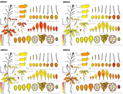
An overview of developmental expression profiles for Arabi-dopsis RSH is available at the Arabidopsis eFP Browser (Schmid et al. 2005; Winter et al. 2007) at the Bio-Array source for Plant Biology (http://bar.utoronto.ca/welcome. htm) and presented here in Fig. 2. Developmental maps of Arabidopsis RSH1, RSH2, RSH3 and CRSH demonstrate highly similar patterns of AtRSH2 and AtRSH3 expression, which reflects the results of in silico sequence and phylo-genetic data analysis; this corroborates the suggestion that true RSH3 is missing from the Arabidopsis lineage, and AtRSH3 is actually the result of RSH2 duplication (Atkin-son et al. 2011). All four RSH are expressed at various stages of embryo development. RSH1 expression fluctuates and reaches its peaks at heart and curled cotyledons stages and CRSH expression peaks in later stages of seed develop-ment (mostly curled and green cotyledons), whereas RSH2 and RSH3 display high expression at late embryo devel-opmental stages (8–10; Fig. 2). While the high RSH2 and RSH3 expression continues in dry seeds, neither RSH1 nor CRSH transcripts appear to be significantly conserved in the organ. Thus, the expression data suggests that RSH1 and CRSH might play a more important role in seed pre-dormancy, whereas RSH2 and RSH3 in seed dormancy or in the imbibing embryo. RSH2 and RSH3 could also pos-sibly regulate plastid development during embryo matura-tion. Arabidopsis embryos display a transient green stage during seed formation. In that time chloroplasts are being formed, whereas in the late embryo development, during seed desiccation, they are de-differentiated into eoplasts that retain the transcriptional apparatus but disassemble the photosynthetic one. Since the primary chloroplast biogen-esis in Arabidopsis embryos is not understood yet, it would be interesting to check whether (p)ppGpp could serve as the initiator/mediator of the chloroplast–eoplast transition. Since ABA is involved in seed maturation, dessication and dormancy and promotes RSH2/3 expression (Yamburenko et al. 2015), it could regulate this process via the RSH2/3-(p)ppGpp module as it was proposed by Pfannschmidt et al. (2015).
and, to some extend in shoot apex, buds as well as flowers (mostly in sepals and petals) and pedicels. CRSH expres-sion is quite distinct, since the highest number of CRSH transcripts is produced in roots and mature pollen, which according to eFP Browser data is not the case for other Arabidopsis RSH. Relatively high CRSH expression also occurs in particular rosette leaves after transition to flower-ing, including cauline and senescent leaves, as well as in inflorescent shoot apex, buds, flowers (mostly sepals) and pedicels. The highest RSH2 and RSH3 expression is reserved for the already mentioned later stages of embryo develop-ment, dry seeds and mature flowers (mostly petals, sepals
and stamens). Similarly to RSH1 and CRSH, very high RSH2 and RSH3 expression also takes place in leaves undergoing senescence (Fig. 2).
The expression levels of RSH in Fig. 2 are not normal-ized. Therefore, while comparing absolute expression values between all AtRSH, we observe that RSH2 and RSH3 tran-scripts are actually the most abundant RSH in Arabidop-sis plants. Their expression in root, hypocotyl, cotyledon, cauline and senescing leaf, flower (sepals, petals, stamens), during seed development as well as in dry seed is higher than the expression of RSH1 and CRSH. Additionally, we also notice higher AtRSH1 and AtRSH3 expression in the first and
Fig. 2 Electronic fluorescent pictographs (eFPs) for Arabidop-sis thaliana RelA/SpoT homolog genes RSH1 (At4g02260), RSH2 (At3g14050), RSH3 (At1g54130) and RSH4 (At1g30850) transcript levels in different plant organs over various developmental stages. Expression levels between RSH are not normalized. High and no expression levels are indicated by red and yellow colors, respec-tively. For later stage siliques (6–10; corresponding to torpedo, walking-stick, curled and green cotyledons stages of embryo devel-opment) only the seeds were collected for analysis—not the siliques
[image:13.595.54.545.54.432.2]second leaves as well as in vegetative rosette in comparison to AtRSH2 and AtCRSH (Arabidopsis eFP Browser).
The other experimental approaches confirm the array data on the expression of all Arabidopsis RSH in flowers and show additionally their expression in pistils. Furthermore, RSH2 and RSH3 expression was shown to fluctuate during flower development. Their expression was shown to be pre-sent in carpels and sepals of juvenile buds and later also in stamens of pollinated flowers. Moreover, their expres-sion was observed also in developing pollen (Masuda et al.
2008a; Mizusawa et al. 2008). One of the reasons for RSH expression in flowers could be their importance for fruit development regulation. Most plastid genes (photosynthe-sis-related, transcription apparatus-coding, tRNAs) as well as RbcL protein in green tomato fruits are strongly down-regulated in comparison to leaves (Kahlau and Bock 2008), suggesting that (p)ppGpp accumulation may trigger fruit development.
The combination of the array and other experimental data clearly demonstrates ubiquitous expression of plant RSH in green tissues. According to the latter ones, Arabi-dopsis RSH1–RSH4 expression occurs in the early stages of seedling development (2–11 days) in the following pattern: AtRSH1 and AtRSH3 genes are highly expressed in hypoco-tyls, leaves, leaf veins and shoot apical meristems, whereas AtRSH2 and AtCRSH are expressed in leaves and shoot apical meristems. However, AtCRSH expression declines 5 days after germination. Interestingly, RSH expression in shoot apical meristems is very pronounced, which is not effectively captured in the array data. In adult plants (50 days old), AtRSH1, AtRSH2 and AtRSH3, but not AtCRSH, are strongly expressed in rosette leaves (Mizusawa et al. 2008). However, Western blot analysis performed with WT plants by use of anti-CRSH antibody also showed CRSH produc-tion in rosette leaves, next to its expression in cauline leaves, siliques, stems and flowers (Masuda et al. 2008a).
According to Mizusawa et al. (2008) only AtRSH2 is expressed in roots of seedlings. The expression of RSH2, and to some extent RSH3, occurs in roots of mature Arabidop-sis plants (Mizusawa et al. 2008), which remains in agree-ment with the array data showing their relatively highest expression in that organ in comparison to other RSH ( Arabi-dopsis eFP Browser). Nevertheless, in other experimental approaches, Arabidopsis RSH2 and RSH3 and partly RSH1 as well as Oryza sativa CRSH1 transcripts were found in roots. Unluckily, in those studies, AtCRSH expression was not tested (Tozawa et al. 2007; Mizusawa et al. 2008; Chen et al. 2014). However, protein expression studies showed no detection of AtCRSH in Arabidopsis roots (Masuda et al.
2008a). Thus, although Arabidopsis CRSH expression in roots is quite pronounced in the absolute array data, it does not appear so in vivo, which is likely due to its lowest rela-tive expression in comparison to other Arabidopsis RSH.
That, along with other AtCRSH expression data, implies that AtCRSH is a rather stress responsive gene.
(p)ppGpp impact on plant growth and development
Chloroplastic phenotypes elicited by different (p)ppGpp lev-els have an impact on plant growth and development. The antagonistic role of Arabidopsis RSH1 vs. RSH2, RSH3 and CRSH in (p)ppGpp production is important for maintain-ing alarmone levels durmaintain-ing vegetative tissue development. Arabidopsis plants overexpressing RSH2 (RSH2oe) are pale and smaller in comparison to WT plants. However, the sur-face of Arabidopsis rsh2 rsh3 or rsh1–4 quadruple mutant plants is also significantly smaller than WT plants. Never-theless, these mutants stay darker than rsh1 or WT plants (Sugliani et al. 2016), underscoring that (p)ppGpp accu-mulation promotes the pale phenotype. AtRSH3oe (in WT) plants, similarly to AtRSH2oe (in WT), are pale and smaller than WT plants (Sugliani et al. 2016). RSH3oe plants in the rsh2 rsh3 background have increased cell number and grow better than WT plants. Nevertheless, these plants also have lower chlorophyll levels and are pale with respect to WT plants, which again shows that (p)ppGpp accumulation invokes pale phenotype (Maekawa et al. 2015). The differ-ences in RSH3 protein-overexpressing lines could be again explained with the fact that the first group overexpressed RSH2–GFP and RSH3–GFP, whereas the latter one native RSH2 and RSH3. Furthermore, as already mentioned, rela-tively lower increment of ppGpp (~threefold higher than WT plants; Maekawa et al. 2015) possibly promotes plant growth, whereas higher (~ 7-fold higher than WT plants; Sugliani et al. 2016) induces opposite effect. Maekawa et al. (2015) proposed that the increased plant size might be the effect of metabolite fitness, provoked by decreased photo-synthesis. Plants of the AtRSH3oe (in rsh2 rsh3 background) line grow also better in nitrogen starvation conditions, what is likely attributed to changes in photosynthetic activities and metabolite balance (Honoki et al. 2017). On the other hand, the results with the AtRSH3oe line (in WT) are supported with the studies on P. patens homologs of AtRSH2/RSH3, PpRSH2a and PpRSH2b, as their overexpressor lines show significant growth suppression in a glucose-concentration-dependent manner (Sato et al. 2015).
The impact of (p)ppGpp on plant growth and develop-ment may result from the regulation of hormone produc-tion, as fatty acids and nucleotides, the levels of which are regulated by (p)ppGpp, are precursors of plant hormones.
Plant senescence, nutrient remobilization and relocation and seed development
et al. 2008b; Sugliani et al. 2016), what correlates with their expression during late plant development in flowers and leaves undergoing senescence (Schmid et al. 2005; Masuda et al. 2008b; Mizusawa et al. 2008) (Fig. 2).
AtRSH1–4 mutant studies showed that RSH2, RSH3 and to some extent CRSH promote plant senescence, whereas RSH1 shows the characteristics of a negative regulator of this process, likely due to their ability to synthetize and hydrolyze (p)ppGpp, respectively. While rsh1 knockout plants manifest accelerated senescence, rsh2 rsh3 mutants as well as other RSH1–RSH4 multiple Arabidopsis mutants carrying insertions in both RSH2 and RSH3 genes display delayed senescence and that effect is bolstered when CRSH is knocked down. Similarly, plants overexpressing RSH1 do not senescence as fast as WT plants. Thus, we can call (p) ppGpp as positive regulators of plant senescence (Sugliani et al. 2016).
Increased levels of (p)ppGpp alone are not enough to trig-ger senescence, as plants that accumulate alarmones do not undergo senescence in vegetative tissues. The phenotype is rather obvious after flowering during the seed filling time. It is known that during senescence nutrients are remobilized from old tissues to reproductive ones (Lim et al. 2007). In RSH mutants with delayed senescence (rsh2 rsh3) or plants with the stay-green phenotype that die while being still green (Arabidopsis quadruple mutant rsh1–4), seed weight is significantly decreased compared to controls. This implies that (p)ppGpp is important for proper seed development and that seeds achieve their weight when alarmone accumula-tion stimulates nutrient remobilizaaccumula-tion from dying vegeta-tive tissues into reproducvegeta-tive organs. Strong expression of Arabidopsis RSH2 and RSH3 during senescence, flower and seed development implies that (p)ppGpp may act as a “push” and/or a “pull” of nutrients from vegetative into reproductive tissues. However, the lines overexpressing RSH2 and RSH3 also produce significantly smaller seeds than WT plants, but that might be the result of overexpression per se (Sugliani et al. 2016).
The accelerated dark-induced senescence phenotype in Arabidopsis rsh1 mutant and RSH3 overexpressing plants, the two lines over accumulating (p)ppGpp, is coupled with accelerated Rubisco degradation, whereas in the line over-expressing RSH1, the Rubisco protein level increases along with darkness exposition time. This implies that (p)ppGpp during senescence promote not only nutrient reallocation but also remobilization by stimulating chlorophyll and Rubisco degradation (Sugliani et al. 2016). Therefore, the plant strin-gent response is not only linked to stress responses in plants but also required for optimal plant growth and development. Nevertheless, (p)ppGpp seem to act as signaling molecules that inform plants about the necessity of nutrient remobiliza-tion from vegetative into generative organs, which resembles their survival functions in bacteria.
Importance of (p)ppGpp production to exert plant senes-cence correlates with low levels of pyrimidine in senescing tissues (Greenberg 1997). As (p)ppGpp inhibit enzymes involved in purine biosynthesis, they may also affect levels of other nucleotides and thus accelerate plant senescence.
Since ABA induces both senescence and AtRSH2 and AtRSH3 gene expression, and gene expression profiles invoked by (p)ppGpp and ABA highly overlap (Maekawa et al. 2015; Yamburenko et al. 2015; Sugliani et al. 2016), we can assume that (p)ppGpp mediates ABA-induced senes-cence in plants.
AtCRSH was found to be important also for proper fer-tilization and silique formation as its knock down leads to abnormal flower development impeding pollination, signifi-cantly smaller than WT siliques and 300-fold reduction of seed quantity (Masuda et al. 2008a). However, lines retain-ing residual AtCRSH expression do not show that phenotype (Sugliani et al. 2016).
Role of RSH proteins in plant stress response
Changes in RSH transcripts and proteins and in (p)ppGpp levels under various stimuli as well as plant RSH expression in E. coli or S. cerevisiae conferring tolerance to different cues suggest that plant RSH play a role in response to abiotic and biotic stress.
In a yeast two-hybrid experiment, A. thaliana RSH1 pro-tein was found to interact with RPP5, encoded by a mem-ber of disease resistance gene class, conferring resistance to pathogens such as Peronospora parasitica. However, no changes on the AtRSH1 transcript level were observed under treatment with Pseudomonas syringae DC3000 or its deriva-tive carrying AvrRps4 as well as under factors known to exert some plant pathogen/insect-induced alike responses or mediate plant responses to pathogens, such as wounding or SA and methyl jasmonate, respectively (Gassmann et al.
1999; van der Biezen et al. 2000; Mizusawa et al. 2008). Moreover, the expression of AtRSH1 was shown to even
Fig. 3 Electronic fluorescent pictographs (eFPs) for Arabidopsis thaliana RSH1 (At4g02260), RSH2 (At3g14050), RSH3 (At1g54130) and RSH4 (At1g30850) transcript levels under different stresses. 18 days after sowing and 3 h after dark/light transition Arabidopsis Col-0 plants treated with cold (plants transferred to ice for rapid cool-ing and kept at 4 °C in the cold room until harvest), 300 mM man-nitol (osmotic stress), 150 mM NaCl (salt stress), drought (plants exposed to air stream for 15 min with loss of approximately 10% fresh weight), 10 μM methyl viologen (oxidative stress), wound-ing (leaves punctuation) or heat (38 °C for 3 h followed by recovery at 25 °C until harvest) conditions were harvested at 1 and 12 h post treatment. Expression levels between RSH are not normalized. High and no expression levels are indicated by red and yellow colors, respectively. More detailed information about the microarray and other studies that are the sources for these stress response maps and further information can be found at Arabidopsis eFP Browser at bar. utoronto.ca, in Winter et al. (2007) and Kilian et al. (2007)