ORIGINAL ARTICLE
Using micro-computed tomography to examine the larynx in cases
of suspected strangulation
—
a comparison of case findings
and control images
Waltraud Baier1&Brian A. Burnett2&Mark Payne3&Jason M. Warnett1&Mark A. Williams1
Received: 4 April 2019 / Accepted: 22 October 2019
#The Author(s) 2019
Abstract
The examination of strangulation is one of the most challenging causes of death diagnoses encountered in forensic pathology. The injuries are often subtle and difficult to detect, especially in cases that lack superficial marks. Fractures of the laryngeal skeleton are commonly regarded as evidence of strangulation but these can be too subtle to be detected during autopsy. Micro-CT is a novel imaging technique that achieves a spatial resolution 1μm or less which lends itself to the examination of small and delicate structures such as the larynx. However, there is little information to date regarding the appearance of the larynx at this scale, thus complicating the interpretation of the micro-CT images. This study therefore uses micro-CT to examine ten larynges from strangulation deaths and to compare them to nineteen samples from donor individuals in order to distinguish between naturally occurring features and actual trauma. It was found that there are several features which mimic damage in the donor group. Using associated case information, initial trends and patterns of different strangulation methods were established.
Keywords Micro-computed tomography . Forensic Imaging . Strangulation . Larynx
Introduction
Strangulation is the third most common homicide method in the UK, although women are even more likely to become a victim due to intimate partner violence [1,2]. Despite the
frequent occurrence of such cases, the diagnosis of strangula-tion in forensic pathology is still predominantly one of exclu-sion, in particular if there is a lack of distinctive signs such as petechial bleeding or bruising of the soft tissues of the neck. Fractures of the laryngeal cartilages or the hyoid bone are considered evidence for strangulation but they can have other causes such as sports injuries, motor vehicle accidents or falls [3]. Such fractures tend to be associated with haemorrhages, but absence thereof can make subtle injuries even more diffi-cult to detect at the risk of potentially missing them during autopsy. In some cases, histological examination is conducted to examine fractures identified at postmortem to assess the timing of the injury in relation to time of death [4]. This pro-cess is destructive, time consuming and costly. With advances in technology, new ways of dealing with this issue have emerged with many researchers advocating the use of com-puted tomography (CT) to study laryngeal trauma [5,6]. Even more recently, initial studies have explored the use of micro-computed tomography (micro-CT) to examine such cases in order to identify micro-fractures which might not be visible on medical CT or postmortem examination [7,8]. Micro-CT has proven successful in a number of different forensic applica-tions such as toolmark analysis [9–11], gunshot wound
* Waltraud Baier
val.baier@warwick.ac.uk
Brian A. Burnett
brian.burnett@uhcw.nhs.uk
Mark Payne
m.payne@west-midlands.pnn.police.uk
Jason M. Warnett
j.m.warnett@warwick.ac.uk
Mark A. Williams
m.a.williams.1@warwick.ac.uk
1 WMG, International Manufacturing Centre, University of Warwick,
Coventry CV4 7AL, UK
2
UHCW NHS Trust, Coventry CV2 2DX, UK
3 West Midlands Police, B4 6NQ, Birmingham, UK
analysis [12,13] and forensic entomology [14]. However, all of the existing literature on using micro-CT for laryngeal trau-ma analysis is based on casework; no comparative studies exist to date to the authors’knowledge. This study aims to address this void as it draws on an unprecedented database of micro-CT scans of larynges from strangulation cases as well as from natural deaths. The injuries observed in the former group were compared with those in the latter control group of uninjured larynges from donor individuals in order to assess which features can occur in the normal population. Studies by Tsai et al. [15] and Baier et al. [16] have compared micro-CT to histology, the current gold standard for forensic injury anal-ysis, and found the method to be reliable for injuries in bone. Method validation is becoming an increasing concern in fo-rensic science and while micro-CT had been validated in in-dustrial settings [17–19] where the method as such has been proven to work, it has not been systematically tested for fo-rensic purposes. This study forms an exploratory study to inform future more structured validation studies.
Materials and methods
Micro-CT
The overall principles of micro-CT are similar to medical CT as it uses a stack of 2D radiographs to examine the internal structure of a sample, but it allows a spatial res-olution, or voxel size, of approximately 40μm for a sam-ple of the size of a larynx. The source and detector of a typical lab-based micro-CT machine are stationary during the scan with the sample on a rotating stage at the centre. Radiographs are acquired at every degree angle through a full 360° rotation and later reconstructed to form the 3D model of the object [20].
All samples were scanned using a Nikon 225/320 LC micro-CT scanner (Nikon Metrology, Tring, UK) and recon-structed using the system’s proprietary software CTPro. Typical scan parameters were
120 kV, 135μA, 500-ms exposure, 24-dB gain, no filtra-tion, creating 3141 projections, although some adjustments were made where necessary. The scan images were visually inspected in VG Studio MAX 2.2 (Volume Graphics, Heidelberg, Germany).
Two groups of larynges were examined. The first consisted of ten samples from forensic strangulation cases. Only cases where strangulation had been confirmed through multiple lines of enquiry (witness statements, confessions, CCTV, forensic post-mortem) were consid-ered in this group; no inconclusive cases were included (age range 20–68 years; male 4, female 6). The second group consisted of nineteen control samples from donor individuals with no recorded history of laryngeal trauma
(age range 46–94 years; male 12, female 7). Only the thyroid cartilage and the hyoid were considered for this study. Full demographic details are provided in Appendix Tables 1 and2.
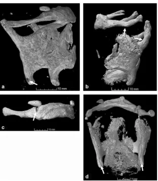
The examination criteria were determined from the control group as potentially misleading features during analysis. These were circular defects within the ossified cartilage, fragmented appearance of ossified material, in-complete fusion of the hyoid elements, abrupt angles and linear grooves within the ossified cartilage, and the pres-ence of triticeous cartilages. Based on the literature, the presence, completeness (incomplete, complete, displaced) and location of fractures were also included as a criterion as they are frequently encountered in strangulation deaths. In order to include all possible fracture origins, they were termed discontinuities in the analysis. An example for each category is shown in Fig.1.
The postmortem findings were also noted for the forensic cases. They were summarised as superficial (petechiae, facial congestion, bruising on the neck, ligature marks) and internal (haemorrhage/bruising on the muscles and soft tissues of the neck). The mode of strangulation (manual, ligature, chokehold) was noted where sufficient evidence was available.
Results
The full results are provided in Appendix Table 1 (stran-gulation group) and Appendix Table 2 (control group). Triticeous cartilages were observed in 30% of cases in the strangulation group (n= 3), and in 47% in the control group (n = 9), their appearance being the same in both groups with smooth cortical bone and a spherical to oval shape. Abrupt angles are seen along the superior thyroid margins, the superior thyroid horns, and the greater horns of the hyoid in 10% of cases in the strangulation group (n
= 1), and 32% of cases in the control group (n = 6). Circular holes within the ossified cartilage occurred in 40% of strangulation cases (n = 4), and 32% of control cases (n = 6), they all display clear and regular margins. Linear grooves were only observed on the posterior aspect (including superior and inferior horns) of the ossified thy-roid cartilage (50% orn = 5 in strangulation group, 32% or n = 6 in control group). They predominantly extend vertically along the posterior margin although some are observed on the lateral aspects of the posterior margin.
displayed a discontinuity left and right of the midline. Seventy percent of the forensic cases (n = 7) displayed a discontinuity on the superior thyroid horns with the base being the most affected area. Of these, six cases displayed bilateral discontinuities (total number of discontinuitiesn
= 13). The inferior thyroid horns were only affected in two cases (20%). The three cases (30%) where the hyoid b o n e w a s f r a c t u r e d w e r e a l l c a u s e d b y l i g a t u r e strangulation.
Discussion
Significance of the results
DiscontinuitiesThe most likely explanation for the fracture-like discontinuity on the inferior thyroid margin, observed in both groups, is a product of the natural ossification process. While they appear like fractures on the volume-rendering, on the 2D sections the edges of the discontinuity appear rounded
Fig. 2 Typical locations of discontinuities identified in this
study (arrows).aDiscontinuity
on the inferior thyroid margin of sample S172809 in the control
group.bFracture at the base of
the superior thyroid horns of the forensic case OP Isengard
Fig. 1 Some of the assessment
features described in this study.a
Circular“hole”within the ossified
cartilage and fragmentary
ossification, S172809.bUnusual
angles of the greater horns (hyoid) and superior horns (thyroid
carti-lage), S173160.cIncomplete
fu-sion of the greater horn and body
of the hyoid, S170345.dLinear
[image:3.595.234.546.49.407.2] [image:3.595.234.545.550.711.2]and uninterrupted. All other discontinuities, exclusively en-countered in the forensic group, are considered true fractures as they display sharper edges with interrupted cortical bone and trabeculae. Hyoid fractures have only been observed in the posterior third of the greater horn where the bone is deli-cate and thin. The greater horn has been observed as a mon fracture location in several other studies of neck com-pression [3,5] and appears to be strong support for compres-sion of the neck. All the fractures of the greater horns of the hyoid occurred in ligature strangulations. This contradicts the review by Dettmeyer et al. [21] who found hyoid fractures to be more common in manual strangulations. The majority of superior thyroid horn fractures occur bilaterally. Only one of those bilateral injuries was not a ligature strangulation. However, there were also cases of ligature strangulation which did not result in bilateral fractures. This is a recurring dilemma in interpreting strangulation deaths that there is much overlap in the injury pattern. Many studies have attempted to classify injuries according to the mode of strangulation [22–24] and while there are some trends there is also overlap which war-rants caution not to solely rely on fracture patterns for the interpretation.
Circular holesSmooth-edged circular holes were only seen in the thyroid cartilage but were not restricted to a specific tion. Their occurrence within the laminae is a common loca-tion for the thyroid foramen which serves as an opening for vessels or nerves [25]. It is possible that the circular openings in other locations fulfil a similar purpose or simply represent ossification irregularities. Their presence in the control group supports an interpretation as a natural cause as opposed to possible fingertip damage.
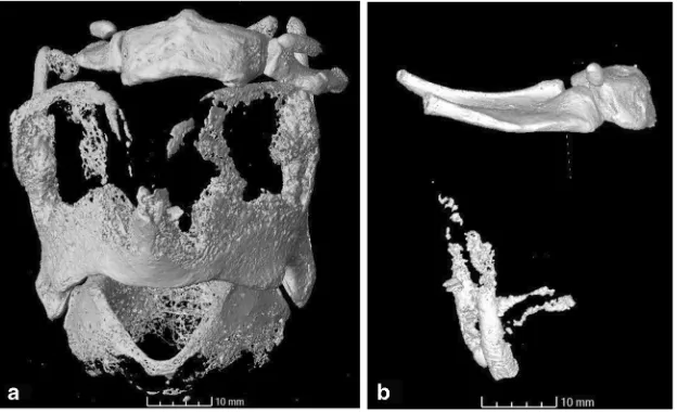
FragmentationThe ossified cartilages in the forensic group appeared more fragmented than those in the control group (Fig. 3). This manifested as an increased number of small pieces of ossified material that did not form a coherent struc-ture. However, this could be explained by younger age of individuals in this group. The more advanced age of the donor individuals in the control group is reflected by the more ad-vanced stages of ossification. Nonetheless, the appearance of the loose fragments was similar in both groups. Individual ossified“nodules”displayed rounded edges. While the major-ity of fragments in the control group are found along the mar-gins of the thyroid laminae, the case examples display frag-ments in all locations. As they are similar in appearance, they are not considered to be compelling evidence of trauma. No other study has examined this feature at this level of detail to the authors’knowledge, further comparison will therefore de-pend on a future expansion of this database.
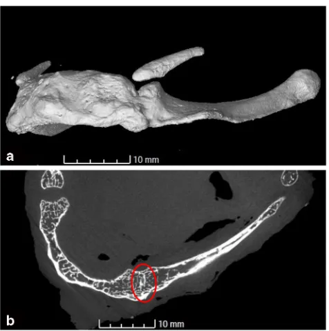
Incomplete fusionThe fusion or non-fusion of the greater horns to the body of the hyoid bone is an easily misinterpreted feature. While it is well-established in the literature that these elements can fuse over time [26,27], in pathological practise hypermobility is sometimes taken as evidence of a hyoid fracture [4]. Depending on the pathologist’s sensitivity and experience, a natural non-union could be mistaken for trauma. Radiologically, a narrow gap between the body and greater horn or incom-plete fusion of the two parts might be mistaken as a frac-ture. The control group has shown several examples of incomplete fusion with a narrow gap still visible at the body-greater horn junction, see Fig. 4 for a transverse section and the volume-rendering of the scan. Both sides
Fig. 3 Different stages of laryngeal cartilage ossification. The control group was composed of generally older individuals than the forensic group, displaying more advanced stages of ossification and therefore a
different pattern of fragmentation.aSample S170405, showing some
fragmentation along the anterior margins of the thyroid laminae.bOP
Aporia, the individual’s younger age (early 20s) is reflected in the limited
[image:4.595.143.455.477.667.2]display an uninterrupted cortical bone surface which only begins to disappear after fusion has completed. No actual fractures were observed at this location in either group.
Abrupt anglesThese were encountered frequently in the control group, often on the superior thyroid horns and along the superior margins of the thyroid laminae. In one control sample, the greater horns of the hyoid were angled upwards in the posterior third. These angles are present in the normal population but from this study it does not become clear whether they are due to normal variation or previous, healed trauma. A study by Maxeiner [28] found that healed fractures of the hyoid and thyroid cartilage can be found amongst alcoholics who are prone to frequent falls or accidents. However, information about the donors’lifestyles was not available in the present study.
Linear grooves These features resemble fine fissures or creases when seen on the 3D volume-rendered scan, shown in Fig.2D. Using the 2D sections helped identify-ing them as superficial. They were frequently observed in the control group which is important to avoid misinterpre-tation as real fractures in the forensic samples. The loca-tion and orientaloca-tion of these creases differed from the actual fractures observed (vertical as opposed to approxi-mately horizontal). This feature in particular has only been made possible to study through the increased detail
of micro-CT and has not been described previously.A sim-ilar feature was a sharp crest running in no particular direction, possibly indicating the ossification “front”. One side of this crest displays a cortical bone surface whereas the other side displays less solidly ossified mate-rial, shown in Fig. 5 for control sample S170452. When examining the volume rendering of a scan, the shadow created by the crest can mimic a fracture line. Having seen this feature in numerous control cases serves as re-assurance that this is a natural feature. This was most commonly observed in the area at the base of the superior thyroid horns. The horns and the posterior margins displayed more densely ossified material compared to the more loosely structured trabeculae at the base of the horns. This contrasting ossification pattern is important to understand as it might impact laryngeal fractures. Saternus et al. [29] argue that the differences in ossifica-tion density create weak links which are then more prone to fracturing. A fracture in these areas would therefore not be unexpected and potentially requires less force than in other areas or if ossification there was more homogenous. The base of the superior thyroid horns was frequently seen as less densely ossified in the control group and fractured in the strangulation group, supporting this theory.
Triticeous cartilages These have occasionally been ob-served in the literature on normal anatomical variation of the larynx [26]. These small spherical ossifications are found in the thyrohyoid membrane which connects the superior thyroid horns to the posterior greater horns of the hyoid. The close proximity to the superior thyroid
Fig. 4 Incomplete fusion of the left greater horn and the body of the hyoid
in control sample S171263.aVolume rendering showing a bony bridge
across the two elements.bTransverse section through the specimen at the
location of the bridge. The area of fusion still displays some of the original cortical bone surfaces which are gradually being resorbed (circled area)
[image:5.595.52.290.50.290.2] [image:5.595.306.546.50.257.2]horns poses the risk of misinterpreting them as fragments of the horns, in particular if the soft tissue structures have not been visualised sufficiently to allow a distinction or if their natural position has been disturbed by the dissection process. The main difference to fragments of the superior thyroid horns is that the triticeous cartilages appear round-ed with a smooth cortical surface whereas horn fragments have at least one irregular aspect.
General discussion
It becomes immediately obvious that the spatial resolu-tion of micro-CT affects the appearance of the ossified laryngeal structures compared to hospital CT. This ap-plies in particular to the volume rendered images, as seen in the illustrations used by Dang-Tran et al. [30] who used medical CT to study the ossification process of the laryngeal cartilages. Micro-CT scans show individual trabeculae and their connective structure while the med-ical CT scans show these structures only as less well-defined rounded surfaces which has the clear potential to miss subtle fractures. This justifies conducting this study in order to understand this new level of detail and the potential for injury detection the method offers. Studies such as the present one also form an important part of the overall method validation which is elementary to the admission of any new method as evidence before a court of law. In the US, the Daubert standards outline that new methods need to be tested (Daubert v Merrell Dow Pharmaceuticals, Inc 509 U.S.579 (1993)); in the UK, this falls under the guidelines set out by the Forensic Science Regulator [31]. The FSR’s role is to ensure that forensic science practitioners adhere to strict quality standards such as using only validated and veri-fied methods, detailed in the Code of Practise [30]. The FSR does not currently hold statutory powers, but these requirements have also been included in the Criminal Procedure Rules (2015) which are legally binding. They serve to direct courts towards more rigorous scrutiny of forensic science evidence in order to reduce the risk of wrongful convictions to which flawed forensic science has contributed significantly in the past [32]. The present study provides initial image data for both injured and uninjured larynges. This does not yet constitute full val-idation but rather a prerequisite to design future valida-tion studies as it provides fundamentals of what to ex-pect from such scans. Ultimately, adding new informa-tion about minute details of the larynx it aims to provide a more solid knowledge foundation onto which expert witnesses can base their interpretation of strangulation deaths, thus increasing the overall evidence quality. However, due to the normal anatomical variation of the human larynx, more control samples can be sourced to
further study these features and establish a rigorous val-idation process. Such a process would include histologi-cal assessment of all the fractures and fracture-mimicking features identified in the control group to verify these. Some of the forensic cases have subsequently been ex-amined histologically as part of the criminal investigation but the features identified on the micro-CT images were not sectioned systematically. Previous research suggests that there is a close match between micro-CT and histol-ogy for samples including larynges [16], adding credibil-ity to the present findings. The lack of a systematic com-parison with histology could be considered a limitation of the present study. This study has identified many fea-tures not previously described in the literature, develop-ing suitable terminology therefore relied solely on the appearance in the micro-CT images.
Conclusion
This study has demonstrated the importance of studying a control population for comparison when introducing a new method into the forensic examination of strangulation deaths. Some of the features observed on the forensic samples, in particular the discontinuities on the inferior thyroid margin, might be misinterpreted as trauma had they not been seen as naturally occurring in the control group. True fractures were only observed on the posterior aspect of the thyroid cartilage and hyoid bone. Misinterpretation of these features could have dramatic consequences if someone was wrongfully convicted of murder based on such findings, thus making future valida-tion studies based on the present results inevitable.
Acknowledgements We would like to express our gratitude to the International Society of Forensic Radiology and Imaging (ISFRI) who provided us with the opportunity to present the preliminary results of this study at their annual conference in Melbourne in May 2018. This presen-tation received the inaugural ISFRI prize for young researchers.
Compliance with ethical standards
Conflict of interest This research was conducted as part of a PhD stu-dentship funded by West Midlands Police. Initial results were presented at the International Society of Forensic Radiology and Imaging annual con-ference, Melbourne May 2018, and received the ISFRI prize.
Ethical approval All procedures performed in studies involving human participants were in accordance with the ethical standards of the institu-tional and/or nainstitu-tional research committee (University of Warwick Biomedical and Scientific Research Council, REGO-2016-1761) and with the 1964 Helsinki declaration and its later amendments or compara-ble ethical standards.
Appendix
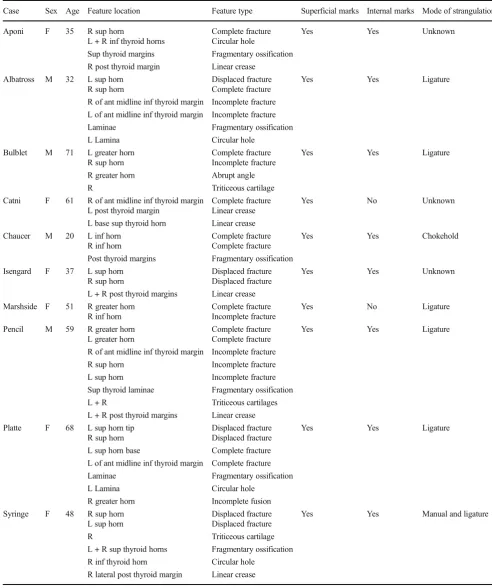
Table 1 Features and their location observed in the suspected strangulation cases along with the autopsy findings
Case Sex Age Feature location Feature type Superficial marks Internal marks Mode of strangulation
Aponi F 35 R sup horn Complete fracture Yes Yes Unknown
L + R inf thyroid horns Circular hole
Sup thyroid margins Fragmentary ossification
R post thyroid margin Linear crease
Albatross M 32 L sup horn Displaced fracture Yes Yes Ligature
R sup horn Complete fracture
R of ant midline inf thyroid margin Incomplete fracture
L of ant midline inf thyroid margin Incomplete fracture
Laminae Fragmentary ossification
L Lamina Circular hole
Bulblet M 71 L greater horn Complete fracture Yes Yes Ligature
R sup horn Incomplete fracture
R greater horn Abrupt angle
R Triticeous cartilage
Catni F 61 R of ant midline inf thyroid margin Complete fracture Yes No Unknown
L post thyroid margin Linear crease
L base sup thyroid horn Linear crease
Chaucer M 20 L inf horn Complete fracture Yes Yes Chokehold
R inf horn Complete fracture
Post thyroid margins Fragmentary ossification
Isengard F 37 L sup horn Displaced fracture Yes Yes Unknown
R sup horn Displaced fracture
L + R post thyroid margins Linear crease
Marshside F 51 R greater horn Complete fracture Yes No Ligature
R inf horn Incomplete fracture
Pencil M 59 R greater horn Complete fracture Yes Yes Ligature
L greater horn Complete fracture
R of ant midline inf thyroid margin Incomplete fracture
R sup horn Incomplete fracture
L sup horn Incomplete fracture
Sup thyroid laminae Fragmentary ossification
L + R Triticeous cartilages
L + R post thyroid margins Linear crease
Platte F 68 L sup horn tip Displaced fracture Yes Yes Ligature
R sup horn Displaced fracture
L sup horn base Complete fracture
L of ant midline inf thyroid margin Complete fracture
Laminae Fragmentary ossification
L Lamina Circular hole
R greater horn Incomplete fusion
Syringe F 48 R sup horn Displaced fracture Yes Yes Manual and ligature
L sup horn Displaced fracture
R Triticeous cartilage
L + R sup thyroid horns Fragmentary ossification
R inf thyroid horn Circular hole
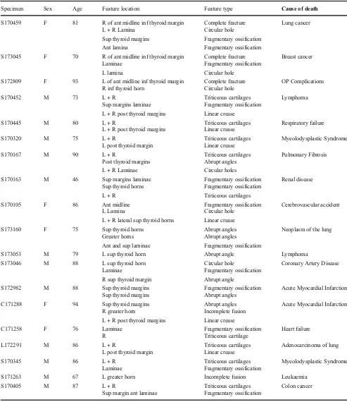
Table 2 Features and their location observed in the control group, along with demographic details and cause of death as provided by the tissue supplier
Specimen Sex Age Feature location Feature type Cause of death
S170459 F 81 R of ant midline inf thyroid margin Complete fracture Lung cancer
L + R Lamina Circular hole
Sup thyroid margins Fragmentary ossification
Ant lamina Fragmentary ossification
S173045 F 70 R of ant midline inf thyroid margin Complete fracture Breast cancer
Laminae Fragmentary ossification
L lamina Circular hole
S172809 F 93 L of ant midline inf thyroid margin Complete fracture OP Complications
R inf thyroid horn Circular hole
S170452 M 73 L + R Triticeous cartilages Lymphoma
Sup margins laminae Fragmentary ossification
L + R post thyroid margins Linear crease
S170445 M 80 L + R Triticeous cartilages Respiratory failure
L + R post thyroid margins Linear crease
S170320 M 75 L + R Triticeous cartilages Myeolodysplastic Syndrome
L post thyroid margin Linear crease
S170167 M 90 L + R Triticeous cartilages Pulmonary Fibrosis
Post thyroid margins Abrupt angles
L + R Laminae Circular holes
S170163 M 46 Sup margins laminae Fragmentary ossification Renal disease
Sup thyroid horns Fragmentary ossification
L + R Triticeous cartilages
S170105 F 86 Ant midline Fragmentary ossification Cerebrovascular accident
L Lamina Circular hole
L + R lateral sup thyroid horns Linear crease
S173160 F 75 Sup thyroid horns Abrupt angles Neoplasm of the lung
Greater horns Abrupt angles
Ant and sup laminae Fragmentary ossification
S173053 M 79 L sup thyroid horn Abrupt angle Lymphoma
S173046 M 88 L sup thyroid horn Circular hole Coronary Artery Disease
Laminae Fragmentary ossification
R sup thyroid margin Abrupt angle
S172982 M 88 Sup thyroid margins Fragmentary ossification Acute Myocardial Infarction
Sup thyroid margins Abrupt angles
C171288 F 94 Sup thyroid margins Abrupt angles Acute Myocardial Infarction
R greater horn Incomplete fusion
L + R post thyroid margins Linear crease
C171258 F 76 Laminae Fragmentary ossification Heart failure
R Triticeous cartilage
L172291 M 86 L + R Triticeous cartilages Adenocarcinoma of lung
L post thyroid margin Linear crease
S170345 M 86 L + R Triticeous cartilages Myeolodysplastic Syndrome
Laminae Fragmentary ossification
S171263 M 67 L greater horn Incomplete fusion Leukaemia
S170405 M 87 L + R Triticeous cartilages Colon cancer
Open AccessThis article is distributed under the terms of the Creative C o m m o n s A t t r i b u t i o n 4 . 0 I n t e r n a t i o n a l L i c e n s e ( h t t p : / / creativecommons.org/licenses/by/4.0/), which permits unrestricted use, distribution, and reproduction in any medium, provided you give appropriate credit to the original author(s) and the source, provide a link to the Creative Commons license, and indicate if changes were made.
References
1. Office for National Statistics (2018) Homicide in England and
Wales: year ending March 2017.https://www.ons.gov.uk/
peoplepopulationandcommunity/crimeandjustice/articles/ homicideinenglandandwales/yearendingmarch2017. Accessed 27 August 2018
2. Strack GB (2007) How to improve your investigation and
prosecu-tion of strangulaprosecu-tion deaths. Naprosecu-tional Family Justice Centre Alliance, San Diego
3. Dunsby AM, Davison AM (2011) Causes of laryngeal cartilage and
hyoid bone fractures found at postmortem.Med Sci Law.51(2):
109–113.https://doi.org/10.1258/msl.2010.010209
4. Davison AM, Williams EJ (2012) Microscopic evidence of
previ-ous trauma to the hyoid bone in a homicide involving pressure to
the neck.Forensic Sci Med Pathol. 8(3):307–311.https://doi.org/
10.1007/s12024-012-9316-3
5. Kempter M, Ross S, Spendlove D, Flach P, Preiss U, Thali M,
Bolliger S (2009) Post-mortem imaging of laryngohyoid fractures
in strangulation incidents: first results.Leg Med.11(6):267–271.
https://doi.org/10.1016/j.legalmed.2009.07.005
6. Becker M, Leuchter I, Platon A, Becker CD, Dulguerov P,
Varoquaux A (2014) Imaging of laryngeal trauma.Eur J Radiol.
83:142–152.https://doi.org/10.1016/j.ejrad.2013.10.021
7. Kettner M, Potente S, Schulz B, Knauff P, Schmidt PH, Ramsthaler
F (2014) Analysis of laryngeal fractures in decomposed bodies
using microfocus computed tomography (mfCT).Forensic Sci
Med Pathol.10(4):607–612. https://doi.org/10.1007/s12024-014-9584-1
8. Fais P, Giraudo C, Viero A, Miotto D, Bortolotti F, Tagliaro F,
Montisci M, Cecchetto G (2016) Micro computed tomography fea-tures of laryngeal fracfea-tures in a case of fatal manual strangulation.
Leg Med.18:85–89.https://doi.org/10.1016/j.legalmed.2016.01. 001
9. Thali MJ, Taubenreuther U, Karolczak M, Braun M, Brueschweiler
W, Kalender WA, Dirnhofer R (2003) Forensic microradiology: micro-computed tomography (Micro-CT) and analysis of patterned
injuries inside of bone.J Forensic Sci.48(6):1336–1342
10. Baier W, Norman DG, Warnett JM, Payne M, Harrison NP, Hunt
NC, Burnett BA, Williams MA (2017) Novel application of
three-dimensional technologies in a case of dismemberment.Forensic Sci
Int.270:139–145.https://doi.org/10.1016/j.forsciint.2016.11.040
11. Norman DG, Watson DG, Burnett BA, Fenne P, Williams MA
(2017) The cutting edge- micro-CT for quantitative toolmark
anal-ysis of sharp force trauma to bone.Forensic Sci Int.283:156–172.
https://doi.org/10.1016/j.forsciint.2017.12.039
12. Fais P, Giraudo C, Viero A, Amagliani A, Viel G, Montisci M,
Miotto D, Cecchetto G (2015) Identification of bullet entrance in different type of intermediate firearm wounds through
micro-computed tomography analysis.J Forensic Radiol Imaging.3(3):
147–152.https://doi.org/10.1016/j.jofri.2015.07.004
13. Cecchetto G, Giraudo C, Amagliani A, Viel G, Fais P, Cavarzeran
F, Feltrin G, Ferrara SD, Montisci M (2011) Estimation of the firing
distance through micro-CT analysis of gunshot wounds.Int J Leg
Med. 125(2):245–251
14. Richards CS, Simonsen TJ, Abel RL, Hall MJR, Schwyn DA,
Wicklein M (2012) Virtual forensic entomology: improving esti-mates of minimum post-mortem interval with 3D micro-computed
tomography.Forensic Sci Int.220(1):251–264.https://doi.org/10.
1016/j.forsciint.2012.03.012
15. Tsai A, McDonald AG, Rosenberg AE, Gupta R, Kleinman PK
(2014) High-resolution CT with histopathological correlates of the
classic metaphyseal lesion of infant abuse.Pediatr Radiol.44(2):
124–140.https://doi.org/10.1007/s00247-013-2813-z
16. Baier W, Mangham C, Warnett JM, Payne M, Painter M, Williams
MA (2019) Using histology to validate micro-CT findings of trau-ma in three post-mortem samples- First steps towards method
val-idation.Forensic Sci Int.297:27–34.https://doi.org/10.1016/j.
forsciint.2019.01.027
17. Brunke O, Santillan J and Suppes A (2010) Preecise 3D
dimension-al metrology using high-resolution x-ray computed tomography
(μCT). In: Stock SR (ed)Proceedings of the SPIE, Volume 7804.
[Online]. SPIE Digital Library.https://www.spie.org/Publications/
Proceedings/Volume/7804?&origin_id= x4323&start_yea r = 2010&end_year = 2010&start_at = 41&SSO = 1. Accessed 17 March 2006
18. Kruth J-P, Bartscher M, Carmignato S, Schmitt R, De Chiffre L,
Weckenmann A (2011) Computed tomography for dimensional
metrology.CIRP Annals-Manufacturing Technology.60(2):821–
842.https://doi.org/10.1016/j.cirp.2011.05.006
19. Lifton JJ, Malcolm AA, McBride JW, and Cross KJ (2013) The
application of voxel size correction in X-ray computed tomography
for dimensional metrology. 2nd Singapore International
Non-destructive Testing Conference and Exhibition, 19-20 July 2013, Singapore
20. Welkenhuyzen F, Kiekens K, Pierlet M, Dewulf W, Bleys P, Kruth
J-P, Voet A (2009) Industrial computer tomography for dimensional measurement: overview of influence factors and improvement strat-egies. In: Dirckx J, Buytaert J (eds) 4th International Conference on Optical Measurement Techniques for Structures and Systems.
Belgium, Shaker, pp 401–410
21. Dettmeyer RB, Verhoff MA, Schütz HF (2014) Forensic medicine:
fundamentals and perspectives. Springer, Heidelberg
22. Maxeiner H, Bockholdt B (2003) Homicidal and suicidal ligature
strangulation- a comparison of the post-mortem findings.Forensic
Sci Int.137(1):60–66. https://doi.org/10.1016/S0379-0738(03) 00279-2
23. Naik SK, Patil D (2005) Fracture of hyoid bone in cases of
asphyxial deaths resulting from constricting force round the neck.
J Indian Academy Forensic Med.27(3):149–153
24. Shaik MM, Chotaliya HJ, Modi AD, Parmar AP, Kalele SD (2013)
A study of gross postmortem findings in cases of hanging and
ligature strangulation.J Indian Academy Forensic Med.35(1):63–
65
25. Raikos A, Paraskevas GK (2013) The thyroid foramen: a systematic
review and surgical considerations.Clinical Anat.26(6):700–708.
https://doi.org/10.1002/ca.22234
26. Di Nunno N, Lombardo S, Costantinides F, Di Nunno C (2004)
Anomalies and alterations of the hyoid-larynx complex in forensic
radiographic studies.American J Forensic Med Pathol.25(1):14–
19.https://doi.org/10.1097/01.paf.0000113931.49721.e4
27. Naimo P, O’Donnell C, Bassed R, Briggs C (2013) The use of
computed tomography in determining developmental changes,
anomalies, and trauma of the thyroid cartilage.Forensic Sci Med
Pathol.9(3):377–385.https://doi.org/10.1007/s12024-013-9457-z
28. Maxeiner H (1999) Healed fractures of the larynx and lingual bone
in forensic autopsy.Archiv fur Kriminologie.203(5-6):175–183
29. Saternus K-S, Maxeiner H, Kernbach-Wighton G, Koebke J (2013)
Traumatology of the superior thyroid horns in suicidal hanging- An
injury analysis.Leg Med.15(3):134–139.https://doi.org/10.1016/j.
30. Dang-Tran KD, Dedouit F, Joffre F, Rougé D, Rousseau H, Telmon N (2010) Thyroid cartilage ossification and multislice computed
tomography examination: a useful tool for age assessment?J
Forensic Sci.55(3):677–683.https://doi.org/10.1111/j.1556-4029. 2010.01318.x
31. Regulator FS (2017) Code of practice and conduct. Forensic
Science Regulator, Birmingham
32. Cole SA (2012) Forensic science and wrongful convictions: from
exposer to contributor to corrector.New Engl Law Review.46(4):
711–736