REVIEW
Disturbed Yin
e
Yang balance: stress increases
the susceptibility to primary and recurrent
infections of herpes simplex virus type 1
Chang Yan
a,b, Zhuo Luo
a,b, Wen Li
a,b, Xue Li
c, Robert Dallmann
d,
Hiroshi Kurihara
a,b,*
, Yi-Fang Li
a,b,*
, Rong-Rong He
a,b,*
a
Guangdong Engineering Research Center of Chinese Medicine & Disease Susceptibility, Jinan University, Guangzhou 510632, China
b
International Cooperative Laboratory of Traditional Chinese Medicine Modernization and Innovative Drug Development of Chinese Ministry of Education (MOE), School of Pharmacy, Jinan University, Guangzhou 510632, China
c
Institute of Atmospheric Environmental Safety and Pollution Control, Jinan University, Guangzhou 510632, China d
Warwick Medical School, University of Warwick, Coventry CV4 7AL, United Kingdom
Received 24 February 2019; received in revised form 27 May 2019; accepted 31 May 2019
Abbreviations:4E-BP, eIF4E-binding protein; AD, Alzheimer’s disease; AKT, protein kinase B; AMPK, AMP-dependent kinase; BCL-2, B-cell lym-phoma 2; cGAS, cyclic GMP-AMP synthase; CNS, central nervous system; CoREST, REST corepressor 1; CORT, corticosterone; CPE, cytopathic effect; CTCF, CCCTC-binding factor; CTL, cytotoxic T lymphocyte; DAMPs, damage-associated molecular patterns; DCs, dendritic cells; DEX, dexamethasone; GREs, GR response elements; GRs, glucocorticoid receptors; H3K9, histone H3 on lysines 9; HCF-1, host cell factor 1; HDACs, histone deacetylases; HPA axis, hypothalamoepituitaryeadrenal axis; HPK, herpetic simplex keratitis; HPT axis, hypothalamicepituitaryethyroid axis; HSV-1, herpes simplex virus
type 1; ICP, infected cell polypeptide; IRF3, interferon regulatory factor 3; KLF15, Kru¨ppel-like transcription factor 15; LAT, latency-associated tran-scripts; LRF, Luman/CREB3 recruitment factor; LSD1, lysine-specific demethylase 1; MAVS, mitochondrial antiviral-signaling protein; MOI, multiplicity of infection; mTOR, mammalian target of rapamycin; ND10, nuclear domains 10; NGF, nerve growth factor; NK cells, natural killer cells; OCT-1, octamer binding protein 1; ORFs, open reading frames; PAMPs, pathogen-associated molecular patterns; PDK1, pyruvate dehydrogenase lipoamide kinase isozyme 1; PI3K, phosphoinositide 3-kinases; PML, promyelocytic leukemia protein; PNS, peripheral nervous system; PRC1, protein regulator of cytokinesis 1; PRRs, pattern-recognition receptors; PTMs, post-translational modifications; RANKL, receptor activator of NF-kB ligands; REST, RE1-silencing tran-scription factor; ROS, reactive oxygen species; SGKs, serum and glucocorticoid-regulated protein kinases; SIRT1, sirtuin 1; sncRNAs, small non-coding RNAs; T3, thyroid hormone; TCM, traditional Chinese medicine; TG, trigeminal ganglia; TK, thymidine kinase; TRIM14, tripartite motif-containing 14;
TRKA, tropomyosin receptor kinase A; TRM, tissue resident memory T cells.
*Corresponding authors. Fax:þ86 20 85221559.
E-mail addresses:hiroshi_kurihara@163.com(H. Kurihara),liyifang706@jnu.edu.cn(Y.-F. Li),rongronghe@jnu.edu.cn(R.-R. He).
Peer review under responsibility of Institute of Materia Medica, Chinese Academy of Medical Sciences and Chinese Pharmaceutical Association.
https://doi.org/10.1016/j.apsb.2019.06.005
2211-3835ª2019 Chinese Pharmaceutical Association and Institute of Materia Medica, Chinese Academy of Medical Sciences. Production and hosting by Elsevier B.V. This is an open access article under the CC BY-NC-ND license (http://creativecommons.org/licenses/by-nc-nd/4.0/).
Chinese Pharmaceutical Association
Institute of Materia Medica, Chinese Academy of Medical Sciences
Acta Pharmaceutica Sinica B
KEY WORDS
Herpes simplex virus type 1;
HSV-1; Susceptibility; Latency; Reactivation; Stress
Abstract Herpes simplex virus type 1 (HSV-1), a neurotropic herpes virus, is able to establish a life-long latent infection in the human host. Following primary replication in mucosal epithelial cells, the vi-rus can enter sensory neurons innervating peripheral tissuesvianerve termini. The viral genome is then transported to the nucleus where it can be maintained without producing infectious progeny, and thus la-tency is established in the cell. YineYang balance is an essential concept in traditional Chinese medicine (TCM) theory. Yin represents stable and inhibitory factors, and Yang represents the active and aggressive factors. When the organism is exposed to stress, especially psychological stress caused by emotional stimulation, the YineYang balance is disturbed and the virus can engage in productive replication,
re-sulting in recurrent diseases. Therefore, a better understanding of the stress-induced susceptibility to HSV-1 primary infection and reactivation is needed and will provide helpful insights into the effective control and treatment of HSV-1. Here we reviewed the recent advances in the studies of HSV-1 suscep-tibility, latency and reactivation. We included mechanisms involved in primary infection and the regula-tion of latency and described how stress-induced changes increase the susceptibility to primary and recurrent infections.
ª2019 Chinese Pharmaceutical Association and Institute of Materia Medica, Chinese Academy of Medical Sciences. Production and hosting by Elsevier B.V. This is an open access article under the CC BY-NC-ND li-cense (http://creativecommons.org/lili-censes/by-nc-nd/4.0/).
1. Introduction
Herpes simplex virus type 1 (HSV-1), a ubiquitous human path-ogen, is a neurotropic virus with a linear double-stranded DNA genome that contains more than 80 open reading frames (ORFs) and is about 152 kilo base-pairs (kbp) long. Upon primary infection, the virus replicates within epithelial cells and undergoes its typical lytic life cycle with a cascade of immediate early (IE), early (E), and late (L) genes. Then, it enters axon terminals of sensory neurons and is retrogradely transported to the corre-sponding sensory ganglia, usually trigeminal ganglia (TG), where a latent infection can be established. In response to a variety of stressors, the latent virus can be reactivated periodically to resume productive replication followed by the formation of infectious virus, which is anterogradely transported back to peripheral tis-sues or infects further neuronal cells to remain in host1,2. During
anterograde transport, both enveloped capsids and non-enveloped capsids are detected3. Primary infection, latency and reactivation
complete the life cycle of the overall HSV-1 infection (Fig. 1). It is also noteworthy that HSV-1 gene expression during latency and reactivation might differ between rodent and mammal models4.
HSV-1 is commonly acquired during early childhood, pri-marily through oral secretions; while sexual transmission is an increasingly common cause of infection in some countries5. Worldwide, the global prevalence of HSV-1 is approximately 90%, and in some rural areas, the seroprevalence is even higher, up to 100%6e11. The success of HSV-1 infection can be attributed to its ability to establish lifelong persistent infection of the host, termed latency. In latent state, HSV-1 maintains the episomal viral genome in neuronal nuclei without producing infectious progeny for long period of time. Eventually, the virus can re-enter the lytic replication program for productive replication, a process known as reactivation. Reactivation ensures long-term persistence and dissemination of the virus to further host cells or new hosts. Furthermore, the lifespan of the latent infected cell is extended, and thus the virus is able to escape immune surveillance.
HSV-1 has raised concerns because it can cause many diseases of various severity. Acute HSV-1 infection can cause herpes labialis (cold sores)12, gingivostomatitis13 and eczema
herpeticum14. It should be noted that, currently, there is no treatment to completely remove HSV-1 once an individual is infected. Reactivation of latently infected HSV-1 can cause recrudescent lesions and is the main reason for herpes viral encephalitis15e17and herpetic keratitis18. Recurrent ocular infec-tion can lead to irreversible corneal scarring and blindness19. Increasing number of studies20,21 have confirmed HSV-1 as pathogen directly related to nervous system degenerative diseases like Alzheimer’s disease (AD). Reactivation of HSV-1 will in-crease the risk of developing AD22. Importantly, an amplified concentration of HSV-1 antibody and the use of antivirals can antagonize the nerve damage of AD, which has also proven that HSV-1 in latent status leads to long term damage to central ner-vous system23,24. It might likewise be the cause of Meniere’s disease, an inner ear disease with spinning sensation, loss of hearing, and pressured feelings in the ear25. Hence, all these
findings have emphasized the importance on the study of HSV-1 latent infection.
As a rapidly developing systematical medical science, tradi-tional Chinese medicine (TCM) is a systematical medicinal science with an application history of over 2000 years in China, widely spread in Asia. At present, a broad range of research in the field of TCM is proceeding. As a treasured resource accumulated by the continuous practice of Chinese people, it has inspired many new discoveries in drug and therapeutic developments26e29. One of the basic theories in the TCM field is the theory of YineYang balance, which is also applied as a philosophical term. Therein, Yin repre-sents the repressive and inhibitory factors, while Yang stands for the active and aggressive factors. The confrontation, homeostasis, and transformation between Yin and Yang compose the YineYang balance. In different contexts and situations, the components of Yin and Yang refer to different elements. For instance, in “Shang-Huo” syndrome, the hyperactivity of Yang, in this case is ““Shang-Huo” (fire), causes increased neuroendocrine activation, hence breaking the YineYang balance30,31. In TCM theory, the disturbance of YineYang balance is an essential cause of diseases.
YineYang balance, stress can increase the susceptibility to in-fectious agents, influence the severity of inin-fectious disease and reactivate latent herpesviruses by significantly modulating the central nervous (CNS), endocrine and immune systems. TCM theory suggests that internal injury caused by excess of seven emotions, named “Qi-Qing Nei-Shang”, also known as emotional stress in modern medical science, disturbs YineYang balance, “Qi-Xue” and viscera balance, inducing the stagnation of Qi32. Chronically, the stagnation transforms into “Shang-Huo” syn-drome, where Yang dominates Yin31. One of the typical symptoms of “Shang-Huo” is heat sore on the face, which is related to the reactivation of latent HSV-1 infection by modern medicine. Here we review the latest insights into the mechanism of stress-induced susceptibility to HSV-1 and its reactivation from latency in order to shed light to future researches on HSV-1 latency and the possible solutions to the effective control of latent HSV-1 infec-tion reactivainfec-tion.
2. Stress increases the susceptibility of HSV-1 infection by disturbing host inner YineYang balance
It has been widely accepted that stress during HSV-1 infections can influence the susceptibility, infection severity and infection types33e36. The host defense against HSV-1 consists of three main parts: innate immunity, adaptive immunity and intrinsic
immunity37. Therein, innate and adaptive immunity are the main defense strategies for most mitotic cells and have been investi-gated more thoroughly38. Under normal condition, the host innate and adaptive immunities perform in a cooperative and mutually restrictive YineYang balance, ensuring adequate level of defense against pathogens. However, when the balance is disturbed by stimulations, the immunity balance begins to wander, which consequently leads to immune compromise.
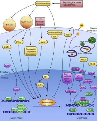
[image:3.595.105.483.60.366.2]and adaptive immune response towards HSV-1 including NK cell activity, HSV-specific CD8þT cell number and activity, immunity related cytokine level and lymphocyte infiltration35. Restraint stress prolongs the length of HSV-1 infection and increases the number of infected neurons, resulting in longer viral progeny and more severe recurrent lesions47. Based on the unpublished data we obtained, using corticosterone (CORT) stress model in normal and glucocorticoid receptor (GR) knocked-down SH-SY5Y cells, as well as restraint-stressed mice models, we have confirmed that stress hormone CORT can enhance HSV-1 susceptibility, and that such increase is largely dependent on GR. Our results further demonstrated that the GR-dependent effect of CORT on HSV-1 susceptibility is related to the inhibition of interferon regulatory factor 3 (IRF3) phosphorylation and the decrease of IFN-b, indicating an inhibitory effect of GR on innate immunity (Fig. 2). Interestingly, it has been found that, different from chronic psy-chological stress and restraint stress, social disruption stress can enhance the innate immune responsed to a primary HSV-1 infection in both cornea and TG of mice by increased
expression of anti-viral cytokines and infiltration of macrophages, ultimately reducing the severity and frequency of future re-currences48. Under HSV-1 infection, tripartite motif-containing 14 (TRIM14) is likely to cleave the ubiquitin chain of cyclic GMP-AMP synthase (cGAS) and prevent it from being degraded through autophagy, ultimately enhancing IFN signaling, thus improving immune responses49.
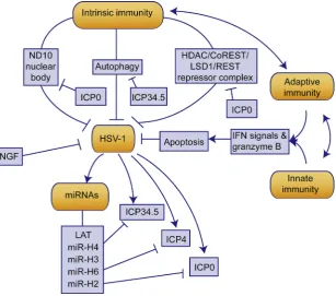
[image:4.595.142.462.297.620.2]Besides innate and adaptive immunity, another currently under researched immunity, intrinsic immunity is closely related to the defense against HSV-1 invasion. To some extent, intrinsic im-munity might play a more important role in neurons than in other cells50. Evidence also shown that neuronal cells are less respon-sive to IFN signaling than other types of cells51. As far as we know, there are three components for host intrinsic defense against HSV-1: autophagy, HSV-1 repressive complex and nuclear do-mains 10 (ND10) nuclear bodies50. As the virus enter the neuron, virions are degraded through xenophagy activated by pathogen-associated molecular patterns (PAMPs), damage-pathogen-associated mo-lecular patterns (DAMPs) and pattern-recognition receptors
(PRRs). During virus replication, viral DNA and proteins are recognized and degraded through autophagy51,52. Autophagy may also be induced by IFN-b during HSV-1 replication53. The increased cGAS mentioned earlier is reported to interact with beclin-1, contributing to the autophagy of viral DNA54. HSV-1 has evolved a confrontational mechanism to counter host autophagic defense through a viral protein, infected cell polypeptide 34.5 (ICP34.5)55e60. It is well understood that autophagy is enhanced under stress61,62. On the one hand, enhanced autophagy may improve the intrinsic defense against HSV-157; on the other hand, however, the increased autophagy may also prolong host cell survival and provide a more advantageous environment for HSV-1 replication63. Besides, whether stress-induced autophagy has the same virus clearance effect as xenophagy, a selective autophagy, remains unknown. Moreover, stress-induced autophagy upregu-lation might increase the degradation of cGAS, casing a loss of IFN signaling49. Therefore, the exact fate of HSV-1 susceptibility under stress-induced autophagy enhancement requires further investigation. The conflict between the facts that stress increases HSV-1 susceptibility and that stress enhances autophagy suggests more complicated mechanisms for stress-induced susceptibility. One possible explanation is that stress-induced autophagy in-creases the degradation of intrinsic defense components, such as promyelocytic leukemia protein (PML) in ND10 nuclear bodies, and hence defecting the intrinsic immune response, which is especially essential for the defense against HSV-1 infection64. Therefore, the stress-induced autophagy of intrinsic immune components may be a possible research direction in the future.
3. The YineYang balance between HSV-1 and host cell defense: the establishment and maintenance of latency
HSV-1 is characterized by establishing latency as a non-integrated, nucleosome-associated episome in neuronal nuclei. In the process of latency establishment, new sets of Yin factors and Yang factors counteract, transform, and ultimately reach a new YineYang balance between the virus and the host. When the new homeostatic YineYang balance is created, the virus enters its latent state in which it resides relatively silently in the nucleus of the infected cells without producing infectious viral progeny. It is hypothesized that neuronal latency is the result of a failure to initiate the lytic cascade, which might be determined by the distinctive architecture of neurons. Therefore, here we introduce the molecular process of normal HSV-1 lytic infection process and illustrate how latency is established. The initiation of IE genes, specifically, ICP0, is the essential trigger for the following expression of E and L genes, indispensable for HSV-1 lytic infection65. Therefore, triggering IE gene expression would be crucial for entering lytic infection. With the cooperation of lysine-specific demethylase 1 (LSD1), the viral protein VP16 recruit octamer binding protein 1 (OCT1) and host cell factor 1 (HCF1) in order to form enough OCT-1/HCF-1/VP16 triplets. The triplets then bind to IE gene promoters and activate IE gene transcrip-tion66. ICP0 can then replace the histone deacetylases (HDACs) in the HDAC/RE1-silencing transcription factor (REST)/REST corepressor 1 (CoREST)/LSD1 repressor complex, after which the previously suppressed E and L gene expression can be triggered67. Only with enough VP16 entering the nucleus can HSV-1 suc-cessfully enter its lytic phase. However, specific characteristics of neurons make it especially suitable for HSV-1 to establish latency. The other two ingredients in the triplet tend to distribute differ-ently in neurons, compared with other cells. HCF1 is more
abundant in the cytoplasm than in the nucleus in unstressed neu-rons, while at the same time, OCT-1 is downregulated in neurons; both lead to less VP16 in the nucleus68. The HSV-1 genome is also closely associated with ND10 nuclear bodies, which consists of main components like PML, death-domain associated protein (Daxx) and SP100 nuclear antigen69. Under the stimulation of IFNs, they are able to bind closely to HSV-1 genome and inhibit its lytic replication70. However, such defense is also countered by viral protein ICP0, which possesses the activity of E3 ubiquitin ligase64.
In fact, the number of viral particles that infect axons is another factor to determine whether the virus enter latency or not. The viral genome was silenced below a threshold multiplicity of infection (MOI) of 0.1; while high MOI infection resulted in productive infections71. To sum up, the special characteristics of neurons which are able to prevent the initiation of the viral lytic cascade and reduce the number of virions from the axons are the two important factors for the establishment of neuronal latency.
In contrast, immune surveillance seems to be dispensable for the establishment of viral latency, though the system is also crit-ical for the early control of viral distribution as well as elimination of the replication of virus. Mice that lack the innate and adaptive immune system are still able to establish HSV-1 latency in TG72. Furthermore, HSV-1 latency can be established in the absence of neuronal IFN signaling73. Evidence in rabbit model showed that latency-associated transcripts (LATs) are able to enhance latency establishment74, which indicates that LAT participate in the establishment of the latent state. However, since latency is still able to establish in the absence of it, and that it also accumulates in productively infected cells post-infection, LAT may not be crucial for latency establishment75.
Whether HSV-1 can establish non-neuronal latency is unclear. However, there are several publications on cornea latent infection, suggesting that the cornea might be a possible site of latency76. There were evidences indicating the potential of HSV-1 latent infection in the human cornea early in the 90s77,78. Further studies have shown that HSV-1 DNA can be present in human corneas even though ocular herpetic disease has not been found in the corneas from animal models including mice79e82and nonhuman primates83. These results are consistent with the findings in rab-bits,i.e., the retrograde migration of HSV-1 DNA occurs from the transplanted corneas of rabbits latently infected with LAT positive HSV-1 to the uninfected rabbits following transcorneal epineph-rine iontophoresis75. In addition, LAT was found in the human cornea80, but no other transcriptional products (RNA) or expres-sion products (protein) were observed. Given the lack of such observations, HSV-1 latency in the cornea is still disputed,i.e., if the virus is truly latent in the cornea or only at a transition point along the exit path from a sensory ganglion. So far, there are three hypotheses for the presence of this DNA in human corneas: (i) operational latency; (ii) a low persistent infection in the cornea and (iii) reactivation from neuronal sites. Hence, operational la-tency in the cornea is very likely to be dependent on novel detection methods, which is more sensitive than present virus detection methods84.
frequently used antiviral treatments for HSV-1 are DNA poly-merase inhibitors including acyclovir, famciclovir, and valaciclo-vir85,86. Attenuated mutations of HSV-1 lower the virulence by deletion ofICP087, deletion ofICP34.588, expression ofICP34.5 complementary miRNAs89, etc. They both act as interventions of the Yang, which attenuate the Yang factors in the YineYang balance and facilitate the maintenance of latency. When the or-ganism experiences stress stimulation, the balance will be inter-rupted, ultimately leading to reactivation.
During latency, viral gene expression is largely suppressed except for the abundant expression of LATs and other non-coding RNAs. LAT and its associated miRNA species have been found to influence viral maintenance by suppressing HSV-1 gene expres-sionin vivoandin vitro90e92. In murine ganglia latently infected by LAT-mutant HSV-1, both the neurovirulence and reactivation frequency are significantly increased93 and the latency is
impaired94, providing further clues for the role of LAT in latency maintenance.
In addition to repressing virus-encoded gene expression, LAT can promote neuronal cell survival by inhibiting apoptosis95, which may contribute to maintaining the latency and increasing the survival of the virus in the host. LAT’s anti-apoptotic function is mediated through multiple ways. On one hand, cells are pro-tected against cold-shock-induced apoptosis mainly by preventing the dephosphorylation of protein kinase B (AKT), a serine/thre-onine protein kinase promoting cell survival96. On the other hand, LAT expressing plasmids are able to inhibit the two major apoptosis pathways in mammals induced by caspase-8, caspase-9,
and caspase-397e99. LAT can prevent infected neurons from being killed by CD8þT cells through anti-apoptosis activity98.
A set of miRNAs derived from LAT are able to target viral transcripts and inhibit HSV-1 gene expression100. HSV-1-miR-H6 can target ICP4 mRNA, inhibiting the expression of the tran-scription factor crucial to E and L gene trantran-scription, blocking virus lytic infection and decreasing IL-6 expression, hence pro-moting HSV-1 latency101,102. HSV-1-miR-H2, which has been associated with the regulation of latency, binds to viral ICP0 mRNA and inhibits its expression. ICP0 plays a major role in both primary and recurrent HSV-1 infections. Its expression triggers the entry of HSV-1 into the replication cycle93,103. HSV1-miR-H4 inhibits the expression of the viral ICP34.5 gene, an important lytic neurovirulence factor indispensable for promoting viral spread from TG cells to non-neuronal cells89,104,105. Two small non-coding RNAs (sncRNAs) derived from LAT also contribute to the decrease of lytic gene expression and apoptosis inhibition91,106.
[image:6.595.152.460.61.332.2]Another strategy employed by HSV-1 is to activate cellular glucose synthesis and glycolysis, in order to increase available energy for virus survival107. The activation of AMP-dependent kinase (AMPK) and sirtuin 1 (SIRT1) pathways is effective in inhibiting productive infection and protecting cells from virus related damage108. The modulation of AMPK/SIRT1 axis is modulated over time to suit the HSV-1 infection process109. An over-expression of Luman/CREB3 recruitment factor (LRF), acts as a repressor in a direct or indirect manner to inhibit essential genes of HSV-1 lytic infection110.
Furthermore, different cytokines have different abilities to support latency and suppress lytic HSV-1 replication, providing a fundamental-level control based on neuronevirus interaction111. IFN-b can achieve control of the infection by enhancing the restriction of HSV-1 replication in neuronal cells104. IFN-b regulates LAT expression, which has a positive effect on neuron survival73. Nerve growth factor (NGF)-responsive receptors and signal transduction pathways are necessary to maintain latency and prevent reactivation. This is consistent with the ability of HSV-1 to establish latency in primary sympathetic neurons and Nd-PC12 cells cultured in the presence of NGF112,113. NGF ablation can induce HSV-1 reactivation in sensory and sympa-thetic neurons in vitro or after anti-NGF treatment in vivo114. Moreover, neurons infected with latent HSV-1 experience reac-tivation in the presence of antibodies to NGF115. The ability of NGF to maintain latency has also been proven when herpetic keratitis was topically treated with NGF116. It turns out that the activation of pyruvate dehydrogenase lipoamide kinase isozyme 1 (PDK1) caused by the reaction of NGF with tropomyosin re-ceptor kinase A (TRKA) is the dominant pathway of NGF to suppress reactivation. Suppression of phosphoinositide 3-kinases (PI3Ks) induces HSV-1 reactivation and activation of AKT, the key component for PI3K pathway, which is particularly critical for the maintenance of latency111,117. This effect of NGF is closely related to mammalian target of rapamycin (mTOR), an important factor in the PI3K/AKT pathway, which controls the population of mRNAs that are actively translated into proteins. These proteins suppress the lytic cycle by sustaining the repressive chromatin state of the viral genome through activating eIF4E-binding protein (4E-BP)118.
During latency, the viral genome associates with nucleosomes by chromatin remodeling. Histone post-translational modifica-tions (PTMs) representative of euchromatin and heterochromatin are found on HSV-1 genes during latency. As a result, the regulation of latent gene expression exists at the level of epige-netic modification. Two activating euchromatin-like modifica-tions that commonly define areas of euchromatin are acetylation of histone H3 on lysines 9 and 14 (H3K9, K14) and dimethyla-tion of histone H3 on lysine 4 (H3K4). Indeed, during lytic infection, acetylation of H3K9 and H3K14 are enriched upon lytic gene promoters; while repressive heterochromatin-like modifications are under-represented119. In contrast, during la-tency, the actively transcribed LAT locus become enriched in acetylated H3K9 and H3K14 at the LAT promoter and enhancer, while these modified histones are absent at theICP0promoter or DNA polymerase gene120. Meanwhile, viral lytic genes are
enriched with repressive heterochromatin-like modifications such as methylated H3K27 and methylated H3K9. These observations correspond with latency feature that the LAT is abundant whilst lytic genes are silent. Furthermore, while HDAC inhibitors induce reactivation in latently infected mice121, inhibitors of LSD1 activities that can specifically block demethylation of the repressive markers, such as H3K9me3, H3K9me2 and H3K27me3, will enhance the epigenetic suppression of the viral genome and reduce the reactivation in cultured neurons122. In
order to prevent the spread of heterochromatin into areas of euchromatin, there must be barriers in place to keep these do-mains separate. CCCTC-binding factor (CTCF), a transcriptional repressor, can bind to the sites in HSV-1 genome as boundary elements123. Thus, the transcriptionally active LAT promoter regions will be segregated from the repressed regions of the genome and the LAT enhancer will be prevented from acting on
the surrounding lytic genes124. These findings have suggested that epigenetic regulation may control the switch between la-tency and reactivation.
Systemically, HSV-1 latent infection is surveyed by the im-mune system through the cooperation of tissue cells and imim-mune cells including CD4þand CD8þ T lymphocytes. These HSV-1 specific T cells belong to tissue resident memory T (TRM) cells. They have a longer lifespan than normal T cells, and establish a long-term residence in TG125. During latency, the lytic genome of HSV-1 is not completely silenced. Instead, limited lytic gene expression is frequently recognized by MHC class I molecules expressing cells, CD4þand CD8þT cells126. For example, local expression of viral proteins such as ICP6 and VP16 is recognized by TG-resident CD4þ and CD8þ T cells127. These facts have
indicated that HSV-1 latency maintenance involves active identi-fication and response for viral gene by host immune system. In TG, both HSV-1 specific and non-specific CD8þT cells exist and cooperate with each other, contributing to the control of la-tency128. When HSV-1 lytic gene expresses at a relatively low level, non-specific CD8þ T cells can inhibit the reactivation through inhibiting ICP0 by IFN-g. A novel autophagic response has been discovered, which is related to the IFN signaling in TG53. For neurons that are refractory to IFN-g, the HSV-1 specific CD8þ T cells can excrete granzyme B to degrade ICP4, and then block viral gene expression129.
The research on HSV-1-specific CD8þT cells is particularly comprehensive. These cells have been shown important for la-tency maintenance130, and the reactivation can be reduced by broadening the repertoire of HSV-1 specific T cell that is resident in TG131. The receptor activator of NF-kB ligands (RANKL) has
the control over the induction of TG-resident CD8þT cells. The activation of RANKL also prevents cell apoptosis132. The bal-ance between the survey of TG-resident HSV-1 specific CD8þT cells and their exhaustion monitored by HSV-1 LAT gene is important for the maintenance of latent status133. Though CD4þ T cells do not have direct effect on latency maintenance, they can perform an indirect function to prevent partial CD8þ T cell exhaustion134. Recently, it has been discovered that CD8aþ dendritic cells (DCs) play a more crucial role in latency main-tenance than CD8þ T cells135. Moreover, CD8aþ DCs are influential in T cell exhaustion, which contributes to latency maintenance136.
4. Stress disrupts the YineYang balance and causes reactivation
oxidational damage, hence inducing the reactivation. As we pre-viously described in our publications, we have developed a suc-cessful disease susceptibility model, restraint-stressed mice model, to simulate the effects of stress on virus infection39e46,138,139. Hence, restraint-stress mice model can be a feasible model for stress-induced HSV-1 reactivation, as illus-trated inFig. 6.
4.1. Stress induces hormonal changes
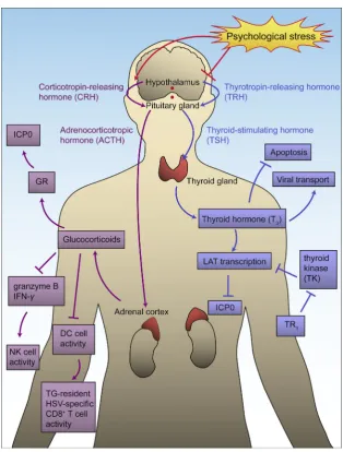
It is well established that stress-induced changes of hormones such as glucocorticoids and thyroid hormone can stimulate HSV-1140,141and drive reactivation from latency (Fig. 7). Stress, e.g., psychological stress and hyperthermia, can activate the hypothalamoepituitaryeadrenal (HPA) axis142and increase the concentration of glucocorticoids in the blood143. Glucocorticoids induce HSV-1 reactivation mainly through two distinct pro-cesses: directly affecting HSV-1 virus and indirectly promoting reactivation. Glucocorticoids activate GRs and directly activate HSV-1 transcription by interacting with the GR response ele-ments (GREs) on virus genome. A recent study showed that when cells are treated with synthetic corticosteroid dexamethasone (DEX), GR and Kru¨ppel-like transcription factor 15 (KLF15) cooperate to stimulate reactivation through the transactivation of ICP0144. Serum and glucocorticoid-regulated protein kinases (SGKs) has been reported to increase after stress stimulation and participate in HSV-1 reactivation from latency145. The increased glucocorticoid level also affects both intrinsic and adaptive im-munities. TG-resident HSV-1-specific CD8þT cells are reduced and those survived CD8þ T cells compromise functionally, which induces HSV-1 reactivation for compromising CD8þ T cell surveillance. The impairment of CD8þT cells may occurvia affecting GRs on DCs that are responsible for priming these HSV-1 specific T cells36,146. Interestingly, however, psycholog-ical stress might not diminish CD8þT cellsviaimpairing DCs. Instead, it could be due to the decrease of T cell stimulative cytokines such as IFN-g and IL-2149. Further effects of gluco-corticoid upregulation by stress are significantly reduced con-centrations of granzyme B and IFN-g142,146,147. Also, stress has
been proven to cause mitochondrial damage and to decrease MAVS expression45. It can decrease NK cell activity in
individuals that are latently infected with HSV-1, which might be due to the reduction of IFN-gand IL-2148.
Thyroid hormone (T3) is able to cause strong suppression of HSV-1 replication149. T3 can activate LAT and consequently repress ICP0 expression150. It also has repressive effect on the HSV-1 thymidine kinase (TK) gene, which is important for viral reactivation149. Psychological stress inhibits the hypothal-amicepituitaryethyroid (HPT) axis and leads to the reduction in T3secretion. Hence, with the decrease of T3and the reduction in its suppression effects, there is an increase of ICP0 expression. It has been shown that the overexpression of thyroid receptorb1 can enhance LAT transcription and recruit H3K9me3 and H3K9me2 to repress theTKgene, leading to an increased virus suppression efficiency. In addition, TG neurons overexpressed with thyroid receptorb1 were less susceptive to the reactivation induced by T3 decrease151. However, it should be noted that the suppression ef-fect of T3only works on neuronal cells, and differential conden-sation of chromosome may be important in this process152. T3also regulates the expression of dynein and modify neuronal outgrowth, suggesting the specific role T3plays in viral transport and anti-apoptosis153.
4.2. Stress reverses chromosomal modification
Under stress, the chromosomal modifications on HSV-1 lytic DNA might be reversed and viral DNA expression might be modified, which lead to induction of lytic replication107. This hypothesis has been supported by a number of studies, which have found that chromatin remodeling around the LAT region and surrounding lytic genes is likely to occurs after a stress stimulus154,155. Loss of CTCF proteins from chromosomal in-sulators through stress-induced phosphorylation increased the accessibility of viral genes for transcriptional activation. Replacement of demethylation of H3K27 on lytic genes with euchromatin that is triggered by displacing protein regulator of cytokinesis 1 (PRC1) complexes has been reported to have the similar function124. As a result, the lytic genes would produc-tively express and eventually reactivate.
[image:8.595.95.491.63.199.2]In fact, a number of transient chromatin modification have been discussed. It has been suggested that histone H3 at serine 10 undergoes a methyl/phospho switch during the first phase of Figure 4 Stress disturbs the YineYang balance between HSV-1 stimulating and inhibiting factors. During HSV-1 latent infection, the HSV-1 stimulating and inhibiting factors form a delicate YineYang balance. Yin factors include virus inhibiting factors such as thyroid hormone,
reactivation, and activation of lytic genes is achieved without the removal of H3K9me3156. It is then followed by VP16 synthesis in the second phase157. When the VP16 promotor is activated, in the absence of other lytic viral gene expressions, the expression of VP16 leads to the exit of latency and the entry of lytic cycle158.
[image:9.595.130.462.80.490.2]Therefore, modification of VP16 is required for reactivation of HSV-1 in neuronal cells159. Administration of an inhibitor for helicase-primase is able to suppress such reactivation, which suggests the chromatin modification mentioned above to be essential160.
4.3. Stress causes oxidational damage and induces apoptosis
Under UV or physical trauma, dendrite mitochondria produce reactive oxygen species (ROS) to inhibit mTOR activity and decrease the expression of B-cell lymphoma 2 (BCL-2), inducing apoptosis161. In order to escape from the soon to be apoptotic cells, HSV-1 spreads among host cells in an attempt to infect new individuals. All viral gene classes will be expressed at the same time and enter full reactivation, followed by the infection of pe-ripheral epithelial and neuronal cells to remain the survival and
spread of HSV-1162. HSV-1 itself may also be a stimulant to apoptosis. HSV-1 protein ICP27 can increase host cell suscepti-bility to apoptosis, which is also probably through the production of higher level of ROS163.
5. Discussion and perspective
[image:10.595.149.458.56.542.2]factors. Stress, especially psychological stress caused by emotional stimulation, is able to increase the susceptibility and severity of infectious diseases and to cause the reactivation from latency, leading to disturbance on life quality. Therefore, reducing the effect of stress on diseases is becoming an urgent and widely concerned topic. Emotional stress causes internal damage called “Qi-Qing Nei-Shang” and consequently disturbs the YineYang balance in the organism, leading to increased susceptibility to HSV-1 and the recurrent lesions. Nevertheless, how emotional stimulation affects the biological pattern and the nature of HSV-1 reactivation susceptibility still needs further systematic study. Moreover, considering that once an individual is infected, no
treatment exists to remove the HSV-1 virus completely from the host, an approach to control latent infection and reduce reac-tivation becomes the crux of the issue.
[image:11.595.136.451.55.470.2]by the immune system. The virus can also deplete T cells to reduce the survey intensity. The YineYang balance between the virus-stimulating and virus-inhibiting factors maintains the la-tency. Under stress, oxidational damage, increased glucocorticoids and decreased thyroid hormones promote the Yang factors and inhibit the Yin factors, consequently disturbing the YineYang balance, leading to productive viral replication.
However, several obstacles remain in the way towards more specific, accurate and coherent understanding of HSV-1 latency and reactivation. Current major animal and cellular models are not sufficient to unravel the mechanistic details. Models of closer genetic similarity with human,e.g.,Rhesus macaques, and newly developed models like tree shrews may be more adequate models to study HSV-1 latency and reactivation4,164. In addition, we still
know very little about the molecular mechanism of other stress hormones such as epinephrine, growth hormone and prolac-tin33,165. More investigations on other stress hormones are required for a better overall understanding on stress and HSV-1 reactivation. Additionally, according to a research by Edgar et al166, herpes virus infection is enhanced under circadian clock disruption stress, suggesting a new possible direction on how stress influences the susceptibility of HSV-1 infection. According to our current understanding on the role of stress in HSV-1 infection and the instruction of TCM theory, many small mole-cules with potential anti-HSV-1 activity have been discovered from TCMs. We have recently published a review specifically focusing on the anti-HSV-1 small molecules originated from different TCMs, and their pharmacodynamics mechanisms167. Therein, three main mechanisms were discussed, including auto-phagy regulation, immunity enhancement, and inhibition of HSV-1 virus replication and infection processes. Numerous sources,e.g., Lychee flower, Houttuynia cordata, and Curcuma longa L., and their effect corresponding mechanisms were dis-cussed in detail. They are promising drug candidates for novel treatments to prevent stress-induced susceptibility to HSV-1 and the following recurrent diseases. Furthermore, the combination of YineYang theory and modern molecular biology may create novel perspectives and approaches in new drug discovery and treatment development. Both Yin and Yang factors we demonstrated above are possible targets for anti-HSV-1 drug and treatment develop-ment, e.g., the enhancement of Yin factors including HSV-1-specific miRNAs, thyroid receptor b1, and ND10 nuclear bodies, and the inhibition of Yang factors including the expression of glucocorticoids, the cooperation of GR and KLF15, the activity of SGKs, the expression of ICP0 and ICP34.5. More systematic researches based on YineYang theory and other TCM theories, combined with genomics, proteomics, metabolomics, high throughputin silicoscreening, etc., may be available approaches to unlock the treasure house of TCM therapy against HSV-1168e170. Further detailed explorations are required for a more thorough understanding of stress-induced HSV-1 susceptibility and reactivation and feasible treatments.
Acknowledgments
This work was supported, in part, by the National Key Research and Development Program of China, China (grant number 2017YFC1700404); Natural Science Foundation of China, China (grant numbers 81622050, 81573675, 81673709, 81560661 and 81873209); the Young Top-notch Talent Support Program of Guangdong Province, China (grant numbers 2014TQ01R229 and
2016TQ03R586); Guangdong Province Ocean and Fisheries Bu-reau-Key Technology Research and Development Program, China (grant number A201701A02); Guangdong Science and Technol-ogy Foundation for Distinguished Young Scholars, China (grant number 2017A030306004) and the Science and Technology Pro-gram of Guangzhou, China (grant numbers 201604046016 and 201610010182).
References
1. Kumar SP, Chandy ML, Shanavas M, Khan S, Suresh KV. Patho-genesis and life cycle of herpes simplex virus infection-stages of primary, latency and recurrence.J Oral Maxillofac Surg Med Pathol
2016;28:350e3.
2. Bigley NJ. Complexity of interferon-gamma interactions with HSV-1.Front Immunol2014;5:15.
3. Antinone SE, Zaichick SV, Smith GA. Resolving the assembly state of herpes simplex virus during axon transport by live-cell imaging.J Virol2010;84:13019e30.
4. Li L, Li Z, Wang E, Yang R, Xiao Y, Han H, et al. Herpes simplex virus 1 infection of tree shrews differs from that of mice in the severity of acute infection and viral transcription in the peripheral nervous system.J Virol2015;90:790e804.
5. Bernstein DI, Bellamy AR, Hook 3rd EW, Levin MJ, Wald A, Ewell MG, et al. Epidemiology, clinical presentation, and antibody response to primary infection with herpes simplex virus type 1 and type 2 in young women.Clin Infect Dis2013;56:344e51.
6. Shen JH, Huang KYA, Chen CY, Chen CJ, Lin TY, Huang YC. Seroprevalence of herpes simplex virus type 1 and 2 in Taiwan and risk factor analysis, 2007.PLoS One2015;10. e0134178.
7. Bradley H, Markowitz LE, Gibson T, McQuillan GM. Seropreva-lence of herpes simplex virus types 1 and 2dUnited States,
1999-2010.J Infect Dis2014;209:325e33.
8. Vilibic-Cavlek T, Kolaric B, Ljubin-Sternak S, Mlinaric-Galinovic G. Herpes simplex virus infection in the Croatian population.Scand J Infect Dis2011;43:918e22.
9. Lin H, He N, Su M, Feng J, Chen L, Gao M. Herpes simplex virus infections among rural residents in eastern China.BMC Infect Dis
2011;11:69.
10. Cunningham AL, Taylor R, Taylor J, Marks C, Shaw J, Mindel A. Prevalence of infection with herpes simplex virus types 1 and 2 in Australia: a nationwide population based survey.Sex Transm Infect
2006;82:164e8.
11. Levett PN. Seroprevalence of HSV-1 and HSV-2 in Barbados.Med Microbiol Immunol2005;194:105e7.
12. Cunningham A, Griffiths P, Leone P, Mindel A, Patel R, Stanberry L, et al. Current management and recommendations for access to antiviral therapy of herpes labialis.J Clin Virol2012;53:6e11.
13. Kolokotronis A, Doumas S. Herpes simplex virus infection, with particular reference to the progression and complications of primary herpetic gingivostomatitis.Clin Microbiol Infect2006;12:202e11.
14. Bussmann C, Peng WM, Bieber T, Novak N. Molecular pathogenesis and clinical implications of eczema herpeticum.Expert Rev Mol Med
2008;10:e21.
15. Whitley RJ. Herpes simplex encephalitis: adolescents and adults.
Antivir Res2006;71:141e8.
16. Steiner I, Kennedy PG, Pachner AR. The neurotropic herpes viruses: herpes simplex and varicella-zoster.Lancet Neurol2007;6:1015e28.
17. Perlejewski K, Popiel M, Laskus T, Nakamura S, Motooka D, Stokowy T, et al. Next-generation sequencing (NGS) in the identi-fication of encephalitis-causing viruses: unexpected detection of human herpesvirus 1 while searching for RNA pathogens.J Virol Methods2015;226:1e6.
18. Shimomura Y. Herpes simplex virus latency, reactivation, and a new antiviral therapy for herpetic keratitis.Nippon Ganka Gakkai Zasshi
19. Toma HS, Murina AT, Areaux Jr RG, Neumann DM, Bhattacharjee PS, Foster TP, et al. Ocular HSV-1 latency, reactivation and recurrent disease.Semin Ophthalmol2008;23:249e73.
20. Harris SA, Harris EA. Herpes simplex virus type 1 and other path-ogens are key causative factors in sporadic Alzheimer’s disease.J Alzheimer’s Dis2015;48:319e53.
21. Lo¨vheim H, Gilthorpe J, Johansson A, Eriksson S, Hallmans G, Elgh F. Herpes simplex infection and the risk of Alzheimer’s disease: a nested case-control study.Alzheimers Dement2015;11:587e92.
22. Lo¨vheim H, Gilthorpe J, Adolfsson R, Nilsson L-G, Elgh F. Reac-tivated herpes simplex infection increases the risk of Alzheimer’s disease.Alzheimers Dement2015;11:593e9.
23. Agostini S, Mancuso R, Baglio F, Cabinio M, Hernis A, Costa AS, et al. High avidity HSV-1 antibodies correlate with absence of amnestic mild cognitive impairment conversion to Alzheimer’s dis-ease.Brain Behav Immun2016;58:254e60.
24. Wozniak MA, Frost AL, Preston CM, Itzhaki RF. Antivirals reduce the formation of key Alzheimer’s disease molecules in cell cultures acutely infected with herpes simplex virus type 1.PLoS One2011;6. e25152.
25. Bartels LJ, Danner CJ, Allen KP. Office-based Meniere’s disease management. Oper Tech Otolayngol Head Neck Surg 2016;27: 225e34.
26. Wang CY, Bai XY, Wang CH. Traditional Chinese medicine: a treasured natural resource of anticancer drug research and develop-ment.Am J Chin Med2014;42:543e59.
27. Wang X, Sun H, Zhang A, Sun W, Wang P, Wang Z. Potential role of metabolomics apporoaches in the area of traditional Chinese medi-cine: as pillars of the bridge between Chinese and Western medicine.
J Pharm Biomed Anal2011;55:859e68.
28. Li S, Zhang B. Traditional Chinese medicine network pharmacology: theory, methodology and application. Chin J Nat Med 2013;11: 110e20.
29. Shen CY, Jiang JG, Yang L, Wang DW, Zhu W. Anti-ageing active ingredients from herbs and nutraceuticals used in traditional Chinese medicine: pharmacological mechanisms and implications for drug discovery.Br J Pharmacol2017;174:1395e425.
30. Zhu SR, Luo X, Li YF, Hiroshi K, He RR. Emotional stress-induced Shanghuo syndrome increases disease susceptibility.China J Chin Mater Med2018;43:1529e35.
31. He RR, Kurihara H. Shanghuo syndrome in traditional Chinese medicine.World Sci Technol2008;10:37e41.
32. Li Y, Qin L, Jiao Y, Zhou Y, Xu L. Relationship of seven emotions and excessive internal heat. J Tradit Complement Med 2017;32: 443e5.
33. Ives AM, Bertke AS. Stress hormones epinephrine and corticosterone selectively modulate herpes simplex virus 1 (HSV-1) and HSV-2 productive infections in adult sympathetic, but not sensory, neu-rons.J Virol2017;91. e00582-17.
34. Perng GC, Osorio N, Jiang X, Geertsema R, Hsiang C, Brown D, et al. Large amounts of reactivated virus in tears precedes recurrent herpes stromal keratitis in stressed rabbits latently infected with herpes simplex virus.Curr Eye Res2016;41:284e91.
35. Ashcraft KA, Bonneau RH. Psychological stress exacerbates primary vaginal herpes simplex virus type 1 (HSV-1) infection by impairing both innate and adaptive immune responses.Brain Behav Immun
2008;22:1231e40.
36. Ashcraft KA, Hunzeker J, Bonneau RH. Psychological stress impairs the local CD8þT cell response to mucosal HSV-1 infection and allows for increased pathogenicity via a glucocorticoid receptor-mediated mechanism.Psychoneuroendocrinology2008;33:951e63.
37. Bieniasz PD. Intrinsic immunity: a front-line defense against viral attack.Nat Immunol2004;5:1109e15.
38. Yordy B, Iwasaki A. Cell type-dependent requirement of autophagy in HSV-1 antiviral defense.Autophagy2013;9:236e8.
39. He RR, Yao XS, Li HY, Dai Y, Duan YH, Li YF, et al. The anti-stress effects of Sarcandra glabra extract on restraint-evoked immuno-compromise.Biol Pharm Bull2009;32:247e52.
40.He RR, Tsoi B, Li YF, Yao XS, Kurihara H. The anti-stress effects of Guangdong herbal tea on immunocompromise in mice loaded with restraint stress.J Health Sci2011;57:255e63.
41.He RR, Wang M, Wang CZ, Chen BT, Lu CN, Yao XS, et al. Pro-tective effect of apple polyphenols against stress-provoked influenza viral infection in restraint mice. J Agric Food Chem 2011;59: 3730e7.
42.Chen H, Jie C, Tang LP, Meng H, Li XB, Li YB, et al. New insights into the effects and mechanism of a classic traditional Chinese me-dicinal formula on influenza prevention. Phytomedicine 2017;27: 52e62.
43.Cao HJ, Tan RR, He RR, Tang LP, Wang XL, Yao N, et al.Sarcandra glabraextract reduces the susceptibility and severity of influenza in restraint-stressed mice.Evid Based Complement Alternat Med2012;
2012:236539.
44.Tang LP, Mao ZF, Li XX, Chen M, Li SB, Tsoi B, et al. ReDuNing, a patented Chinese medicine, reduces the susceptibility to H1N1 influenza of mice loaded with restraint stress.Eur J Integr Med2014;
6:637e45.
45.Cai Y, Li YF, Tang LP, Tsoi B, Chen M, Chen H, et al. A new mechanism of vitamin C effects on A/FM/1/47(H1N1) virus-induced pneumonia in restraint-stressed mice. BioMed Res Int2015;2015: 675149.
46.Jie C, Luo Z, Chen H, Wang M, Yan C, Mao ZF, et al. Indirubin, a bisindole alkaloid fromIsatis indigotica, reduces H1N1 susceptibil-ity in stressed mice by regulating MAVS signaling.Oncotarget2017;
8:105615e29.
47.Ortiz GC, Sheridan JF, Marucha PT. Stress-induced changes in pathophysiology and interferon gene expression during primary HSV-1 infection.Brain Behav Immun2003;17:329e38.
48.Dong-Newsom P, Powell ND, Bailey MT, Padgett DA, Sheridan JF. Repeated social stress enhances the innate immune response to a primary HSV-1 infection in the cornea and trigeminal ganglia of Balb/c mice.Brain Behav Immun2010;24:273e80.
49.Chen M, Meng Q, Qin Y, Liang P, Tan P, He L, et al. TRIM14 in-hibits cGAS degradation mediated by selective autophagy receptor p62 to promote innate immune responses.Mol Cell2016;64:105e19.
50.Enquist LW, Leib DA. Intrinsic and innate defenses of neurons: de´tente with the herpesviruses.J Virol2017;91:e01200e16.
51.Yordy B, Iijima N, Huttner A, Leib D, Iwasaki A. A neuron-specific role for autophagy in antiviral defense against herpes simplex virus.
Cell Host Microbe2012;12:334e45.
52.Ma Y, Galluzzi L, Zitvogel L, Kroemer G. Autophagy and cellular immune responses.Immunity2013;39:211e27.
53.Katzenell S, Leib DA. Herpes simplex virus and interferon signaling induce novel autophagic clusters in sensory neurons.J Virol2016;90: 4706e19.
54.Liang Q, Seo GJ, Choi YJ, Kwak MJ, Ge J, Rodgers MA, et al. Crosstalk between the cGAS DNA sensor and Beclin-1 autophagy protein shapes innate antimicrobial immune responses. Cell Host Microbe2014;15:228e38.
55.Su C, Zhan G, Zheng C. Evasion of host antiviral innate immunity by HSV-1, an update.Virol J2016;13:38.
56.Paludan SR, Bowie AG, Horan KA, Fitzgerald KA. Recognition of herpesviruses by the innate immune system.Nat Rev Immunol2011;
11:143e54.
57.O’Connell D, Liang C. Autophagy interaction with herpes simplex virus type-1 infection.Autophagy2016;12:451e9.
58.Rosato PC, Leib DA. Neurons versus herpes simplex virus: the innate immune interactions that contribute to a host-pathogen standoff.
Future Virol2015;10:699e714.
59.Leib DA, Alexander DE, Cox D, Yin J, Ferguson TA. Interaction of ICP34.5 with Beclin 1 modulates herpes simplex virus type 1 path-ogenesis through control of CD4þT-cell responses.J Virol2009;83: 12164e71.
61.Ogata M, Hino SI, Saito A, Morikawa K, Kondo S, Kanemoto S, et al. Autophagy is activated for cell survival after endoplasmic re-ticulum stress.Mol Cell Biol2006;26:9220e31.
62.Murrow L, Debnath J. Autophagy as a stress-response and quality-control mechanism: implications for cell injury and human disease.
Annu Rev Pathol2013;8:105e37.
63.Shintani T, Klionsky DJ. Autophagy in health and disease: a double-edged sword.Science2004;306:990e5.
64.Wang S, Long J, Zheng CF. The potential link between PML NBs and ICP0 in regulating lytic and latent infection of HSV-1.Protein Cell2012;3:372e82.
65.Kukhanova MK, Korovina AN, Kochetkov SN. Human herpes sim-plex virus: life cycle and development of inhibitors.Biochemistry (Mosc)2014;79:1635e52.
66.Zhou G, Du T, Roizman B. The role of the CoREST/REST repressor complex in herpes simplex virus 1 productive infection and in la-tency.Viruses2013;5:1208e18.
67.Roizman B. The checkpoints of viral gene expression in productive and latent infection: the role of the HDAC/CoREST/LSD1/REST repressor complex.J Virol2011;85:7474e82.
68.Hafezi W, Lorentzen EU, Eing BR, Mu¨ller M, King NJ, Klupp B, et al. Entry of herpes simplex virus type 1 (HSV-1) into the distal axons of trigeminal neurons favors the onset of nonproductive, silent infection.PLoS Pathog2012;8:e1002679.
69.Xu P, Mallon S, Roizman B. PML plays both inimical and beneficial roles in HSV-1 replication.Proc Natl Acad Sci U S A 2016;113: E3022e8.
70.Xu P, Roizman B. The SP100 component of ND10 enhances accu-mulation of PML and suppresses replication and the assembly of HSV replication compartments.Proc Natl Acad Sci U S A2017;114: E3823e9.
71.Koyuncu OO, Song R, Greco TM, Cristea IM, Enquist LW. The number of alphaherpesvirus particles infecting axons and the axonal protein repertoire determines the outcome of neuronal infection.
mBio2015;6. e00276-15.
72.Ellison AR, Yang L, Voytek C, Margolis TP. Establishment of latent herpes simplex virus type 1 infection in resistant, sensitive, and immunodeficient mouse strains.Virology2000;268:17e28.
73.Rosato PC, Katzenell S, Pesola JM, North B, Coen DM, Leib DA. Neuronal IFN signaling is dispensable for the establishment of HSV-1 latency.Virology2016;497:323e7.
74.Perng GC, Slanina SM, Yukht A, Ghiasi H, Nesburn AB, Wechsler SL. The latency-associated transcript gene enhances establishment of herpes simplex virus type 1 latency in rabbits. J Virol2000;74:1885e91.
75.Zheng X, Marquart ME, Loustch JM, Shah P, Sainz B, Ray A, et al. HSV-1 migration in latently infected and naive rabbits after penetrating keratoplasty. Investig Ophthalmol Vis Sci 1999;40: 2490e7.
76.Al-Dujaili LJ, Clerkin PP, Clement C, McFerrin HE, Bhattacharjee PS, Varnell ED, et al. Ocular herpes simplex virus: how are latency, reactivation, recurrent disease and therapy interre-lated?.Future Microbiol2011;6:877e907.
77.Shimomura Y, Mori Y, Inoue Y, Kiritooshi A, Ohashi Y, Manabe R. Herpes simplex virus latency in human cornea. Jpn J Ophthalmol
1993;37:318e24.
78.Pavan-Langston D, Rong BL, Dunkel EC. Extraneuronal herpetic latency: animal and human corneal studies.Acta Ophthalmol Suppl
1989;192:135e41.
79.Kaye SB, Baker K, Bonshek R, Maseruka H, Grinfeld E, Tullo A, et al. Human herpesviruses in the cornea.Br J Ophthalmol2000;84: 563e71.
80.Higaki S, Fukuda M, Shimomura Y. Virological and molecular bio-logical evidence supporting herpes simplex virus type 1 corneal la-tency.Jpn J Ophthalmol2015;59:131e4.
81.Kaufman HE, Azcuy AM, Varnell ED, Sloop GD, Thompson HW, Hill JM. HSV-1 DNA in tears and saliva of normal adults.Investig Ophthalmol Vis Sci2005;46:241e7.
82. Fukuda M, Deai T, Higaki S, Hayashi K, Shimomura Y. Presence of a large amount of herpes simplex virus genome in tear fluid of herpetic stromal keratitis and persistent epithelial defect patients. Semin Ophthalmol2008;23:217e20.
83. Kennedy DP, Clement C, Arceneaux RL, Bhattacharjee PS, Huq TS, Hill JM. Ocular herpes simplex virus type 1: is the cornea a reservoir for viral latency or a fast pit stop?.Cornea2011;30:251e9.
84. Kennedy PG, Cohrs RJ. Varicelzoster virus human ganglionic la-tency: a current summary.J Neurovirol2010;16:411e8.
85. Kimberlin DW, Whitley RJ. Antiviral therapy of HSV-1 and-2. In: Arvin AC-FG, Mocarski E, et al., editors.Human herpesviruses: biology, therapy, and immunoprophylaxis. Cambridge: Cambridge University Press; 2007. p. 1153e74.
86. Snoeck R. Antiviral therapy of herpes simplex. Int J Antimicrob Agents2000;16:157e9.
87. Stow ND, Stow EC. Isolation and characterization of a herpes sim-plex virus type 1 mutant containing a deletion within the gene encoding the immediate early polypeptide Vmw110. J Gen Virol
1986;67:2571e85.
88. Valyi-Nagy T, Fareed MU, O’Keefe JS, Gesser RM, MacLean AR, Brown SM, et al. The herpes simplex virus type 1 strain 17þgamma 34.5 deletion mutant 1716 is avirulent in SCID mice.J Gen Virol
1994;75:2059e63.
89. Flores O, Nakayama S, Whisnant AW, Javanbakht H, Cullen BR, Bloom DC. Mutational inactivation of herpes simplex virus 1 microRNAs identifies viral mRNA targets and reveals phenotypic effects in culture.J Virol2013;87:6589e603.
90. Umbach JL, Kramer MF, Jurak I, Karnowski HW, Coen DM, Cullen BR. MicroRNAs expressed by herpes simplex virus 1 during latent infection regulate viral mRNAs.Nature2008;454:780e3.
91. Shen W, Sa e Silva M, Jaber T, Vitvitskaia O, Li S, Henderson G, et al. Two small RNAs encoded within the first 1.5 kilobases of the herpes simplex virus type 1 latency-associated transcript can inhibit productive infection and cooperate to inhibit apoptosis.J Virol2009;
83:9131e9.
92. Nicoll MP, Hann W, Shivkumar M, Harman LE, Connor V, Coleman HM, et al. The HSV-1 latency-associated transcript func-tions to repress latent phase lytic gene expression and suppress virus reactivation from latently infected neurons.PLoS Pathog2016;12. e1005539.
93. Jiang X, Brown D, Osorio N, Hsiang C, BenMohamed L, Wechsler SL. Increased neurovirulence and reactivation of the herpes simplex virus type 1 latency-associated transcript (LAT)-negative mutant dLAT2903 with a disrupted LAT miR-H2.J Neurovirol2016;
22:38e49.
94. Nicoll MP, Proenca JT, Connor V, Efstathiou S. Influence of herpes simplex virus 1 latency-associated transcripts on the establishment and maintenance of latency in the ROSA26R reporter mouse model.
J Virol2012;86:8848e58.
95. You Y, Cheng AC, Wang MS, Jia RY, Sun KF, Yang Q, et al. The suppression of apoptosis by alpha-herpesvirus.Cell Death Dis2017;
8:e2749.
96. Carpenter D, Hsiang C, Jiang X, Osorio N, BenMohamed L, Jones C, et al. The herpes simplex virus type 1 (HSV-1) latency-associated transcript (LAT) protects cells against cold-shock-induced apoptosis by maintaining phosphorylation of protein kinase B (AKT).J Neurovirol2015;21:568e75.
97. Jones C. Bovine herpes virus 1 (BHV-1) and herpes simplex virus type 1 (HSV-1) promote survival of latently infected sensory neurons, in part by inhibiting apoptosis.J Cell Death2013;6:1e16.
98. Jiang XZ, Chentoufi AA, Hsiang CH, Carpenter D, Osorio N, BenMohamed L, et al. The herpes simplex virus type 1 latency-associated transcript can protect neuron-derived C1300 and Neu-ro2A cells from granzyme B-induced apoptosis and CD8 T-cell killing.J Virol2011;85:2325e32.
laddering following cold shock induced apoptosis. Virology2007;
369:12e8.
100. Piedade D, Azevedo-Pereira JM. The role of microRNAs in the pathogenesis of herpesvirus infection.Viruses2016;8:156. 101. Duan F, Ni S, Nie Y, Huang Q, Wu K. Small interfering RNA
tar-geting for infected-cell polypeptide 4 inhibits herpes simplex virus type 1 replication in retinal pigment epithelial cells.Clin Exp Oph-thalmol2012;40:195e204.
102. Duan F, Liao J, Huang Q, Nie Y, Wu K. HSV-1 miR-H6 inhibits HSV-1 replication and IL-6 expression in human corneal epithelial cells in vitro.Clin Dev Immunol2012;2012:192791.
103. Jiang X, Brown D, Osorio N, Hsiang C, Li L, Chan L, et al. A herpes simplex virus type 1 mutant disrupted for microRNA H2 with increased neurovirulence and rate of reactivation.J Neurovirol2015;
21:199e209.
104. Rosato PC, Leib DA. Neuronal interferon signaling is required for protection against herpes simplex virus replication and pathogenesis.
PLoS Pathog2015;11. e1005028.
105. Mattila RK, Harila K, Kangas SM, Paavilainen H, Heape AM, Mohr IJ, et al. An investigation of herpes simplex virus type 1 latency in a novel mouse dorsal root ganglion model suggests a role for ICP34.5 in reactivation.J Gen Virol2015;96:2304e13.
106. Gupta A, Gartner JJ, Sethupathy P, Hatzigeorgiou AG, Fraser NW. Anti-apoptotic function of a microRNA encoded by the HSV-1 la-tency-associated transcript.Nature2006;442:82e5.
107. Sanchez EL, Lagunoff M. Viral activation of cellular metabolism.
Virology2015;479e480:609e18.
108. Leyton L, Hott M, Acun˜a F, Caroca J, Nun˜ez M, Martin C, et al. Nutraceutical activators of AMPK/Sirt1 axis inhibit viral production and protect neurons from neurodegenerative events triggered during HSV-1 infection.Virus Res2015;205:63e72.
109. Martin C, Leyton L, Arancibia Y, Cuevas A, Zambrano A, Concha MI, et al. Modulation of the AMPK/Sirt1 axis during neuronal infection by herpes simplex virus type 1.J Alzheimer’s Dis
2014;42:301e12.
110. Audas TE, Hardy-Smith PW, Penney J, Taylor T, Lu R. Character-ization of nuclear foci-targeting of Luman/CREB3 recruitment factor (LRF/CREBRF) and its potential role in inhibition of herpes simplex virus-1 replication.Eur J Cell Biol2016;95:611e22.
111. Camarena V, Kobayashi M, Kim JY, Roehm P, Perez R, Gardner J, et al. Nature and duration of growth factor signaling through receptor tyrosine kinases regulates HSV-1 latency in neurons. Cell Host Microbe2010;8:320e30.
112. Wilcox CL, Smith RL, Freed CR, Johnson Jr EM. Nerve growth factor-dependence of herpes simplex virus latency in peripheral sympathetic and sensory neurons in vitro. J Neurosci 1990;10: 1268e75.
113. Danaher RJ, Jacob RJ, Miller CS. Establishment of a quiescent herpes simplex virus type 1 infection in neurally-differentiated PC12 cells.J Neurovirol1999;5:258e67.
114. Hill JM, Garza Jr HH, Helmy MF, Cook SD, Osborne PA, Johnson Jr EM, et al. Nerve growth factor antibody stimulates reactivation of ocular herpes simplex virus type 1 in latently infected rabbits.J Neurovirol1997;3:206e11.
115. Zhou G, Du T, Roizman B. HSV carrying WT REST establishes latency but reactivates only if the synthesis of REST is suppressed.
Proc Natl Acad Sci U S A2013;110:E498e506.
116. Lambiase A, Coassin M, Costa N, Lauretti P, Micera A, Ghinelli E, et al. Topical treatment with nerve growth factor in an animal model of herpetic keratitis.Graefes Arch Clin Exp Ophthalmol2008;246: 121e7.
117. Liu X, Cohen JI. The role of PI3K/Akt in human herpesvirus infection: from the bench to the bedside.Virology2015;479e480:
568e77.
118. Kobayashi M, Wilson AC, Chao MV, Mohr I. Control of viral latency in neurons by axonal mTOR signaling and the 4E-BP translation repressor.Genes Dev2012;26:1527e32.
119.Huang J, Kent JR, Placek B, Whelan KA, Hollow CM, Zeng PY, et al. Trimethylation of histone H3 lysine 4 by Set1 in the lytic infection of human herpes simplex virus 1. J Virol 2006;80: 5740e6.
120.Kubat NJ, Amelio AL, Giordani NV, Bloom DC. The herpes simplex virus type 1 latency-associated transcript (LAT) enhancer/rcr is hyperacetylated during latency independently of LAT transcription.J Virol2004;78:12508e18.
121.Clement C, Bhattacharjee PS, Kumar M, Foster TP, Thompson HW, Hill JM. Upregulation of mouse genes in HSV-1 latent TG after butyrate treatment implicates the multiple roles of the LAT-ICP0 locus.Investig Ophthalmol Vis Sci2011;52:1770e9.
122.Hill JM, Quenelle DC, Cardin RD, Vogel JL, Clement C, Bravo FJ, et al. Inhibition of LSD1 reduces herpesvirus infection, shedding, and recurrence by promoting epigenetic suppression of viral genomes.
Sci Transl Med2014;6:265ra169.
123.Guo J, Li N, Han J, Pei F, Wang T, Lu D, et al. DNA recognition patterns of the multi-zinc-finger protein CTCF: a mutagenesis study.
Acta Pharm Sin B2018;8:900e8.
124.Bloom DC, Giordani NV, Kwiatkowski DL. Epigenetic regulation of latent HSV-1 gene expression. Biochim Biophys Acta 2010;1799: 246e56.
125.Egan KP, Wu S, Wigdahl B, Jennings SR. Immunological control of herpes simplex virus infections.J Neurovirol2013;19:328e45.
126.Ma JZ, Russell TA, Spelman T, Carbone FR, Tscharke DC. Lytic gene expression is frequent in HSV-1 latent infection and correlates with the engagement of a cell-intrinsic transcriptional response.PLoS Pathog2014;10. e1004237.
127.van Velzen M, Jing L, Osterhaus AD, Sette A, Koelle DM, Verjans GM. Local CD4 and CD8 T-cell reactivity to HSV-1 antigens documents broad viral protein expression and immune competence in latently infected human trigeminal ganglia. PLoS Pathog 2013;9. e1003547.
128.Sheridan BS, Cherpes TL, Urban J, Kalinski P, Hendricks RL. Reevaluating the CD8 T-cell response to herpes simplex virus type 1: involvement of CD8 T cells reactive to subdominant epitopes.J Virol
2009;83:2237e45.
129.Knickelbein JE, Khanna KM, Yee MB, Baty CJ, Kinchington PR, Hendricks RL. Noncytotoxic lytic granule-mediated CD8þ T cell inhibition of HSV-1 reactivation from neuronal latency. Science
2008;322:268e71.
130.Held K, Derfuss T. Control of HSV-1 latency in human trigeminal ganglia-current overview.J Neurovirol2011;17:518e27.
131.Ghiasi H, St Leger AJ, Jeon S, Hendricks RL. Broadening the repertoire of functional herpes simplex virus type 1-specific CD8þT cells reduces viral reactivation from latency in sensory ganglia. J Virol2013;191:2258e65.
132.Finsterbusch K, Piguet V. Down-RANKing the threat of HSV-1: RANKL upregulates MHC-Class-I-restricted anti-viral immunity in herpes simplex virus infection. J Investig Dermatol 2015;135: 2565e7.
133.Srivastava R, Dervillez X, Khan AA, Chentoufi AA, Chilukuri S, Shukr N, et al. The herpes simplex virus latency-associated transcript gene is associated with a broader repertoire of virus-specific exhausted CD8þT cells retained within the trigeminal ganglia of latently infected HLA transgenic rabbits.J Virol2016;90:3913e28.
134.Frank GM, Lepisto AJ, Freeman ML, Sheridan BS, Cherpes TL, Hendricks RL. Early CD4þT cell help prevents partial CD8þT cell exhaustion and promotes maintenance of herpes simplex virus 1 la-tency.J Immunol2010;184:277e86.
135.Mott KR, Gate D, Matundan HH, Ghiasi YN, Town T, Ghiasi H. CD8þT cells play a bystander role in mice latently infected with herpes simplex virus 1.J Virol2016;90:5059e67.
137.Kennedy PG, Rovnak J, Badani H, Cohrs RJ. A comparison of herpes simplex virus type 1 and varicella-zoster virus latency and reac-tivation.J Gen Virol2015;96:1581e602.
138.Jiang CY, Huang JW, Jie C, Yan C, Li YT, Kurihara H, et al. Wan-glaoji herbal tea protects against influenza-induced pneumonia in restraint-stressed miceviaits anti-inflammatory effects.Int J Phar-macol2018;14:342e51.
139.Li YF, He RR, Tsoi B, Li XD, Li WX, Abe K, et al. Anti-stress effects of carnosine on restraint-evoked immunocompromise in mice through spleen lymphocyte number maintenance.PLoS One2012;7. e33190.
140.Sinani D, Cordes E, Workman A, Thunuguntia P, Jones C. Stress-induced cellular transcription factors expressed in trigeminal ganglionic neurons stimulate the herpes simplex virus 1 ICP0 pro-moter.J Virol2013;87:13042e7.
141.Kushnir AS, Davido DJ, Schaffer PA. Role of nuclear factor Y in stress-induced activation of the herpes simplex virus type 1 ICP0 promoter.J Virol2010;84:188e200.
142.Curtin NM, Boyle NT, Mills KH, Connor TJ. Psychological stress suppresses innate IFN-gamma productionviaglucocorticoid receptor activation: reversal by the anxiolytic chlordiazepoxide.Brain Behav Immun2009;23:535e47.
143.Noisakran S, Halford WP, Veress L, Carr DJ. Role of the hypotha-lamic pituitary adrenal axis and IL-6 in stress-induced reactivation of latent herpes simplex virus type 1.J Immunol1998;160:5441e7.
144.Ostler JB, Harrison KS, Schroeder K, Thunuguntla P, Jones C. The glucocorticoid receptor (GR) stimulates herpes simplex virus 1 productive infection, in part because the infected cell protein 0 (ICP0) promoter is cooperatively transactivated by the GR and Kru¨ppel-like transcription factor 15.J Virol2019;93. e02063-18. 145.Kook I, Jones C. The serum and glucocorticoid-regulated protein
kinases (SGK) stimulate bovine herpesvirus 1 and herpes simplex virus 1 productive infection.Virus Res2016;222:106e12.
146.Elftman MD, Hunzeker JT, Mellinger JC, Bonneau RH, Norbury CC, Truckenmiller ME. Stress-induced glucocorticoids at the earliest stages of herpes simplex virus-1 infection suppress subsequent antiviral immunity, implicating impaired dendritic cell function. J Immunol2010;184:1867e75.
147.Sommershof A, Basler M, Riether C, Engler H, Groettrup M. Attenuation of the cytotoxic T lymphocyte response to lymphocytic choriomeningitis virus in mice subjected to chronic social stress.
Brain Behav Immun2011;25:340e8.
148.Marketon JIW, Glaser R. Stress hormones and immune function.Cell Immunol2008;252:16e26.
149.Figliozzi RW, Chen F, Balish M, Ajavon A, Hsia SV. Thyroid hormone-dependent epigenetic suppression of herpes simplex virus-1 gene expression and viral replication in differentiated neuroendocrine cells.J Neurol Sci2014;346:164e73.
150.Bedadala GR, Pinnoji RC, Palem JR, Hsia SC. Thyroid hormone controls the gene expression of HSV-1 LAT and ICP0 in neuronal cells.Cell Res2010;20:587e98.
151.Chen F, Figliozzi RW, Bedadala G, Palem J, Hsia SV. Overexpression of thyroid hormone receptor beta1 altered thyroid hormone-mediated regulation of herpes simplex virus-1 replication in differentiated cells.J Neurovirol2016;22:555e63.
152.Chen F, Palem J, Balish M, Figliozzi R, Ajavon A, Hsia SV. A novel thyroid hormone mediated regulation of HSV-1 gene expression and replication is specific to neuronal cells and associated with disruption of chromatin condensation.SOJ Pharm Pharm Sci2014;1:7.
153. Hsia SC, Bedadala GR, Balish MD. Effects of thyroid hormone on HSV-1 gene regulation: implications in the control of viral latency and reactivation.Cell Biosci2011;1:24.
154. Neumann DM, Bhattacharjee PS, Giordani NV, Bloom DC, Hill JM.
In vivochanges in the patterns of chromatin structure associated with the latent herpes simplex virus type 1 genome in mouse trigeminal ganglia can be detected at early times after butyrate treatment.J Virol
2007;81:13248e53.
155. Wang QY, Zhou C, Johnson KE, Colgrove RC, Coen DM, Knipe DM. Herpesviral latency-associated transcript gene promotes assembly of heterochromatin on viral lytic-gene promoters in latent infection.Proc Natl Acad Sci U S A2005;102:16055e9.
156. Avgousti DC, Weitzman MD. Stress flips a chromatin switch to wake up latent virus.Cell Host Microbe2015;18:639e41.
157. Kim JY, Mandarino A, Chao MV, Mohr I, Wilson AC. Transient reversal of episome silencing precedes VP16-dependent transcription during reactivation of latent HSV-1 in neurons.PLoS Pathog2012;8: e1002540.
158. Thompson RL, Preston CM, Sawtell NM.De novosynthesis of VP16 coordinates the exit from HSV latencyin vivo.PLoS Pathog2009;5. e1000352.
159. Danaher RJ, Cook RK, Wang C, Triezenberg SJ, Jacob RJ, Miller CS. C-terminal trans-activation sub-region of VP16 is uniquely required for forskolin-induced herpes simplex virus type 1 reactivation from quiescently infected-PC12 cells but not for replication in neuronally differentiated-PC12 cells.J Neurovirol2013;19:32e41.
160. Kaufman HE, Varnell ED, Gebhardt BM, Thompson HW, Atwal E, Rubsamen-Waigmann H, et al. Efficacy of a helicase-primase in-hibitor in animal models of ocular herpes simplex virus type 1 infection.J Ocul Pharmacol Ther2008;24:34e42.
161. Alexander A, Cai SL, Kim J, Nanez A, Sahin M, MacLean KH, et al. ATM signals to TSC2 in the cytoplasm to regulate mTORC1 in response to ROS.Proc Natl Acad Sci U S A2010;107:4153e8.
162. Du T, Zhou G, Roizman B. Induction of apoptosis accelerates reac-tivation of latent HSV-1 in ganglionic organ cultures and replication in cell cultures.Proc Natl Acad Sci U S A2012;109:14616e21.
163. Kim JC, Choi SH, Kim JK, Kim Y, Kim HJ, Im JS, et al. Herpes simplex virus type 1 ICP27 induces apoptotic cell death by increasing intracellular reactive oxygen species. Mol Biol (Mosk)
2008;42:470e7.
164. Aravantinou M, Frank I, Arrode-Bruses G, Szpara M, Grasperge B, Blanchard J, et al. A model of genital herpes simplex virus type 1 infection in rhesus macaques.J Med Primatol2017;46:121e8.
165. Glaser R, Kiecolt-Glaser JK. Stress-induced immune dysfunction: implications for health.Nat Rev Immunol2005;5:243e51.
166. Edgar RS, Stangherlin A, Nagy AD, Nicoll MP, Efstathiou S, O’Neill JS, et al. Cell autonomous regulation of herpes and influenza virus infection by the circadian clock.Proc Natl Acad Sci U S A
2016;113:10085e90.
167. Li W, Wang X-H, Luo Z, Liu L-F, Yan C, Yan C-Y, et al. Traditional Chinese medicine as a potential source for HSV-1 therapy by acting on virus or the susceptibility of host.Int J Mol Sci2018;19:3266. 168. Gu P, Chen H. Modern bioinformatics meets traditional Chinese
medicine.Briefings Bioinf2014;15:984e1003.
169. Zhao J, Jiang P, Zhang W. Molecular networks for the study of TCM pharmacology.Briefings Bioinf2010;11:417e30.
170. Li P, Yang LP, Gong YW. Application of systems biology technology in research of traditional Chinese medicine.J Tradit Chin Med2009;