warwick.ac.uk/lib-publications
Original citation:
Kardooni, Hoda, Gonzalez-Gualda, Estela, Stylianakis, Emmanouil, Saffaran, Sina, Waxman,
Jonathan and Kypta, Robert M. (2018) CRISPR-mediated reactivation of DKK3 expression
attenuates TGF-β signaling in prostate cancer. Cancers, 10 (6). 165.
doi:10.3390/cancers10060165
Permanent WRAP URL:
http://wrap.warwick.ac.uk/103139
Copyright and reuse:
The Warwick Research Archive Portal (WRAP) makes this work of researchers of the
University of Warwick available open access under the following conditions.
This article is made available under the Creative Commons Attribution 4.0 International
license (CC BY 4.0) and may be reused according to the conditions of the license. For more
details see:
http://creativecommons.org/licenses/by/4.0/
A note on versions:
The version presented in WRAP is the published version, or, version of record, and may be
cited as it appears here.
Article
CRISPR-Mediated Reactivation of DKK3 Expression
Attenuates TGF-
β
Signaling in Prostate Cancer
Hoda Kardooni1 ID, Estela Gonzalez-Gualda1, Emmanouil Stylianakis1, Sina Saffaran2 ID, Jonathan Waxman1and Robert M. Kypta1,3,*ID
1 Department of Surgery and Cancer, Imperial College London, London W12 0NN, UK;
h.kardooni@imperial.ac.uk (H.K.); eg460@cam.ac.uk (E.G.-G.); e.stylianakis17@imperial.ac.uk (E.S.); j.waxman@imperial.ac.uk (J.W.)
2 School of Engineering, University of Warwick, Coventry CV4 7AL, UK; S.Saffaran.1@warwick.ac.uk 3 Centre for Cooperative Research in Biosciences, CIC bioGUNE, 48160 Derio, Spain
* Correspondence: r.kypta@imperial.ac.uk or rkypta@cicbiogune.es; Tel.: +44-207-594-1928 or +34-94-406-1321
Received: 9 May 2018; Accepted: 24 May 2018; Published: 28 May 2018
Abstract:TheDKK3gene encodes a secreted protein, Dkk-3, that inhibits prostate tumor growth and metastasis. DKK3is downregulated by promoter methylation in many types of cancer, including prostate cancer. Gene silencing studies have shown that Dkk-3 maintains normal prostate epithelial cell homeostasis by limiting TGF-β/Smad signaling. While ectopic expression of Dkk-3 leads to prostate cancer cell apoptosis, it is unclear if Dkk-3 has a physiological role in cancer cells. Here, we show that treatment of PC3 prostate cancer cells with the DNA methyltransferase (DNMT) inhibitor decitabine demethylates the DKK3 promoter, induces DKK3 expression, and inhibits TGF-β/Smad-dependent transcriptional activity. Direct induction of DKK3 expression using CRISPR-dCas9-VPR also inhibited TGF-β/Smad-dependent transcription and attenuated PC3 cell migration and proliferation. These effects were not observed in C4-2B cells, which do not respond to TGF-β. TGF-βsignals can regulate gene expression directly via SMAD proteins and indirectly by increasing DNMT expression, leading to promoter methylation. Analysis of genes downregulated by promoter methylation and predicted to be regulated by TGF-βfound that DKK3 induction increased expression of PTGS2, which encodes cyclooxygenase-2. Together, these observations provide support for using CRISPR-mediated induction of DKK3 as a potential therapeutic approach for prostate cancer and highlight complexities in Dkk-3 regulation of TGF-βsignaling.
Keywords:prostate cancer; gene promoter methylation; TGF-βsignaling; Dickkopf-3; PTGS2
1. Introduction
Prostate cancer (PCa) is the most commonly diagnosed cancer and the second cause of cancer-related deaths among men in the U.S. [1]. Survival rates in the last five years have improved as a result of improved earlier diagnosis and the implementation of prostate specific antigen (PSA) test screening. First line treatment includes androgen receptor (AR)-directed therapies, since most tumors express AR and depend on its signaling. However, many patient tumors become resistant after hormone-therapy and relapse within 24 months, developing castration-resistant PCa (CRPC) [2], highlighting the need for novel therapies and treatments targeting alternative signaling pathways involved in PCa progression. Recent studies have demonstrated that epigenetic alterations, particularly changes in gene promoter methylation, are frequent during PCa tumorigenesis [3]. Cancer genomics studies have identified recurrently mutated genes and mutation hotspots in a number of cancer types. However, such studies in prostate adenocarcinomas have identified no genes recurrently mutated in more than a seventh of cases [4]. The biological, and hence clinical behavior, of prostate
adenocarcinomas may therefore be determined by variations in the levels of expression of a range of genes, regulated by epigenetic mechanisms. Epigenetic aberrations play an important role in the pathogenesis of the majority of cancers, and one of the most widespread and powerful mechanisms is gene silencing through DNA methylation [5]. DNA methylation is a chemical process defined as the covalent addition of a methyl group to the 50carbon of a cytosine nucleotide, leading to the formation of 5-methylcytosine within the so-called CpG dinucleotides [6]. CpG dinucleotides are usually located in clusters named CpG islands, commonly found in the promoter regions of genes, and their hypermethylation results in the repression of gene transcription by further direct and indirect mechanisms [5,7,8]. The detection of DNA methylation changes in cancer genomes can highlight genes that may provide evidence of deregulated signaling networks and pathways, as well as potential candidates for biomarker development. The process of DNA methylation is tightly regulated by DNA methyltransferases (DNMTs) and DNA demethylases, respectively [9]. The DNMT family includes DNMT1, DNMT3 (DNMT3A, DNMT3B), and DNMT3L [8]. DNMT3A and DNMT3B are de novo methyltransferases that function independently of replication and initiate methylation at CpG sites on unmethylated and/or hemi-methylated DNA [10]. On the other hand, DNMT1 is a maintenance methyltransferase that acts during replication and has a preference for hemi-methylated DNA [11]. Demethylation is carried out by the ten-eleven translocation (TET) family of DNA hydroxylases, which regulate demethylation by oxidizing 5-methylcytosine to 5-hydroxymethylcytosine and further derivatives and are deregulated in many cancer, including prostate cancer [12].
To date, over 860 genes have been described as hypermethylated in PCa [3]. Of note, the Dickkopf-related protein 3 (DKK3) gene, also known as ‘reduced expression in immortalized cells’ (REIC), is remarkably silenced by promoter methylation in many types of cancer, including PCa [13–15], particularly in high Gleason grade tumors [16]. The DKK3 gene product, Dickkopf-3 (Dkk-3), plays a role in prostate gland architecture [17] and displays potent tumor suppressor activity [18]. Ectopic expression of DKK3 in PCa cells leads to apoptosis [13,19] and inhibits proliferation, migration [15,20], and metastasis [19]. Moreover, injection of a DKK3-expressing adenovirus into the tumors of a patient with incurable PCa reduced metastatic tumor growth [21], and an early stage clinical trial found that intra-prostatic injection of DKK3 adenovirus significantly improved survival of patients with high-risk localized PCa [22]. Thus, it may be possible to develop new therapies that re-activate Dkk-3 expression or that activate the Dkk-3 signal in cancer cells.
Dkk-3 is a member of a family of secreted glycoproteins first identified as inhibitors of Wnt/β-catenin signaling, through their ability to bind to LRP5/6 Wnt co-receptors [23]. Unlike the other family members, however, Dkk-3 does not associate with LRP5/6 and its effect on Wnt/β-catenin activity remains controversial [21]. We and others have found that Dkk-3 regulates the response to transforming growth factor-β(TGF-β) in prostate epithelial, stromal, and PCa cells [20,24,25]. TGF-β
signaling can have pro- and anti-tumorigenic effects, depending on cellular context and stage of progression [26]. Binding of TGF-βto its receptors leads to phosphorylation of Smad2/3, resulting in transcriptional regulation of target genes. In early-stage cancers, this leads to cell cycle arrest and/or apoptosis, but is pro-tumorigenic in advanced cancers, promoting tumor cell motility, invasion, and metastasis [27]. The molecular drivers for the switch from anti- to pro-tumorigenic effects are attractive targets for cancer drug development and are under intense research [28–31]. In addition, TGF-βincreases DNMT expression in PCa [32], providing an indirect route to regulation of gene expression. Indeed, many genes found to be hypermethylated in PCa are linked to TGF-βsignaling [33].
system is attractive, as it can be used to target genes precisely. CRISPR-Cas9 genome editing uses the endonuclease Cas9 to induce targeted indels and precise sequence changes. A guide RNA (gRNA) molecule binds to Cas9 and directs it to a complementary target sequence, where the protein performs a DNA double-stranded break. Several groups have developed modified versions of Cas9 for applications that go beyond genome editing. Bikard et al. engineered dCas9 by mutating the Cas9 catalytic site [37], and Chavez et al. fused dCas9 to a tripartite transcriptional activation domain (dCas9-VPR) to induce expression of gRNA target genes [38]. Here, we use dCas9-VPR and gRNAs targeting the DKK3 gene promoter to examine the consequences of re-activating endogenous DKK3 expression on PCa cell physiology and on the expression of genes regulated by promoter methylation. Our results demonstrate that CRISPR-dCas9-VPR induction of Dkk-3 is sufficient to inhibit the response to TGF-βand alter the expression of PTGS2, a TGF-βregulated gene that is methylated in PCa. 2. Results
2.1. Decitabine Treatment of PC3 Cells Increases DKK3 Expression and Inhibits TGF-β-Dependent Gene Reporter Activity
In order to confirm thatDKK3mRNA expression is repressed by promoter methylation, PC3 cells were analyzed using Combined Bisulfite Restriction Analysis (CoBRA) to determine the extent of
DKK3gene methylation at the promoter CpG island. This confirmed a high level of methylation that was reduced by treatment of cells with decitabine (Figure1A), consistent with previous studies [39]. Moreover, analysis of DKK3 gene expression by q-RT-PCR indicated that decitabine treatment increased
DKK3mRNA levels, both in PC3 cells, as previously reported [39], and in C4-2B cells (Figure1B). Given the links between Dkk-3 and TGF-β-signaling [20,24,40], gene reporter assays were carried out to determine the effects of decitabine on TGF-β-dependent transcription using pGL3-CAGA12, which encodes the luciferase gene fused to 12 repeats of a Smad-binding site, and renilla, to control for transfection efficiency. Decitabine reduced TGF-β-dependent gene reporter activity in PC3 cells but not in C4-2B cells (Figure1C), consistent with Dkk-3 inhibition of TGF-βsignaling in PC3 cells and not in C4-2B cells, which do not express TGFBR2 [41].
Cancers 2018, 10, x 3 of 19
attractive, as it can be used to target genes precisely. CRISPR-Cas9 genome editing uses the endonuclease Cas9 to induce targeted indels and precise sequence changes. A guide RNA (gRNA) molecule binds to Cas9 and directs it to a complementary target sequence, where the protein performs a DNA double-stranded break. Several groups have developed modified versions of Cas9 for applications that go beyond genome editing. Bikard et al. engineered dCas9 by mutating the Cas9 catalytic site [37], and Chavez et al. fused dCas9 to a tripartite transcriptional activation domain (dCas9-VPR) to induce expression of gRNA target genes [38]. Here, we use dCas9-VPR and gRNAs targeting the DKK3 gene promoter to examine the consequences of re-activating endogenous DKK3 expression on PCa cell physiology and on the expression of genes regulated by promoter methylation. Our results demonstrate that CRISPR-dCas9-VPR induction of Dkk-3 is sufficient to inhibit the response to TGF-β and alter the expression of PTGS2, a TGF-β regulated gene that is methylated in PCa.
2. Results
2.1. Decitabine Treatment of PC3 Cells Increases DKK3 Expression and Inhibits TGF-β-Dependent Gene Reporter Activity
[image:4.595.129.465.477.725.2]In order to confirm that DKK3 mRNA expression is repressed by promoter methylation, PC3 cells were analyzed using Combined Bisulfite Restriction Analysis (CoBRA) to determine the extent of DKK3 gene methylation at the promoter CpG island. This confirmed a high level of methylation that was reduced by treatment of cells with decitabine (Figure 1A), consistent with previous studies [39]. Moreover, analysis of DKK3 gene expression by q-RT-PCR indicated that decitabine treatment increased DKK3 mRNA levels, both in PC3 cells, as previously reported [39], and in C4-2B cells (Figure 1B). Given the links between Dkk-3 and TGF-β-signaling [20,24,40], gene reporter assays were carried out to determine the effects of decitabine on TGF-β-dependent transcription using pGL3-CAGA12, which encodes the luciferase gene fused to 12 repeats of a Smad-binding site, and renilla, to control for transfection efficiency. Decitabine reduced TGF-β-dependent gene reporter activity in PC3 cells but not in C4-2B cells (Figure 1C), consistent with Dkk-3 inhibition of TGF-β signaling in PC3 cells and not in C4-2B cells, which do not express TGFBR2 [41].
Figure 1. Decitabine treatment of PC3 cells increases DKK3 expression and inhibits TGF-β-dependent gene reporter activity. (A) Combined bisulfite restriction analysis (CoBRA) of DKK3 gene promoter methylation in PC3 cells after treatment with decitabine (2 µM, 3 days); gel shows undigested (U) and BstUI-digested (D) PCR products, * promoter de-methylation. (B) Q-RT-PCR analysis of DKK3 mRNA levels in untreated and 5-aza-2′deoxycytidine (5-aza-dC)-treated PC3 and C4-2B cells; * p < 0.05, 2-tailed Student’s t-test versus untreated. (C) TGF-β-dependent gene reporter activity (luciferase/renilla), relative to control, in PC3 and C4-2B cells transfected with CAGA12-luciferase and renilla and either untreated (control) or treated with decitabine (Aza) and/or TGF-β for 24 h. Data are shown as means ± SEM (n = 3). * p < 0.05, ANOVA and two-tailed Student’s t-test.
2.2. CRISPR-Mediated Activation of the DKK3 Promoter Increases DKK3 mRNA and Dkk-3 Protein Levels
Decitabine is likely to have global effects on gene expression, so its effect on TGF-β-dependent gene reporter activity could be unrelated to DKK3 mRNA expression. Therefore, in order to activate endogenous DKK3 expression specifically, we used CRISPR to target the transcriptional activator dCas9-VPR to the DKK3 gene promoter. Five guide RNAs (gRNAs) targeting different sites of the DKK3 promoter were designed (Figure 2A). Plasmids expressing these gRNAs, individually or in combination were co-transfected with dCas9-VPR plasmid and DKK3 mRNA expression was measured by q-RT-PCR after 48 h. The results showed a remarkable increase of DKK3 mRNA levels, the extent of which depended on the gRNA used and the cell line transfected. In PC3 cells, gRNA-1, -2 and -4 significantly increased DKK3 gene expression, as did the combination of all five gRNAs (gAll) (Figure 2B). The same gRNAs increased DKK3 expression in C4-2B cells, with gRNA-4 being the most effective, achieving a >400-fold increase (Figure 2C). Next, western blotting analysis was used to determine if the increases in DKK3 mRNA expression were sufficient to lead to detectable levels of Dkk-3 protein. Analysis of cell extracts and cell-conditioned media (CM) five days after transfection revealed that transfection of dCas9-VPR with the combination of all five gRNAs led to detectable levels of Dkk-3 protein in cell extracts and in cell CM, both in PC3 (Figure 2D) and C4-2B (Figure 2E) cells. ELISA analysis of cell CM at 72 h found that the amounts of Dkk-3 in CM from PC3 cells transfected with control gRNA and DKK3 gRNAs were 0.027 ± 0.033 and 0.105 ± 0.084 ng/mL (mean and SD, n = 3), respectively. In C4-2B cell CM, this was 0.035 and 1.3 ng/ml, respectively. These results indicate that CRISPR/dCas9-VPR induction of DKK3 mRNA expression leads to low but measurable levels of secretion of Dkk-3 protein. In order to determine if DKK3 expression could be
induced by targeted demethylation of the DKK3 promoter, we used an expression vector encoding
dCas9 fused to the catalytic domain of the demethylase Tet1 (dCas9-Tet1CD) [42]. Expression of
DKK3 gRNAs with dCas9-Tet1CD significantly increased DKK3 mRNA expression in PC3 cells and
C4-2B cells, and this was not observed using an inactive mutant of Tet1 (Figure 2F). However, we were unable to detect Dkk-3 protein at the time point used in either cell line.
0 5 10 15 20 C A G A lu c/ R en ill a ac ti vi ty PC3 0 0.2 0.4 0.6 0.8 1 C A G A lu c/ R en ill a ac ti vi ty C4-2B * C
Figure 1.Decitabine treatment of PC3 cells increases DKK3 expression and inhibits TGF-β-dependent gene reporter activity. (A) Combined bisulfite restriction analysis (CoBRA) ofDKK3gene promoter methylation in PC3 cells after treatment with decitabine (2µM, 3 days); gel shows undigested (U) and BstUI-digested (D) PCR products, * promoter de-methylation. (B) Q-RT-PCR analysis ofDKK3mRNA levels in untreated and 5-aza-20deoxycytidine (5-aza-dC)-treated PC3 and C4-2B cells; *p< 0.05, 2-tailed Student’st-test versus untreated. (C) TGF-β-dependent gene reporter activity (luciferase/renilla), relative to control, in PC3 and C4-2B cells transfected with CAGA12-luciferase and renilla and either untreated (control) or treated with decitabine (Aza) and/or TGF-βfor 24 h. Data are shown as means±SEM (n= 3). *p< 0.05, ANOVA and two-tailed Student’st-test.
2.2. CRISPR-Mediated Activation of the DKK3 Promoter Increases DKK3 mRNA and Dkk-3 Protein Levels
Decitabine is likely to have global effects on gene expression, so its effect on TGF-β-dependent gene reporter activity could be unrelated toDKK3mRNA expression. Therefore, in order to activate endogenous DKK3 expression specifically, we used CRISPR to target the transcriptional activator dCas9-VPR to the DKK3 gene promoter. Five guide RNAs (gRNAs) targeting different sites of the DKK3 promoter were designed (Figure2A). Plasmids expressing these gRNAs, individually or in combination were co-transfected with dCas9-VPR plasmid and DKK3 mRNA expression was measured by q-RT-PCR after 48 h. The results showed a remarkable increase ofDKK3mRNA levels, the extent of which depended on the gRNA used and the cell line transfected. In PC3 cells, gRNA-1, -2 and -4 significantly increased DKK3 gene expression, as did the combination of all five gRNAs (gAll) (Figure2B). The same gRNAs increased DKK3 expression in C4-2B cells, with gRNA-4 being the most effective, achieving a >400-fold increase (Figure2C). Next, western blotting analysis was used to determine if the increases inDKK3mRNA expression were sufficient to lead to detectable levels of Dkk-3 protein. Analysis of cell extracts and cell-conditioned media (CM) five days after transfection revealed that transfection of dCas9-VPR with the combination of all five gRNAs led to detectable levels of Dkk-3 protein in cell extracts and in cell CM, both in PC3 (Figure2D) and C4-2B (Figure2E) cells. ELISA analysis of cell CM at 72 h found that the amounts of Dkk-3 in CM from PC3 cells transfected with control gRNA and DKK3 gRNAs were 0.027±0.033 and 0.105±0.084 ng/mL (mean and SD,
Figure 2. CRISPR-mediated induction of DKK3. (A) DKK3 gene promoter with the positions of gRNAs, PAM sequences (underlined) and translation start (blue). (B,C) Q-RT-PCR analysis of DKK3 mRNA expression in PC3 (B) and C4-2B (C) cells 48 h after transfection with dCas9-VPR and the indicated gRNAs; graphs show mean ± SD of triplicate wells, n = 5. * p < 0.05 versus control gRNA, one-way ANOVA and two-tailed Student’s t-test. (D,E) Western blots of cell lysates (extracts) and conditioned media (CM) from PC3 (D) and C4-2B (E) cells 5 d after transfection with dCas9-VPR and control gRNA (gRNA-C) or DKK3 gRNAs 1, 2, 3, 4 and 5 together (All-gRNAs) and treated for the first two days with 1 μg/mL puromycin, were probed for Dkk-3 and GAPDH. (F) DKK3 mRNA expression in PC3 and C4-2B cells after transfection with dCas9-Tet1CD (Tet1) or the inactive mutant (Tet1mut); graphs show mean ± SD of triplicate wells, n = 4; * p < 0.05 versus control gRNA, two-tailed Student’s t-test.
2.3. CRISPR-Mediated Induction of Dkk-3 Inhibits TGF-βSignaling in PC3 Cells
In order to determine if CRISPR induction of Dkk-3 inhibits TGF-β-dependent signaling, gene reporter assays were carried out using cells transfected with luciferase reporters and dCas9-VPR and either control or DKK3 gRNAs. TGF-βtreatment increased CAGA luciferase activity in PC3 cells transfected with control gRNA, as expected (Figure3A). This activity was reduced by transfection of DKK3 gRNAs, indicating that CRISPR-mediated induction of Dkk-3 inhibits TGF-β-dependent signaling. In contrast, induction of Dkk-3 in C4-2B cells had no effect on CAGA luciferase activity (Figure3B), consistent with the negligible effects of TGF-βin these cells, which do not express TGF-β
receptors [41].
2.3. CRISPR-Mediated Induction of Dkk-3 Inhibits TGF-β Signaling in PC3 Cells
In order to determine if CRISPR induction of Dkk-3 inhibits TGF-β-dependent signaling, gene reporter assays were carried out using cells transfected with luciferase reporters and dCas9-VPR and either control or DKK3 gRNAs. TGF-β treatment increased CAGA luciferase activity in PC3 cells transfected with control gRNA, as expected (Figure 3A). This activity was reduced by transfection of DKK3 gRNAs, indicating that CRISPR-mediated induction of Dkk-3 inhibits TGF-β-dependent signaling. In contrast, induction of Dkk-3 in C4-2B cells had no effect on CAGA luciferase activity (Figure 3B), consistent with the negligible effects of β in these cells, which do not express TGF-β receptors [41].
Figure 3. CRISPR-mediated induction of Dkk-3 inhibits TGF-β signaling in PC3 cells. (A,B) Gene reporter assays using PC3 (A) and C4-2B (B) cells transfected with CAGA12-luciferase, renilla, dCas9-VPR and either gRNA-control (gRNA-C) or DKK3 gRNAs (gRNA-all). TGF-β1 (2 ng/mL) was added 24 h after transfection and reporter activity was measured after 24 h. Graphs show mean CAGA luciferase/renilla activity, relative to untreated gRNA-control cells. * p < 0.05, two-tailed Student’s t -test, n = 3.
2.4. CRISPR-Mediated Induction of Dkk-3 Reduces PC3 Cell Number
Ectopic expression of Dkk-3 rapidly induces apoptosis in prostate cancer cells [13]. To determine if CRISPR-mediated induction of DKK3 affects prostate cancer cell number, cells were transfected with dCas9-VPR and control or DKK3 gRNAs and cultured for two days and five days, after which cells were fixed and stained using crystal violet to provide a measure of cell number. Compared to control gRNA, transfection of dCas9-VPR with DKK3 gRNAs did not significantly affect cell number at two days (Figure 4A). After five days, however, there was a notable and significant reduction in cell number. On the other hand, there was no effect on C4-2B cell number at either time point (Figure 4B). These results indicate that CRISPR activation of DKK3 expression has a limited and delayed effect on cell number, in contrast to what has been observed using ectopically expressed DKK3.
0 5 10 15 20 25
TGFβ - TGFβ +
C ag al u c /R e n ill a ac ti vi ty PC3 0 5 10 15 20 25
TGFβ - TGFβ +
[image:7.595.118.483.231.420.2]C ag al u c/ R e n ill a ac ti vi ty C42B gRNA-control gRNA-all * A B
Figure 3. CRISPR-mediated induction of Dkk-3 inhibits TGF-βsignaling in PC3 cells. (A,B) Gene reporter assays using PC3 (A) and C4-2B (B) cells transfected with CAGA12-luciferase, renilla,
dCas9-VPR and either gRNA-control (gRNA-C) or DKK3 gRNAs (gRNA-all). TGF-β1 (2 ng/mL)
was added 24 h after transfection and reporter activity was measured after 24 h. Graphs show mean CAGA luciferase/renilla activity, relative to untreated gRNA-control cells. *p< 0.05, two-tailed Student’st-test,n= 3.
2.4. CRISPR-Mediated Induction of Dkk-3 Reduces PC3 Cell Number
Figure 4. CRISPR-mediated induction of Dkk-3 reduces PC3 but not C4-2B cell number. (A,B) Crystal violet assays for PC3 (A) and C4-2B (B) cells transfected with dCas9-VPR and control (gRNA-C) or DKK3 (All-gRNAs) gRNAs after two or five days. Values shown represent the average fold change, relative to gRNA-C cells at each time point, ± SD of triplicate wells (n = 3). * p < 0.05, two-tailed Student’s t-test.
2.5. CRISPR-Mediated Induction of Dkk-3 Reduces PC3 Cell Migration
Given the effect of CRISPR induction of DKK3 on TGF-β-dependent transcriptional activity, we wished to determine if it also had an impact on TGF-β-dependent prostate cancer cell migration. To this end, cells were transfected, cultured for one or four days and then plated in inserts in serum-free media with or without TGF-β, and with medium containing 10% FCS in the lower chamber. After 24 h, migrated cells were stained and counted. CRISPR induction of DKK3 did not significantly affect the migration of PC3 (Figure 5A) or C4-2B (Figure 5B) cells when they were cultured for one day prior to plating for migration assays. However, when transfected cells were cultured for four days prior to plating, expression of dCas9-VPR and DKK3 gRNAs significantly reduced TGF-β-dependent migration (Figure 5A). A reduction was also observed both in the absence of exogenous TGF-β, possibly reflecting inhibition of autocrine TGF-β signaling. In contrast, there was no significant effect on C4-2B cell migration (Figure 5B). Equal numbers of viable cells were plated in each case, so the effect on cell migration was not a result of a reduction in cell number. These results indicate that activation of endogenous DKK3 expression inhibits PC3 but not C4-2B cell migration, in keeping with its effects on TGF-β signaling in these cell lines.
(A)
0 0.3 0.6 0.9 1.2 1.5 Fo ld in cr ea se in c el l n u m b er PC3 0 0.3 0.6 0.9 1.2 1.5 Fo ld in cr ea se in c e ll n u m b er
2 days 5 days
C42B gRNA-C
All-gRNAs
*
2 days 5 days
[image:8.595.117.484.563.731.2]A B
Figure 4.CRISPR-mediated induction of Dkk-3 reduces PC3 but not C4-2B cell number. (A,B) Crystal violet assays for PC3 (A) and C4-2B (B) cells transfected with dCas9-VPR and control (gRNA-C) or DKK3 (All-gRNAs) gRNAs after two or five days. Values shown represent the average fold change, relative to gRNA-C cells at each time point,±SD of triplicate wells (n= 3). *p< 0.05, two-tailed Student’st-test.
2.5. CRISPR-Mediated Induction of Dkk-3 Reduces PC3 Cell Migration
Given the effect of CRISPR induction of DKK3 on TGF-β-dependent transcriptional activity, we wished to determine if it also had an impact on TGF-β-dependent prostate cancer cell migration. To this end, cells were transfected, cultured for one or four days and then plated in inserts in serum-free media with or without TGF-β, and with medium containing 10% FCS in the lower chamber. After 24 h, migrated cells were stained and counted. CRISPR induction of DKK3 did not significantly affect the migration of PC3 (Figure5A) or C4-2B (Figure5B) cells when they were cultured for one day prior to plating for migration assays. However, when transfected cells were cultured for four days prior to plating, expression of dCas9-VPR and DKK3 gRNAs significantly reduced TGF-β-dependent migration (Figure5A). A reduction was also observed both in the absence of exogenous TGF-β, possibly reflecting inhibition of autocrine TGF-βsignaling. In contrast, there was no significant effect on C4-2B cell migration (Figure5B). Equal numbers of viable cells were plated in each case, so the effect on cell migration was not a result of a reduction in cell number. These results indicate that activation of endogenous DKK3 expression inhibits PC3 but not C4-2B cell migration, in keeping with its effects on TGF-βsignaling in these cell lines.
Cancers 2018, 10, x 7 of 19
Figure 4. CRISPR-mediated induction of Dkk-3 reduces PC3 but not C4-2B cell number. (A,B) Crystal violet assays for PC3 (A) and C4-2B (B) cells transfected with dCas9-VPR and control (gRNA-C) or DKK3 (All-gRNAs) gRNAs after two or five days. Values shown represent the average fold change, relative to gRNA-C cells at each time point, ± SD of triplicate wells (n = 3). * p < 0.05, two-tailed Student’s t-test.
2.5. CRISPR-Mediated Induction of Dkk-3 Reduces PC3 Cell Migration
Given the effect of CRISPR induction of DKK3 on TGF-β-dependent transcriptional activity, we wished to determine if it also had an impact on TGF-β-dependent prostate cancer cell migration. To this end, cells were transfected, cultured for one or four days and then plated in inserts in serum-free media with or without TGF-β, and with medium containing 10% FCS in the lower chamber. After 24 h, migrated cells were stained and counted. CRISPR induction of DKK3 did not significantly affect the migration of PC3 (Figure 5A) or C4-2B (Figure 5B) cells when they were cultured for one day prior to plating for migration assays. However, when transfected cells were cultured for four days prior to plating, expression of dCas9-VPR and DKK3 gRNAs significantly reduced TGF-β-dependent migration (Figure 5A). A reduction was also observed both in the absence of exogenous TGF-β, possibly reflecting inhibition of autocrine TGF-β signaling. In contrast, there was no significant effect on C4-2B cell migration (Figure 5B). Equal numbers of viable cells were plated in each case, so the effect on cell migration was not a result of a reduction in cell number. These results indicate that activation of endogenous DKK3 expression inhibits PC3 but not C4-2B cell migration, in keeping with its effects on TGF-β signaling in these cell lines.
(A)
0 0.3 0.6 0.9 1.2 1.5 Fo ld in cr ea se in c el l n u m b er PC3 0 0.3 0.6 0.9 1.2 1.5 Fo ld in cr ea se in c e ll n u m b er
2 days 5 days
C42B gRNA-C
All-gRNAs
*
2 days 5 days
A B
(B)
Figure 5. CRISPR-mediated induction of Dkk-3 reduces PC3 cell migration. PC3 and C4-2B cells transfected for one or four days with dCas9-VPR and either control (gRNA-control) or DKK3 (All gRNAs) gRNAs were plated in transwell inserts in serum-free medium ± 2 ng/mL TGF-β1 and allowed to migrate for 24 h. (A,B) Graphs show average fold change in cell migration (± TGF-β) for PC3 (A) and C4-2B (B) cells, 2 d or 5 d after transfection, relative to migration of untreated gRNA-control cells, ± SD of duplicate wells (n = 3). * p < 0.05, two-tailed Student’s t-test. Representative images (5d) show migrated cells in the inserts stained with crystal violet, 200× magnification.
2.6. Effect of Activation of Endogenous DKK3 on Gene Expression
TGF-β regulates the expression of DNMT1, whose expression predicts disease recurrence [32], and many genes hypermethylated in PCa are linked to TGF-β signaling [33]. We therefore wished to determine if DKK3 has an impact on genes that are methylated in PCa and potentially regulated by TGF-β. To identify such genes, we re-analyzed data for differential DNA methylation of genes in PCa and normal (benign) prostate [3], focusing on the 168 genes that were identified as hypermethylated in at least 3 of the 17 studies examined (Figure S1). Defining probes with β-values > 0.5 and < 0.35 as methylated and un-methylated, respectively, provided a list of 49 genes that were differentially methylated in benign prostate and PCa (Figure 6A). The differential mRNA expression of these genes in PCa, compared to in normal prostate was then analyzed using PCa datasets from Oncomine (https://www.oncomine.org) and cBioPortal for Cancer Genomics (http://www.cbioportal.org) and the genes were ranked according to their downregulated expression in PCa (Figure S2). Finally, ToppGene (https://toppgene.cchmc.org/) was used to identify genes with significant links to TGF-β signaling. Among these genes, we selected PTSG2, ANXA2, HFE, SLC16A5, and CYP27A1 for further analysis. In addition, we analyzed DKK3 and other genes potentially regulated by DKK3 [43], by TGF-β (FZD8 [44]) or by Wnt signaling (NKD1).
Analysis of the methylation status of the promoters of these genes in PC3 cells using Gene Expression Omnibus (GEO) (ncbi.nlm.nih.gov/geo/) indicated that DKK3, SLC16A5, PTGS2, HFE, and
TGFBI had β-values > 0.5, while CYP27A1, ANXA2, and FZD8 had β-values < 0.35. For the DKK3
promoter (Figure S3) the average β-values were 0.86 and 0.18 in PC3 and RWPE1 cells, respectively (Figure 6B). Analysis of these genes in patient samples from the TGCA dataset using the MethHC database (methhc.mbc.nctu.edu.tw) confirmed significant hypermethylation of the DKK3, PTGS2,
SLC16A5, HFE, and CYP27A1 promoters in prostate tumors, compared to normal prostate. ACTG2
[image:9.595.110.488.88.272.2]and ECM1 were also more highly methylated in tumors than in normal prostate, but from a high basal level, while TGFBI and ANXA2 were more highly methylated in tumors, but from a low basal level in normal prostate. NKD1 and FZD8 were only very weakly methylated both in patient tumors and in normal prostate (Figure 6C).
Figure 5. CRISPR-mediated induction of Dkk-3 reduces PC3 cell migration. PC3 and C4-2B cells transfected for one or four days with dCas9-VPR and either control (gRNA-control) or DKK3 (All gRNAs) gRNAs were plated in transwell inserts in serum-free medium±2 ng/mL TGF-β1 and allowed to migrate for 24 h. (A,B) Graphs show average fold change in cell migration (±TGF-β) for PC3 (A) and C4-2B (B) cells, 2 d or 5 d after transfection, relative to migration of untreated gRNA-control cells,±SD of duplicate wells (n= 3). *p< 0.05, two-tailed Student’st-test. Representative images (5d) show migrated cells in the inserts stained with crystal violet, 200×magnification.
2.6. Effect of Activation of Endogenous DKK3 on Gene Expression
TGF-βregulates the expression of DNMT1, whose expression predicts disease recurrence [32], and many genes hypermethylated in PCa are linked to TGF-βsignaling [33]. We therefore wished to determine if DKK3 has an impact on genes that are methylated in PCa and potentially regulated by TGF-β. To identify such genes, we re-analyzed data for differential DNA methylation of genes in PCa and normal (benign) prostate [3], focusing on the 168 genes that were identified as hypermethylated in at least 3 of the 17 studies examined (Figure S1). Defining probes withβ-values > 0.5 and < 0.35 as methylated and un-methylated, respectively, provided a list of 49 genes that were differentially methylated in benign prostate and PCa (Figure 6A). The differential mRNA expression of these genes in PCa, compared to in normal prostate was then analyzed using PCa datasets from Oncomine (https://www.oncomine.org) and cBioPortal for Cancer Genomics (http://www.cbioportal.org) and the genes were ranked according to their downregulated expression in PCa (Figure S2). Finally, ToppGene (https://toppgene.cchmc.org/) was used to identify genes with significant links to TGF-β
signaling. Among these genes, we selectedPTSG2,ANXA2,HFE,SLC16A5, andCYP27A1for further analysis. In addition, we analyzedDKK3and other genes potentially regulated by DKK3 [43], by TGF-β(FZD8[44]) or by Wnt signaling (NKD1).
Analysis of the methylation status of the promoters of these genes in PC3 cells using Gene Expression Omnibus (GEO) (ncbi.nlm.nih.gov/geo/) indicated thatDKK3,SLC16A5,PTGS2,HFE, andTGFBIhadβ-values > 0.5, whileCYP27A1,ANXA2, andFZD8hadβ-values < 0.35. For theDKK3
promoter (Figure S3) the averageβ-values were 0.86 and 0.18 in PC3 and RWPE1 cells, respectively (Figure6B). Analysis of these genes in patient samples from the TGCA dataset using the MethHC database (methhc.mbc.nctu.edu.tw) confirmed significant hypermethylation of theDKK3,PTGS2,
SLC16A5,HFE, andCYP27A1promoters in prostate tumors, compared to normal prostate.ACTG2
C
Figure 6. Hypermethylation of the DKK3 promoter region (A) Dendrogram of two-dimensional hierarchical clustering analysis of 49 genes that are more highly methylated in tumor samples than in normal prostate, selected by comparison of average promoter methylation (beta-values) in multiple studies of prostate. (B) Average methylation (beta-values) of DKK3 promoter region in PC3 and RWPE1 cells from GEO (GSM1323600 and GSM1323601, respectively). (C) Methylation status (percentage) in the CpG sites of the promoter regions for the indicated genes in normal prostate and prostate cancer, extracted from the TGCA dataset using the MethHC database.
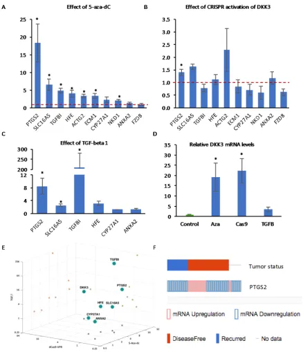
[image:10.595.96.500.82.400.2]In order to determine the extent to which the expression of these genes is regulated by gene promoter methylation in PCa, q-RT-PCR analysis was carried out to measure mRNA expression in PC3 cells treated with decitabine. Most of the genes were significantly upregulated after 24 h, with PTGS2 being the most highly-induced (Figure 7A), while there was no effect on ANXA2 and FZD8, consistent with their low levels of methylation. We then examined the effects of CRISPR-mediated induction of DKK3 on the expression of the same genes. Induction of DKK3 resulted in a significant increase in the expression of PTGS2 and trends for increased expression of SLC16A5 and for reduced expression of TGFBI and FZD8 (Figure 7B). Finally, we examined the effects of TGF-β on the expression of some of the genes. As expected, TGFBI was highly-induced by TGF-β treatment of PC3 cells (Figure 7C). In addition, TGF-β treatment resulted in significant increases in the expression of PTGS2 and SLC16A5 and a trend for increased expression of HFE. CRISPR induction of DKK3 (22-fold) was similar to its induction by decitabine (19-(22-fold), while there was a trend for increased DKK3 expression in TGF-β-treated cells (Figure 7D). Comparison of the effects of decitabine, CRISPR-mediated DKK3 activation, and TGF-β suggested that, among the genes examined, the regulation of PTGS2 expression matched most closely to that of DKK3 (Figure 7E). To determine the clinical relevance of PTGS2 expression, cBioPortal was used to look for correlations with disease recurrence, which found PTGS2 levels altered >1.5-fold in 24% of the 150 sequenced patient tumors from the Taylor et al. MSKCC dataset [45] (Figure 7F). Importantly, among the recurrent tumors, only reduction in expression was observed (10 cases), whereas among the disease-free tumors, PTGS2 was reduced in 11 and increased in 9 cases.
Figure 6. Hypermethylation of the DKK3 promoter region (A) Dendrogram of two-dimensional hierarchical clustering analysis of 49 genes that are more highly methylated in tumor samples than in normal prostate, selected by comparison of average promoter methylation (beta-values) in multiple studies of prostate. (B) Average methylation (beta-values) of DKK3 promoter region in PC3 and RWPE1 cells from GEO (GSM1323600 and GSM1323601, respectively). (C) Methylation status (percentage) in the CpG sites of the promoter regions for the indicated genes in normal prostate and prostate cancer, extracted from the TGCA dataset using the MethHC database.
In order to determine the extent to which the expression of these genes is regulated by gene promoter methylation in PCa, q-RT-PCR analysis was carried out to measure mRNA expression in PC3 cells treated with decitabine. Most of the genes were significantly upregulated after 24 h, with
PTGS2being the most highly-induced (Figure7A), while there was no effect onANXA2andFZD8, consistent with their low levels of methylation. We then examined the effects of CRISPR-mediated induction ofDKK3on the expression of the same genes. Induction ofDKK3resulted in a significant increase in the expression ofPTGS2and trends for increased expression ofSLC16A5and for reduced expression ofTGFBIandFZD8(Figure7B). Finally, we examined the effects of TGF-βon the expression of some of the genes. As expected,TGFBI was highly-induced by TGF-βtreatment of PC3 cells (Figure7C). In addition, TGF-βtreatment resulted in significant increases in the expression ofPTGS2
andSLC16A5and a trend for increased expression ofHFE. CRISPR induction ofDKK3(22-fold) was similar to its induction by decitabine (19-fold), while there was a trend for increased DKK3 expression in TGF-β-treated cells (Figure7D). Comparison of the effects of decitabine, CRISPR-mediated DKK3 activation, and TGF-βsuggested that, among the genes examined, the regulation ofPTGS2expression matched most closely to that ofDKK3(Figure7E). To determine the clinical relevance ofPTGS2
expression, cBioPortal was used to look for correlations with disease recurrence, which foundPTGS2
observed (10 cases), whereas among the disease-free tumors, PTGS2 was reduced in 11 and increased in 9 cases.
[image:11.595.79.518.124.634.2]Cancers 2018, 10, x 10 of 19
Figure 7. Q-RT-PCR analysis of the expression of the indicated genes in PC3 cells (A) treated with
5_Aza-dC for 72 h, (B) transfected with dCas9-VPR and control gRNA or DKK3 gRNAs for 48 h and
(C) treated with TGF-β1 for 24 h. (D). Q-RT-PCR analysis of the expression of DKK3 in PC3 cells under
the indicated conditions. Graphs show mean expression relative to control (dashed red lines indicate
control level set to 1) ± SD, n = 3 to 5, * p < 0.05 two-tailed Student’s t-test. (E) 3D plot illustrating the
effects of decitabine, CRISPR induction of DKK3 and TGF-β1 on the indicated genes. (F) Comparison
of tumors with changes in PTGS2 expression (up, red; down, blue) and disease-free status of patients, from cBioPortal using the MSKCC 2010 dataset [45].
3. Discussion
Epigenetic silencing of the tumor suppressor DKK3 by promoter methylation is a common event in many solid tumors and is more prevalent in advanced stages of the disease. Advanced PCa continues to have a poor survival rate due to acquired resistance to the treatments currently available,
3. Discussion
Epigenetic silencing of the tumor suppressorDKK3by promoter methylation is a common event in many solid tumors and is more prevalent in advanced stages of the disease. Advanced PCa continues to have a poor survival rate due to acquired resistance to the treatments currently available, emphasizing the need for finding new targets for more effective therapies. When designing strategies to prevent cancer progression, gene hypermethylation presents the advantage of being readily reversible, in contrast to permanent alterations in DNA, such as point mutations or gene deletions. Considering the previous reports describing the tumor suppressive roles of DKK3 [13,15,19,20], its inhibitory effect on malignant TGF-βsignaling [20,24], and its potential in recent clinical trials [21,22], we have focused on reactivating endogenousDKK3expression using a CRISPR-based approach. This has advantages over ectopic expression of DKK3, which can be toxic [46], and over using recombinant Dkk-3, which has little apparent activity in many contexts [25]. Moreover, by directly targeting theDKK3genomic locus, the CRISPR-based approach should result in expression of all variants. This is particularly important when considering the effects of Dkk-3 isoforms, for example, the intracellular form of Dkk-3, Dkk-3b [47]. CRISPR activation of DKK3 also provides specificity, unlike demethylating drugs [34]. In this study, we used dCas9-VPR to induce DKK3 expression to a level that was sufficient to affect PC3 cell number and migration. The VPR transcriptional activation domain proved essential in this context, since induction of DKK3 mRNA by dCas9-Tet1CD did not lead to increased Dkk-3 protein levels. However, we anticipate that targeted demethylation can be improved by using lentiviral delivery and allowing more time for induction to take place. A general limitation of the CRISPR approach is that it requires a means of delivery, for example using viral vectors (reviewed in [48]). However, a modified CRISPR/Cas9 system was recently used with success for gene activationin vivo[49], indicating the emerging importance of this technology in translational applications.
A major goal of this study was to investigate the effect of DKK3 re-expression on TGF-β/Smad signaling, since overexpression of this pathway is frequently observed in advanced metastatic PCa patients and is associated with poor survival. Our results indicate a significant inhibition of the response to TGF-β1 when DKK3 is re-expressed in PC3 cells. This outcome is in agreement with previous studies reporting links between DKK3 and TGF-βsignaling [20,24].
One of the tumor suppressive mechanisms described for DKK3 is its inhibition of cell proliferation [18]. Our results using PC3 cells are consistent with these previous studies and indicate that CRISPR activation of DKK3 has a negative effect on cell number. Importantly, although there was a small reduction in cell proliferation two days after transfection, it was only at the later time point when this reduction was significant. This highlights an important aspect of the approach used, in which time is required for induction of endogenous Dkk-3 to reach a level where it has a measurable physiological effect on cells. In contrast to PC3 cells, DKK3 induction had no effect on C4-2B cell number at the time points studied. This might imply a link between the effects of Dkk-3 on prostate cancer cell proliferation and on TGF-βsignaling. However, if this is the case, the mechanism remains unclear, as TGF-βdoes not affect PC3 cell proliferation [50]. In LNCaP cells, the parental line for C4-2B, adenoviral expression of DKK3 reduces CD147 levels, concomitant with a reduction in cell growth [51]. However, we did not find significant differences in the expression of the genes encoding CD147 or its accessory proteins MCT1/4 after CRISPR-mediated induction of DKK3 in PC3 cells [52]. Given the limited effects of Dkk-3 induction on prostate cancer cell proliferation, it would be interesting to explore its effects when used in combination with other therapies. In this context, downregulation of DKK3 gene expression by DNA promoter methylation in non-small cell lung cancer has been reported to increase resistance to docetaxel [53]. Since docetaxel is the standard chemotherapy drug used to treat castrate-resistant prostate cancer, it would be interesting to determine the effects of combining CRISPR-mediated activation of DKK3 with docetaxel and of restoring DKK3 expression in docetaxel-resistant prostate cancer cells.
DKK3 was only substantial at the later time point. Interestingly, we also detected a significant inhibition in migration in the absence of exogenous TGF-β, which may reflect inhibition of migration driven by autocrine TGF-βsignals [50]. TGF-βis a well-established promoter of the epithelial-mesenchymal transition (EMT) in prostate epithelial and prostate cancer cells [54]. This response to TGF-β in prostate epithelial cells is, in part, mediated by increased methylation of the E-cadherin gene (CDH1) promoter [55]. Also, as prostate cancer progresses, it can take on a neuroendocrine (NE)-like phenotype. It is possible, therefore, that induction of DKK3 reverses the EMT or NE-like phenotype. However, preliminary analysis of RNA-Seq data from PC3 cells transiently expressing dCas9-VPR and either control gRNA and DKK3 gRNA did not find significant differences in the expression levels of mesenchymal genes (VIM, SNAI1, SNAI2, SNAI3, ZEB1, ZEB2, TWIST1, TWIST2, and CDH2), epithelial genes (CLD1 and CDH1), or neuroendocrine (NE) genes (ENO2 and CHGA), apart from ZEB2, which increased slightly (1.27-fold,p= 0.047) [52]. This suggests that Dkk-3 has a different mechanism of action. Alternatively, the lack of effect on EMT/NE genes may be because PC3 cells express very high levels of EMT and NE genes that are difficult to reverse. Future studies will need to address this question in prostate cancer cell lines with stronger epithelial features and that respond to TGF-β.
Despite strong evidence implicating TGF-βsignaling in driving the methylation of several genes in PCa and the inhibition of TGF-βsignaling by DKK3, we only observed significant differences in the expression ofPTGS2. It is important to note that these analyses were performed 48 h after transfection, and therefore, if DKK3 inhibits TGF-βsignaling indirectly, more significant changes in gene expression may only be detected at later time points. It remains to be determined if PTGS2 upregulation plays a role in the tumor-suppressive effects of DKK3. This may seem unlikely, asPTGS2
encodes cyclooxygenase-2 (COX-2), which is upregulated in PCa [56]. However, reduced expression of
PTGS2mRNA by promoter hypermethylation has been reported to be an indicator of poor prognosis in PCa [57] and the COX-2 inhibitor celecoxib does not improve PCa patient survival [58]. Moreover, our analysis also found a correlation between lowPTGS2expression and tumor recurrence (Figure7F). TGFBI, in contrast, is strongly induced by TGF-β and can promote prostate tumor growth and metastasis [59]. DKK3 silencing increases TGFBI levels in prostate epithelial cells [43], so we anticipated that DKK3 re-expression might inhibitTGFBI expression in PC3 cells, and a trend for inhibition was observed.
We were intrigued to note that CRISPR activation ofDKK3, which inhibited TGF-β-dependent gene reporter activity (Figure3A), had opposite effects onPTGS2andFZD8expression, despite both
PTGS2andFZD8being induced by TGF-β. SincePTGS2but notFZD8was upregulated by decitabine (Figure7A), we considered the possibility that differences in promoter methylation might account for their differential responses to DKK3. The ability of decitabine to function as an indirect inhibitor of TGF-βsignaling has been reported in other contexts. For example, decitabine reverses TGF-β-induced EMT in gastric cancer cells [60] and in PC9 non-small cell lung cancer cells [61] by reducing aberrant TGF-β-induced hypermethylation of the miR-200 gene promoter. At low doses, decitabine induces the proteasomal degradation of DNMT1, reducing proliferation and inducing differentiation [62]. Thus, decitabine might be described as an indirect inhibitor of TGF-βsignaling.
TGF-β has been reported to increase DNMT gene expression in prostate cancer [32], which could lead to induction of genes via promoter demethylation. Analysis of DNMT family gene expression in PC3 cells confirmed that TGF-βtreatment increased expression of DNMT1, DNMT3A, and DNMT3B (Figure S4A). Interestingly, CRISPR induction of DKK3 resulted in a small but significant reduction in the expression of DNMT1 and trends for increased expression of DNMT3A and DNMT3B (Figure S4B). DNMT1 was the most abundant of the DNMTs (average Ct values for DNMT1, DNMT3A, and DNMT3B were approximately 24, 28, and 28, respectively). Thus, by reducingDNMT1expression, DKK3 may facilitate promoter demethylation, resulting in increased expression ofPTGS2, whose expression was found to be most sensitive to decitabine (Figure7A). In contrast, DKK3 inhibitedFZD8
small number of genes for analysis. Future studies using whole-genome approaches to measure the effects of DKK3 on gene promoter methylation and transcription should help test this hypothesis and take us closer to determining the mechanism of action of DKK3.
4. Materials and Methods
4.1. Cell Lines and Cell Culture
The PC3 human prostatic small cell carcinoma cell line [63] and the C4-2B cell line, which originated from the human adenocarcinoma LNCaP cell line after selection for metastasis to bone in mouse xenografts [64], were used in this study. Both cell lines were kindly provided by Charlotte Bevan (Imperial College London, London, UK). Cells were maintained in complete medium (RPMI 1640 with GlutaMAXTM(Sigma-Aldrich Company Ltd., Gillingham, UK) supplemented with 10% Fetal Calf Serum (FCS) and 100 units/mL of penicillin and streptomycin (P/S)). Cells were cultured at 37◦C and 5% CO2.
4.2. Plasmid Vectors and gRNA Design
The sequences of the DKK3 gene promoter region containing the CpG islands were extracted using the UCSC genome browser. Potential gRNA sequences, predicted using sgRNA Designer (www.portals.broadinistitute.org), were selected to bind between 1 and 1000 bp upstream of the transcriptional start site (TSS). Five gRNAs targeting different DKK3 sites and a scrambled gRNA (gRNA-control) were cloned into pRP[gRNA]-Puro-U6 (Cyagen Biosciences Inc., Santa Clara, CA, USA). dCas9-VPR (#63798), dCas9-Tet1CD (#84475), and dCas9TetCD_IM (#84479) were from Addgene LGC Standards (Teddington, UK). Sequences for gRNAs are listed in Table S1.
4.3. Transient Transfection
Transfections were performed using the Lipofectamine®LTX and Plus Reagent (Life Technologies Limited, Paisley, UK) according to the manufacturer’s protocol. Cells were transfected in 6-well plates seeded with 5×10⁵cells per well. For experiments where gRNAs were transfected together, the amount of each gRNA was scaled down to equalize the total amount of gRNA used for each condition and the dCas9-VPR to gRNA ratio was 3:1.
4.4. RNA Extraction, cDNA Synthesis, and qPCR
RNA was isolated using TRIZOL reagent (Invitrogen, Carlsbad, CA, USA), according to manufacturer’s instructions. Quantitative real-time PCR (q-RT-PCR) was performed using the SYBR Green PCR Master Mix (Life Technologies Limited) and a 7900HT Fast Real-Time PCR thermal cycler (Fisher Scientific UK Ltd., Loughborough, UK), using the amplification parameters: 50◦C for 2 min and 95◦C for 10 min, followed by 40 cycles at 95◦C for 15 s and 60◦C for 1 min. Raw count values obtained with SDS 2.0 (Applied Biosystems, Foster City, CA, USA) were imported into Excel to calculate the fold changes, normalized to expression of the gene36B4in the same sample. Relative quantification was carried out using the 2-∆∆Ct method. The primer sequences and the concentrations used for PCR are in Table S2.
4.5. Demethylation Using 5-Aza-20Deoxycytidine (5-Aza-dC)
PC3 cells were plated in 6-well plates for 24 h and treated with 1µM 5-Aza-dC for three days, by which time cells were 80% confluent and then harvested. 5-Aza-dC was prepared fresh in ddH2O and filter-sterilized (Sigma-Aldrich Company Ltd.).
4.6. Genomic DNA Extraction, Bisulfite Conversion, and Combined Bisulfite Restriction Analysis (CoBRA)
controls was carried out using the EZ DNA methylation kit (Zymo Research, Cambridge, UK) according to manufacturer’s instructions. PCR primers for the DKK3 promoter region were designed using MethPrimer software; primer sequences are in the Supplementary Data. The PCR amplification method was designed for bisulfite-modified DNA using Touch-Up gradient PCR [65]. A 10-cycle loop, consisting of 95◦C for 30 s, 48◦C for 30 s (−0.5◦C every cycle), and 72◦C for 1 min, was carried out and repeated five times (50 cycles). CoBRA amplified products were digested with BstUI (Life Technologies Limited) using 10µL PCR product (0.1–0.5µg of DNA) in a final volume of 25µL with distilled water for 6 h at 37◦C.
4.7. Protein Extraction, Western Blotting, and ELISAs
Two days after cell transfection, media were changed to normal growth medium for cell extracts or serum-free medium for collection of cell-conditioned media (CM), supplemented with 1µg/mL puromycin. Three days later, CM was collected from the cells cultured in serum-free media, and cells cultured in normal growth medium were washed with cold PBS and lysed in 300µl RIPA (0.5% Na deoxycholate, 1% Triton X-100, 20 mM Tris pH 8.0, 0.1% SDS, 150 mM NaCl), with 1 mM EDTA, cOmpleteTMEDTA-free Protease Inhibitor Cocktail (Sigma-Aldrich Company Ltd.) and 1× PhosSTOPTMPhosphatase Inhibitor Cocktail (Sigma-Aldrich Company Ltd.). Cells were then collected by scraping, transferred into a cold centrifuge tube and incubated on ice for 15 min. Lysates were centrifuged at 4◦C (15,000×g) for 15 min and the supernatant collected. Samples were separated by 6–8% SDS-PAGE and transferred to nitrocellulose or PVDF membranes (Bio-Rad Laboratories Ltd., Watford, UK). Membranes were blocked in 5% milk powder (Sigma) or 5% BSA (Sigma) in 1× Tris buffered saline, 0.1% Tween (TBST) for 1 h at room temperature. Blots were probed overnight with goat anti-DKK3 (1:500) at 4◦C. Membranes washed again 3×10 min in TBST and incubated with secondary antibodies (1:5000) for 1 h at room temperature, washed 3×5 min in TBST and then exposed to ECL Clarity reagent (BioRad), exposed to X-ray film, which were developed using an OptiMax film processor. Membranes were also probed with anti-human GAPDH (1:5,000) for 1 h at room temperature, and donkey anti-mouse-HRP linked secondary antibody (1:15,000) for 45 min. ELISAs were performed for CM from transfected cells using the Human Dkk-3 DuoSet kit (R&D Systems Ltd., Abingdon, UK), according to the manufacturer’s instructions. The absorbance of each standard and sample was determined using an OptiMax tunable microplate reader (Molecular Devices, San Jose, CA, USA) set to 450 nm and 540 nm. Samples were quantified based on a standard curve generated using recombinant purified Dkk-3 (R&D Systems) and a four-parameter logistic curve-fit.
4.8. Gene Reporter, Proliferation, and Migration Assays
of 200,000 cells/well for the short time-point (2 d) or 50,000 cells/well for the long time-point (5 d). The day after plating, cells were transfected as above. For the short time point, cells were trypsinized after 24 h, while for the long time-point this was after 4 d. Cells were centrifuged at 500×gfor 5 min and resuspended in 2 mL serum-free RPMI medium±2 ng/mL TGF-β1. 50,000 cells were plated on 8µm pore Falcon Transwell®polycarbonate membrane inserts (Scientific Laboratory Supplies Ltd., Nottingham, UK) in a 24-well plate, in duplicate for each treatment and transfection condition. 800µL of complete medium was added to the outside of each insert. Cells were allowed to migrate for 24 h at 37◦C, and those that had not migrated were removed with a cotton bud. The inserts were washed in PBS and cells fixed in cold methanol (1 mL/insert) for 20 min, allowed to dry for 30 min and then stained with 0.2% crystal violet in 20% methanol (600µL/insert) for 30 min. Inserts were washed in distilled water and seven pictures were taken for each insert at 200×magnification using a Nikon Eclipse TE200-U and QCapture software (Qimaging Inc., Surrey, BC, Canada). Migrated cells were counted using Image J and the average numbers of migrated cells calculated for each insert. This value was normalized by dividing the number of migrated cells by the OD reading at 595 nm from additional wells of cells that had been plated in parallel without inserts to control for effects on cell number.
4.9. Bioinformatics Analysis
MSKCC mRNA expression data were extracted from cBIO (www.cbioportal.org/) for selected genes usingz-score vs normal with threshold±2. Oncomine (www.oncomine.org/) was also used to compare mRNA expression in prostate cancer datasets to determine the extent of their downregulation across multiple prostate cancer datasets. The differential expression of each gene between PCa and normal samples was analyzed for fold-change and statistical significance. ToppGene (www.toppgene.
cchmc.org/) was used to investigate the relevance of the genes to the TGF-βsignaling pathway, based
on the functional similarity between the seed list and training gene list.
4.10. Statistical Analysis
All data are presented as mean and standard error of mean (SEM) or standard deviation (SD). Microsoft Excel was used to perform the two sample, paired Student’st-test to calculate the statistical significance and to perform one-way ANOVA to compare multiple experimental conditions. Ap-value < 0.05 was considered to be statistical significant. *p< 0.05. For qPCR analysis,∆∆Ct values were log-transformed before the test.
5. Conclusions
In conclusion, in this study, we demonstrate that CRISPR-mediated activation of DKK3 expression is feasible, providing further evidence for potential therapeutic application of this technology, and have generated results strengthening the link between Dkk-3 and TGF-βregulation of gene expression. Supplementary Materials:The following are available online athttp://www.mdpi.com/2072-6694/10/6/165/s1. Figure S1: Methodological workflow of analyses performed; Figure S2: Analysis of the expression levels of the indicated genes in PCa datasets; Figure S3: Location of CpG probes in the DKK3 promoter; Figure S4: Effects
of TGF-βand CRISPR induction of DKK3 onDNMTmRNA expression; Table S1: Sequences and genomic
coordinates of gRNAs used to target the DKK3 promoter; Table S2: Primer sequences used for qPCR AND CoBRA
Author Contributions:R.M.K., J.W., and H.K. conceived and designed the experiments; H.K., E.G.-G., and E.S. performed the experiments; R.M.K., H.K., E.G.-G., E.S., and S.S. analyzed the data; R.M.K., H.K., E.G.-G., and E.S. wrote the paper.
Funding:This research was principally funded by the Harris Family Harvey Spack Memorial Fund.
Conflicts of Interest:The authors declare no conflict of interest. The funders had no role in the design of the study; in the collection, analyses, or interpretation of data; in the writing of the manuscript, or in the decision to publish the results.
References
1. Siegel, R.L.; Miller, K.D.; Jemal, A. Cancer statistics, 2018. CA Cancer J. Clin. 2018,68, 7–30. [CrossRef] [PubMed]
2. Sharifi, N.; Dahut, W.L.; Steinberg, S.M.; Figg, W.D.; Tarassoff, C.; Arlen, P.; Gulle, J.L. A retrospective study of the time to clinical endpoints for advanced prostate cancer.BJU Int.2005,96, 985–989. [CrossRef] [PubMed]
3. Massie, C.E.; Mills, I.G.; Lynch, A.G. The importance of DNA methylation in prostate cancer development. J. Steroid Biochem. Mol. Biol.2016. [CrossRef] [PubMed]
4. Barbieri, C.E.; Baca, S.C.; Lawrence, M.S.; Demichelis, F.; Blattner, M.; Theurillat, J.-P.; White, T.A.; Stojanov, P.; Van Allen, E.; Stransky, N.; et al. Exome sequencing identifies recurrent SPOP, FOXA1 and MED12 mutations in prostate cancer.Nat. Genet.2012,44, 685–689. [CrossRef] [PubMed]
5. Jones, P.A.; Baylin, S.B. The fundamental role of epigenetic events in cancer.Nat. Rev. Genet.2002,3, 415–428. [CrossRef] [PubMed]
6. Noushmehr, H.; Weisenberger, D.J.; Diefes, K.; Phillips, H.S.; Pujara, K.; Berman, B.P.; Pan, F.; Pelloski, C.E.; Sulman, E.P.; Bhat, K.P.; et al. Identification of a CpG Island Methylator Phenotype that Defines a Distinct Subgroup of Glioma.Cancer Cell2010,17, 510–522. [CrossRef] [PubMed]
7. Baylin, S.B. DNA methylation and gene silencing in cancer. Nat. Clin. Pract. Oncol. 2005, 2, S4–S11. [CrossRef] [PubMed]
8. Esteller, M.; Herman, J.G. Cancer as an epigenetic disease: DNA methylation and chromatin alterations in human tumours.J. Pathol.2002,196, 1–7. [CrossRef] [PubMed]
9. Rhee, I.; Bachman, K.E.; Park, B.H.; Jair, K.-W.; Yen, R.-W.C.; Schuebel, K.E.; Cui, H.; Feinberg, A.P.; Lengauer, C.; Kinzler, K.W.; Baylin, S.B.; Vogelstein, B. DNMT1 and DNMT3b cooperate to silence genes in human cancer cells.Nature2002,416, 552–556. [CrossRef] [PubMed]
10. Okano, M.; Bell, D.W.; Haber, D.A.; Li, E. DNA methyltransferases DNMT3A and DNMT3B are essential for de novo methylation and mammalian development.Cell1999,99, 247–257. [CrossRef]
11. Robert, M.-F.; Morin, S.; Beaulieu, N.; Gauthier, F.; Chute, I.C.; Barsalou, A.; MacLeod, A.R. DNMT1 is required to maintain CpG methylation and aberrant gene silencing in human cancer cells.Nat. Genet.2003, 33, 61–65. [CrossRef] [PubMed]
12. Smeets, E.; Lynch, A.G.; Prekovic, S.; Van den Broeck, T.; Moris, L.; Helsen, C.; Joniau, S.; Claessens, F.; Massie, C.E. The role of TET-mediated DNA hydroxymethylation in prostate cancer.Mol. Cell. Endocrinol. 2018,462, 41–55. [CrossRef] [PubMed]
13. Abarzua, F. Adenovirus-Mediated Overexpression of REIC/Dkk-3 Selectively Induces Apoptosis in Human Prostate Cancer Cells through Activation of c-Jun-NH2-Kinase.Cancer Res.2005,65, 9617–9622. [CrossRef] [PubMed]
14. Lodygin, D.; Epanchintsev, A.; Menssen, A.; Diebold, J.; Hermeking, H. Functional epigenomics identifies genes frequently silenced in prostate cancer.Cancer Res.2005,65, 4218–4227. [CrossRef] [PubMed] 15. Kawano, Y.; Kitaoka, M.; Hamada, Y.; Walker, M.; Waxman, J.; Kypta, R. Regulation of prostate cell growth
and morphogenesis by Dickkopf-3.Oncogene2006,25, 6528–6537. [CrossRef] [PubMed]
16. Zenzmaier, C.; Untergasser, G.; Hermann, M.; Dirnhofer, S.; Sampson, N.; Berger, P. Dysregulation of Dkk-3 expression in benign and malignant prostatic tissue.Prostate2008,68, 540–547. [CrossRef] [PubMed] 17. Romero, D.; Kypta, R. Dickkopf-3 function in the prostate: implications for epithelial homeostasis and tumor
progression.Bioarchitecture2013,3, 42–44. [CrossRef] [PubMed]
18. Veeck, J.; Dahl, E. Targeting the Wnt pathway in cancer: The emerging role of Dickkopf-3.Biochim. Biophys. Acta Rev. Cancer2012,1825, 18–28. [CrossRef] [PubMed]
20. Romero, D.; Kawano, Y.; Bengoa, N.; Walker, M.M.; Maltry, N.; Niehrs, C.; Waxman, J.; Kypta, R. Downregulation of Dickkopf-3 disrupts prostate acinar morphogenesis through TGF-β/Smad signaling. J. Cell Sci.2013,6, 1208. [CrossRef]
21. Kumon, H.; Sasaki, K.; Ariyoshi, Y.; Sadahira, T.; Ebara, S.; Hiraki, T.; Kanazawa, S.; Yanai, H.; Watanabe, M.; Nasu, Y. Ad-REIC gene therapy: Promising results in a patient with metastatic CRPC following chemotherapy. Clin. Med. Insights Oncol.2015,9, 31–38. [CrossRef] [PubMed]
22. Kumon, H.; Ariyoshi, Y.; Sasaki, K.; Sadahira, T.; Araki, M.; Ebara, S.; Yanai, H.; Watanabe, M.; Nasu, Y. Adenovirus vector carrying REIC/DKK-3 gene: neoadjuvant intraprostatic injection for high-risk localized prostate cancer undergoing radical prostatectomy.Cancer Gene Ther.2016, 1–10. [CrossRef] [PubMed] 23. Niehrs, C. Function and biological roles of the Dickkopf family of Wnt modulators. Oncogene2006,25,
7469–7481. [CrossRef] [PubMed]
24. Romero, D.; Al-Shareef, Z.; Gorroño-Etxebarria, I.; Atkins, S.; Turrell, F.; Chhetri, J.; Bengoa-Vergniory, N.; Zenzmaier, C.; Berger, P.; Waxman, J.; et al. Dickkopf-3 regulates prostate epithelial cell acinar morphogenesis and prostate cancer cell invasion by limiting TGF-β-dependent activation of matrix metalloproteases. Carcinogenesis2016,37, 18–29. [CrossRef] [PubMed]
25. Zenzmaier, C.; Sampson, N.; Plas, E.; Berger, P. Dickkopf-related protein 3 promotes pathogenic stromal remodeling in benign prostatic hyperplasia and prostate cancer.Prostate2013,73, 1441–1452. [CrossRef] [PubMed]
26. Massagué, J. TGFβsignalling in context.Nat. Rev. Mol. Cell Biol.2012,13, 616–630. [CrossRef] [PubMed] 27. Neuzillet, C.; Tijeras-Raballand, A.; Cohen, R.; Cros, J.; Faivre, S.; Raymond, E.; De Gramont, A. Targeting
the TGF?? pathway for cancer therapy.Pharmacol. Ther.2015,147, 22–31. [CrossRef] [PubMed]
28. Chung, S.-W.; Cooper, C.R.; Farach-Carson, M.C.; Ogunnaike, B.A. A control engineering approach to understanding the TGF- paradox in cancer.J. R. Soc. Interface2012,9, 1389–1397. [CrossRef] [PubMed] 29. Li, Q.; Wang, Y.; Xiao, H.; Li, Y.; Kan, X.; Wang, X.; Wang, Z.; Yang, Q.; Chen, X.; Weng, X.; et al.
Chamaejasmenin B, a novel candidate, inhibits breast tumor metastasis by rebalancing TGF-βparadox. Oncotarget2016,7. [CrossRef] [PubMed]
30. Wendt, M.K.; Tian, M.; Schiemann, W.P. Deconstructing the mechanisms and consequences of TGF-β-induced EMT during cancer progression.Cell Tissue Res.2012,347, 85–101. [CrossRef] [PubMed]
31. Zhang, Q.; Yu, N.; Lee, C. Vicious cycle of TGF-β signaling in tumor progression and metastasis. Am. J. Clin. Exp. Urol.2014,2, 149–155. [PubMed]
32. Zhang, Q.; Chen, L.; Helfand, B.T.; Jang, T.L.; Sharma, V.; Kozlowski, J.; Kuzel, T.M.; Zhu, L.J.; Yang, X.J.; Javonovic, B.; et al. TGF-βregulates DNA methyltransferase expression in prostate cancer, correlates with aggressive capabilities, and predicts disease recurrence.PLoS ONE2011,6, 1–13. [CrossRef] [PubMed] 33. Lee, C.; Zhang, Q.; Zi, X.; Dash, A.; Soares, M.B.; Rahmatpanah, F.; Jia, Z.; McClelland, M.; Mercola, D.
TGF-beta mediated DNA methylation in prostate cancer.Transl. Androl. Urol.2012,1, 78–88. [CrossRef] [PubMed]
34. Jones, P.A.; Issa, J.-P.J.; Baylin, S. Targeting the cancer epigenome for therapy. Nat. Rev. Genet. 2016,17, 630–641. [CrossRef] [PubMed]
35. Baumgart, S.J.; Haendler, B. Exploiting Epigenetic Alterations in Prostate Cancer.Int. J. Mol. Sci.2017,18. [CrossRef] [PubMed]
36. Marques-Magalhães,Â.; Graça, I.; Henrique, R.; Jerónimo, C. Targeting DNA methyltranferases in urological tumors.Front. Pharmacol.2018,9. [CrossRef] [PubMed]
37. Bikard, D.; Jiang, W.; Samai, P.; Hochschild, A.; Zhang, F.; Marraffini, L.A. Programmable repression and activation of bacterial gene expression using an engineered CRISPR-Cas system.Nucleic Acids Res.2013,41, 7429–7437. [CrossRef] [PubMed]
38. Chavez, A.; Scheiman, J.; Vora, S.; Pruitt, B.W.; Tuttle, M.; Iyer, E.P.; Lin, S.; Kiani, S.; Guzman, C.D.; Wiegand, D.J.; et al. Highly efficient Cas9-mediated transcriptional programming.Nat. Methods2015,12, 326–328. [CrossRef] [PubMed]