Crystal structure of (4
E
)-4-(8-methoxy-2
H
-chromen-2-ylidene)-3-methyl-1-phenyl-1
H
-pyrazol-5(4
H
)-one
Muhammad Salim,aMunawar Ali Munawar,a
Muhammad Nawaz Tahir,b* Muhammad Shahidaand Khizar Iqbal Malika
a
Department of Chemistry, University of the Punjab, Lahore, Punjab, Pakistan, and
b
Department of Physics, University of Sargodha, Sargodha, Punjab, Pakistan. *Correspondence e-mail: dmntahir_uos@yahoo.com
Received 6 May 2015; accepted 18 May 2015
Edited by W. Imhof, University Koblenz-Landau, Germany
In the title compound, C20H16N2O3, the phenyl substituent attached to the pyrazole ring makes a dihedral angle of 4.87 (7) with the rest of the molecule. In the crystal,
molecules are connected into inversion dimers of theR2 2
(14) type by pairs of C—H O interactions.–interactions exist between the benzene and pyrazole rings at a distance of 3.701 (1) A˚ . Similarly, – interactions are present at a centroid–centroid distance of 3.601 (1) A˚ between the oxygen-containing heterocyclic ring and methoxy substituted aromatic ring of a neighbouring molecule. Additional C— H and C O interactions are also observed.
Keywords:crystal structure; pyrazolone;–interactions.
CCDC reference:1401584
1. Related literature
For related structures, see: Chaudhryet al.(2012); Holzeret al. (1999); Maliket al.(2009).
2. Experimental
2.1. Crystal data
C20H16N2O3 Mr= 332.35
Monoclinic,C2=c a= 28.179 (5) A˚
b= 4.7108 (8) A˚
c= 23.819 (5) A˚
= 92.957 (7)
V= 3157.7 (10) A˚3 Z= 8
MoKradiation
= 0.10 mm1 T= 296 K
0.400.220.18 mm
2.2. Data collection
Bruker Kappa APEXII CCD diffractometer
Absorption correction: multi-scan (SADABS; Bruker, 2005)
Tmin= 0.961,Tmax= 0.985
13056 measured reflections 3419 independent reflections 2389 reflections withI> 2(I)
Rint= 0.033
2.3. Refinement
R[F2> 2(F2)] = 0.042 wR(F2) = 0.127
S= 1.06 3419 reflections
229 parameters
H-atom parameters constrained
max= 0.22 e A˚ 3
[image:1.610.313.566.565.629.2]min=0.16 e A˚ 3
Table 1
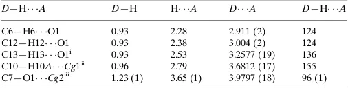
Hydrogen-bond geometry and C—H and C O interactions (A˚ ,
).
Cg1 andCg2 are the centroids of the N1/N2/C7–C9 and C11–C14/C19/O2 rings, respectively.
D—H A D—H H A D A D—H A
C6—H6 O1 0.93 2.28 2.911 (2) 124
C12—H12 O1 0.93 2.38 3.004 (2) 124
C13—H13 O1i
0.93 2.53 3.2577 (19) 136
C10—H10A Cg1ii
0.96 2.79 3.6812 (17) 155
C7—O1 Cg2iii
1.23 (1) 3.65 (1) 3.9797 (18) 96 (1)
Symmetry codes: (i)x;yþ2;z; (ii)x;y1;z; (iii)x;yþ1;z.
Data collection: APEX2 (Bruker, 2007); cell refinement: SAINT (Bruker, 2007); data reduction:SAINT; program(s) used to solve structure:SHELXS97(Sheldrick, 2008); program(s) used to refine structure: SHELXL2014 (Sheldrick, 2015); molecular graphics: ORTEP-3 for Windows(Farrugia, 2012) andPLATON(Spek, 2009); software used to prepare material for publication:WinGX(Farrugia, 2012) andPLATON.
data reports
o414
Salimet al. doi:10.1107/S2056989015009445 Acta Cryst.(2015).E71, o414–o415Acknowledgements
The authors acknowledge the provision of funds for the purchase of the diffractometer and encouragement by Dr Muhammad Akram Chaudhary, Vice Chancellor, University of Sargodha, Pakistan.
Supporting information for this paper is available from the IUCr electronic archives (Reference: IM2465).
References
Bruker (2005).SADABS. Bruker AXS Inc., Madison, Wisconsin, USA. Bruker (2007).APEX2andSAINT. Bruker AXS Inc., Madison, Wisconsin,
USA.
Chaudhry, F., Tahir, M. N., Khan, M. A., Ather, A. Q. & Asif, N. (2012).Acta Cryst.E68, o2044.
Farrugia, L. J. (2012).J. Appl. Cryst.45, 849–854.
Holzer, W., Mereiter, K. & Plagens, B. (1999).Heterocycles,50, 799–818. Malik, K. I., Munawar, M. A., Khan, M. A., Nadeem, S. & Mukhtar-ul-Hassan
(2009).Acta Cryst.E65, o3046.
supporting information
sup-1
Acta Cryst. (2015). E71, o414–o415
supporting information
Acta Cryst. (2015). E71, o414–o415 [doi:10.1107/S2056989015009445]
Crystal structure of (4
E
)-4-(8-methoxy-2
H
-chromen-2-ylidene)-3-methyl-1-phenyl-1
H
-pyrazol-5(4
H
)-one
Muhammad Salim, Munawar Ali Munawar, Muhammad Nawaz Tahir, Muhammad Shahid and
Khizar Iqbal Malik
S1. Comment
The crystal structures of 5-methyl-2-phenyl-4-((E)-3-phenyl-2-hydroxy- prop-2-enylidene)-1,2-dihydro-3H
-pyrazol-3-one (Holzer et al., 1999), (4Z)-4-((2E)-1-hydroxy-3-(4-methoxyphenyl)prop-2-en-1- ylidene)-3-methyl-1-phenyl-1H
-pyrazol-5(4H)-one (Malik et al., 2009) and (4Z)-4-((2E)-1-hydroxy-3-(3-nitrophenyl)prop-
2-en-1-ylidene)-3-methyl-1-(4-methylphenyl)-1H-pyrazol-5(4H)-one (Chaudhry, et al., 2012) have been published which are related to the
title compound (I, Fig. 1). No crystal structure has been found containing a 8-methoxy-2H-chromene subunit. (I) is
synthesized for the biological studies etc.
In (I), the benzene ring A (C1–C6) and the
(4E)-4-(8-methoxy-2H-chromen-2-ylidene)-5-methyl-2,4-dihydro-3H-pyrazol-3-one (C7 –C20/N1/N2/O1/O2/O3) part of the molecule are planar with r. m. s. deviations of 0.0053 and 0.0108
Å, respectively. The dihedral angle between A/B is 4.87 (7)°. The molecules are dimerized due to C—H···O interactions
completing R22 (14) Molecules are connected to inversion dimers dimerized of the R22 (14) type by C—H···O interactions.
There exist π–π interactions at a distance of 3.7011 (12) Å between the centeroids of Cg1—Cg3i and Cg3—Cg1ii [i = x, -
1 + y, z and ii = x, 1 + y, z], where Cg1 and Cg3 are the centroids of heterocyclic ring C (N1/N2/C7/C8/C9) and benzene
ring A, respectively. Similarly, there exist π–π interaction at a distance of 3.6012 (11) Å between the centeroids of Cg2—
Cg4ii and Cg4—Cg2i [i = x, - 1 + y, z and ii = x, 1 + y, z], where Cg2 and Cg4 are the centroids of heterocyclic ring D
(C11—C14/C19/O2) and methoxy containing benzene ring E (C14—C19), respectively. There exist C—H···π and
C=O···π interactions (Table 1).
S2. Experimental
For the preparation of title compound (I), a mixture of 4-acetyl-3-methyl-1-phenyl-5-hydroxy pyrazole (0.218 g, 1
mmoL), 2-methoxybenzaldehyde (0.205 g, 1.5 mmoL) was refluxed for 8 h in glacial acetic acid (15 ml) and
concentrated sulfuric acid (0.2 ml). The reaction mixture was diluted with distilled water (60 ml). The precipitate was
filtered, washed with methanol and dried. The crude product was purified by column chromatography using n-hexane and
ethyl acetate mixtures as eluents. The product was recrystallized using n-hexane to afford red needles (yield = 63%, m.p.
503 K)
S3. Refinement
H-atoms were positioned geometrically (C–H = 0.93–0.96 Å) and refined as riding with Uiso(H) = xUeq(C, O), where x =
Figure 1
View of the title compound with the atom numbering scheme. Thermal ellipsoids are drawn at the 50% probability level.
H-atoms are shown by small circles of arbitrary radii.
Figure 2
[image:4.610.135.478.292.647.2]supporting information
sup-3
Acta Cryst. (2015). E71, o414–o415
(4E)-4-(8-Methoxy-2H-chromen-2-ylidene)-3-methyl-1-phenyl-1H-pyrazol-5(4H)-one
Crystal data
C20H16N2O3 Mr = 332.35
Monoclinic, C2/c a = 28.179 (5) Å
b = 4.7108 (8) Å
c = 23.819 (5) Å
β = 92.957 (7)°
V = 3157.7 (10) Å3 Z = 8
F(000) = 1392
Dx = 1.398 Mg m−3
Mo Kα radiation, λ = 0.71073 Å Cell parameters from 2389 reflections
θ = 2.9–27.0°
µ = 0.10 mm−1 T = 296 K Needle, red
0.40 × 0.22 × 0.18 mm
Data collection
Bruker Kappa APEXII CCD diffractometer
Radiation source: fine-focus sealed tube Graphite monochromator
Detector resolution: 7.70 pixels mm-1 ω scans
Absorption correction: multi-scan
(SADABS; Bruker, 2005)
Tmin = 0.961, Tmax = 0.985
13056 measured reflections 3419 independent reflections 2389 reflections with I > 2σ(I)
Rint = 0.033
θmax = 27.0°, θmin = 2.9° h = −35→35
k = −6→5
l = −24→30
Refinement
Refinement on F2
Least-squares matrix: full
R[F2 > 2σ(F2)] = 0.042 wR(F2) = 0.127 S = 1.06 3419 reflections 229 parameters 0 restraints
Primary atom site location: structure-invariant direct methods
Secondary atom site location: difference Fourier map
Hydrogen site location: inferred from neighbouring sites
H-atom parameters constrained
w = 1/[σ2(F
o2) + (0.0648P)2 + 0.6123P]
where P = (Fo2 + 2Fc2)/3
(Δ/σ)max < 0.001
Δρmax = 0.22 e Å−3
Δρmin = −0.16 e Å−3
Extinction correction: SHELXL2014 (Sheldrick, 2015), Fc*=kFc[1+0.001xFc2λ3/sin(2θ)]-1/4
Extinction coefficient: 0.0021 (4)
Special details
Geometry. All e.s.d.'s (except the e.s.d. in the dihedral angle between two l.s. planes) are estimated using the full
covariance matrix. The cell e.s.d.'s are taken into account individually in the estimation of e.s.d.'s in distances, angles and torsion angles; correlations between e.s.d.'s in cell parameters are only used when they are defined by crystal symmetry. An approximate (isotropic) treatment of cell e.s.d.'s is used for estimating e.s.d.'s involving l.s. planes.
Refinement. Refinement of F2 against ALL reflections. The weighted R-factor wR and goodness of fit S are based on F2,
conventional R-factors R are based on F, with F set to zero for negative F2. The threshold expression of F2 > σ(F2) is used
only for calculating R-factors(gt) etc. and is not relevant to the choice of reflections for refinement. R-factors based on F2
are statistically about twice as large as those based on F, and R-factors based on ALL data will be even larger.
Fractional atomic coordinates and isotropic or equivalent isotropic displacement parameters (Å2)
x y z Uiso*/Ueq
N1 0.14466 (4) 0.9876 (2) 0.09568 (5) 0.0407 (3) N2 0.18451 (4) 0.8288 (2) 0.08158 (6) 0.0417 (3) C1 0.14967 (6) 1.1800 (3) 0.14142 (6) 0.0410 (4) C2 0.19424 (6) 1.2193 (3) 0.16813 (7) 0.0501 (4) H2 0.2202 1.1180 0.1563 0.060* C3 0.19992 (7) 1.4086 (4) 0.21225 (8) 0.0575 (5) H3 0.2298 1.4329 0.2301 0.069* C4 0.16197 (8) 1.5617 (4) 0.23019 (8) 0.0593 (5) H4 0.1661 1.6913 0.2595 0.071* C5 0.11808 (7) 1.5206 (4) 0.20427 (8) 0.0619 (5) H5 0.0923 1.6222 0.2164 0.074* C6 0.11134 (7) 1.3295 (3) 0.16006 (7) 0.0541 (5) H6 0.0812 1.3025 0.1431 0.065* C7 0.10494 (5) 0.9304 (3) 0.06052 (7) 0.0410 (4) C8 0.12183 (5) 0.7169 (3) 0.02175 (6) 0.0372 (4) C9 0.17098 (5) 0.6713 (3) 0.03861 (7) 0.0370 (4) C10 0.20563 (5) 0.4738 (3) 0.01377 (7) 0.0451 (4) H10A 0.1932 0.2840 0.0139 0.068* H10B 0.2352 0.4800 0.0356 0.068* H10C 0.2108 0.5304 −0.0242 0.068* C11 0.09468 (5) 0.5939 (3) −0.02107 (6) 0.0368 (4) C12 0.04599 (5) 0.6554 (3) −0.03528 (7) 0.0455 (4) H12 0.0304 0.7917 −0.0149 0.055* C13 0.02230 (6) 0.5199 (3) −0.07759 (7) 0.0492 (4) H13 −0.0096 0.5601 −0.0856 0.059* C14 0.04580 (5) 0.3138 (3) −0.11063 (7) 0.0435 (4) C15 0.02414 (6) 0.1660 (4) −0.15629 (8) 0.0562 (5) H15 −0.0078 0.1959 −0.1664 0.067* C16 0.05019 (7) −0.0228 (4) −0.18596 (8) 0.0583 (5) H16 0.0357 −0.1184 −0.2164 0.070* C17 0.09766 (6) −0.0735 (3) −0.17137 (7) 0.0490 (4) H17 0.1146 −0.2024 −0.1921 0.059* C18 0.12003 (5) 0.0664 (3) −0.12619 (7) 0.0404 (4) C19 0.09317 (5) 0.2600 (3) −0.09639 (6) 0.0373 (4) C20 0.19362 (6) −0.1685 (4) −0.13639 (8) 0.0533 (5) H20A 0.1797 −0.3531 −0.1328 0.080* H20B 0.2255 −0.1704 −0.1201 0.080* H20C 0.1942 −0.1191 −0.1754 0.080*
Atomic displacement parameters (Å2)
U11 U22 U33 U12 U13 U23
supporting information
sup-5
Acta Cryst. (2015). E71, o414–o415
C2 0.0550 (11) 0.0476 (9) 0.0477 (10) −0.0008 (7) 0.0044 (8) −0.0021 (8) C3 0.0697 (13) 0.0533 (10) 0.0496 (11) −0.0105 (9) 0.0030 (9) −0.0022 (9) C4 0.0898 (16) 0.0451 (9) 0.0432 (11) −0.0025 (9) 0.0052 (10) −0.0037 (8) C5 0.0816 (15) 0.0547 (10) 0.0501 (11) 0.0199 (9) 0.0104 (10) −0.0039 (9) C6 0.0592 (11) 0.0539 (9) 0.0491 (11) 0.0147 (8) 0.0022 (9) −0.0044 (9) C7 0.0380 (9) 0.0383 (7) 0.0472 (10) 0.0053 (6) 0.0059 (7) 0.0001 (7) C8 0.0347 (8) 0.0346 (7) 0.0427 (9) 0.0040 (6) 0.0070 (7) 0.0010 (7) C9 0.0369 (8) 0.0331 (7) 0.0413 (9) 0.0045 (6) 0.0063 (7) 0.0037 (7) C10 0.0390 (9) 0.0433 (8) 0.0531 (10) 0.0080 (6) 0.0040 (7) −0.0034 (7) C11 0.0342 (8) 0.0340 (7) 0.0430 (9) 0.0046 (6) 0.0084 (7) 0.0028 (7) C12 0.0368 (9) 0.0446 (8) 0.0554 (11) 0.0090 (7) 0.0066 (8) 0.0007 (8) C13 0.0324 (9) 0.0537 (9) 0.0609 (11) 0.0080 (7) −0.0020 (8) 0.0042 (8) C14 0.0364 (9) 0.0443 (8) 0.0493 (10) 0.0028 (6) −0.0020 (7) 0.0039 (7) C15 0.0417 (10) 0.0619 (10) 0.0634 (12) 0.0029 (8) −0.0131 (9) 0.0000 (9) C16 0.0561 (11) 0.0632 (11) 0.0541 (12) −0.0012 (9) −0.0139 (9) −0.0080 (9) C17 0.0504 (10) 0.0507 (9) 0.0457 (10) 0.0016 (7) 0.0004 (8) −0.0065 (8) C18 0.0358 (9) 0.0419 (8) 0.0434 (9) 0.0002 (6) 0.0017 (7) 0.0006 (7) C19 0.0366 (8) 0.0362 (7) 0.0388 (9) −0.0014 (6) 0.0000 (7) 0.0013 (7) C20 0.0453 (10) 0.0585 (10) 0.0565 (11) 0.0111 (8) 0.0060 (8) −0.0096 (9)
Geometric parameters (Å, º)
O1—C7 1.2299 (17) C8—C9 1.438 (2) O2—C11 1.3584 (17) C9—C10 1.4931 (19) O2—C19 1.3766 (17) C10—H10A 0.9600 O3—C18 1.3578 (18) C10—H10B 0.9600 O3—C20 1.4244 (18) C10—H10C 0.9600 N1—C7 1.389 (2) C11—C12 1.426 (2) N1—N2 1.4046 (16) C12—C13 1.341 (2) N1—C1 1.419 (2) C12—H12 0.9300 N2—C9 1.3048 (19) C13—C14 1.433 (2) C1—C6 1.382 (2) C13—H13 0.9300 C1—C2 1.390 (2) C14—C19 1.384 (2) C2—C3 1.381 (2) C14—C15 1.404 (2) C2—H2 0.9300 C15—C16 1.372 (2) C3—C4 1.376 (3) C15—H15 0.9300 C3—H3 0.9300 C16—C17 1.385 (2) C4—C5 1.367 (3) C16—H16 0.9300 C4—H4 0.9300 C17—C18 1.385 (2) C5—C6 1.391 (2) C17—H17 0.9300 C5—H5 0.9300 C18—C19 1.401 (2) C6—H6 0.9300 C20—H20A 0.9600 C7—C8 1.462 (2) C20—H20B 0.9600 C8—C11 1.371 (2) C20—H20C 0.9600
C7—N1—C1 129.21 (13) O2—C11—C8 116.14 (13) N2—N1—C1 118.35 (12) O2—C11—C12 118.14 (14) C9—N2—N1 106.59 (12) C8—C11—C12 125.72 (14) C6—C1—C2 119.19 (15) C13—C12—C11 121.15 (15) C6—C1—N1 121.59 (15) C13—C12—H12 119.4 C2—C1—N1 119.22 (14) C11—C12—H12 119.4 C3—C2—C1 120.04 (16) C12—C13—C14 120.59 (14) C3—C2—H2 120.0 C12—C13—H13 119.7 C1—C2—H2 120.0 C14—C13—H13 119.7 C4—C3—C2 120.87 (18) C19—C14—C15 118.31 (15) C4—C3—H3 119.6 C19—C14—C13 117.17 (14) C2—C3—H3 119.6 C15—C14—C13 124.52 (15) C5—C4—C3 118.99 (17) C16—C15—C14 119.84 (16) C5—C4—H4 120.5 C16—C15—H15 120.1 C3—C4—H4 120.5 C14—C15—H15 120.1 C4—C5—C6 121.25 (18) C15—C16—C17 121.19 (16) C4—C5—H5 119.4 C15—C16—H16 119.4 C6—C5—H5 119.4 C17—C16—H16 119.4 C1—C6—C5 119.64 (18) C16—C17—C18 120.50 (16) C1—C6—H6 120.2 C16—C17—H17 119.7 C5—C6—H6 120.2 C18—C17—H17 119.7 O1—C7—N1 125.57 (15) O3—C18—C17 125.90 (14) O1—C7—C8 130.78 (15) O3—C18—C19 116.25 (13) N1—C7—C8 103.65 (12) C17—C18—C19 117.84 (14) C11—C8—C9 129.57 (13) O2—C19—C14 121.56 (13) C11—C8—C7 124.98 (13) O2—C19—C18 116.12 (13) C9—C8—C7 105.45 (13) C14—C19—C18 122.31 (14) N2—C9—C8 111.87 (13) O3—C20—H20A 109.5 N2—C9—C10 119.63 (13) O3—C20—H20B 109.5 C8—C9—C10 128.50 (14) H20A—C20—H20B 109.5 C9—C10—H10A 109.5 O3—C20—H20C 109.5 C9—C10—H10B 109.5 H20A—C20—H20C 109.5 H10A—C10—H10B 109.5 H20B—C20—H20C 109.5
supporting information
sup-7
Acta Cryst. (2015). E71, o414–o415
N2—N1—C7—O1 −179.78 (15) C20—O3—C18—C17 −2.8 (2) C1—N1—C7—O1 −0.6 (3) C20—O3—C18—C19 177.68 (14) N2—N1—C7—C8 0.35 (16) C16—C17—C18—O3 −179.84 (15) C1—N1—C7—C8 179.58 (13) C16—C17—C18—C19 −0.3 (2) O1—C7—C8—C11 0.0 (3) C11—O2—C19—C14 −0.5 (2) N1—C7—C8—C11 179.86 (14) C11—O2—C19—C18 178.69 (12) O1—C7—C8—C9 179.92 (17) C15—C14—C19—O2 179.76 (14) N1—C7—C8—C9 −0.22 (15) C13—C14—C19—O2 0.2 (2) N1—N2—C9—C8 0.20 (16) C15—C14—C19—C18 0.6 (2) N1—N2—C9—C10 −179.44 (12) C13—C14—C19—C18 −178.94 (14) C11—C8—C9—N2 179.93 (14) O3—C18—C19—O2 0.4 (2) C7—C8—C9—N2 0.02 (17) C17—C18—C19—O2 −179.14 (13) C11—C8—C9—C10 −0.5 (3) O3—C18—C19—C14 179.59 (14) C7—C8—C9—C10 179.61 (14) C17—C18—C19—C14 0.0 (2) C19—O2—C11—C8 −179.97 (12)
Hydrogen-bond geometry (Å, º)
Cg1 and Cg2 are the centroids of the N1/N2/C7–C9 and C11–C14/C19/O2 rings, respectively.
D—H···A D—H H···A D···A D—H···A
C6—H6···O1 0.93 2.28 2.911 (2) 124 C12—H12···O1 0.93 2.38 3.004 (2) 124 C13—H13···O1i 0.93 2.53 3.2577 (19) 136
C10—H10A···Cg1ii 0.96 2.79 3.6812 (17) 155
C7—O1···Cg2iii 1.23 (1) 3.65 (1) 3.9797 (18) 96 (1)