Crystal structure of (
E
)-2-[4-(4-hydroxy-
phenyl)butan-2-ylidene]hydrazine-1-carbothioamide
Adriano Bof de Oliveira,a* Johannes Beck,bChristian Landvogt,bBa´rbara Regina Santos Feitosaaand Fillipe Vieira Rochac
aDepartamento de Quı´mica, Universidade Federal de Sergipe, Av. Marechal Rondon s/n, 49100-000 Sa˜o Cristo´va˜o-SE, Brazil,b
Institut fu¨r Anorganische Chemie, Universita¨t Bonn, Gerhard-Domagk-Strasse 1, D-53121 Bonn, Germany, and cInstituto de Quı´mica, Universidade Estadual Paulista, Rua Francisco Degni s/n, 14801-970 Araraquara-SP, Brazil. *Correspondence e-mail:
adriano@daad-alumni.de
Received 24 November 2014; accepted 1 December 2014
Edited by H. Stoeckli-Evans, University of Neuchaˆtel, Switzerland
The title compound, C11H15N3OS, is a thiosemicarbazone
derivative of the raspberry ketone rheosmin [systematic name: 4-(4-hydroxyphenyl)butane-2-one]. The molecule deviates from planarity, with the bridging C—C—C N torsion angle equal to101.3 (2). The maximum deviation from the mean plane of the non-H atoms of the thiosemicarbazone fragment [C N—N—C( S)—N] is 0.085 (5) A˚ for the Schiff base N atom, and the dihedral angle between this mean plane and the aromatic ring is 50.31 (8). In the crystal, molecules are linked by N—H O, N—H S and O—H S hydrogen bonds, forming a three-dimensional structure, with the molecules stacked along [011].
Keywords:crystal structure; thiosemicarbazone; raspberry ketone; hydrogen bonding; three-dimensional.
CCDC reference:1036979
1. Related literature
For one of the first reports of thiosemicarbazone derivatives synthesis, see: Freund & Schander (1902). For a report concerning the synthesis of the raspberry ketone, see: Hoff-mann & Degner (1981). For the biological properties of thiosemicarbazone compounds as well as for their importance in coordination chemistry, see: Lobanaet al.(2009).
2. Experimental
2.1. Crystal data
C11H15N3OS Mr= 237.32 Monoclinic,P21=c a= 13.5604 (7) A˚ b= 9.7578 (6) A˚ c= 9.3079 (4) A˚
= 95.194 (3)
V= 1226.56 (11) A˚3 Z= 4
MoKradiation
= 0.25 mm1 T= 293 K
0.170.130.09 mm
2.2. Data collection
Nonius KappaCCD diffractometer Absorption correction: multi-scan
(Blessing, 1995) Tmin= 0.929,Tmax= 0.994
12737 measured reflections 2806 independent reflections 1587 reflections withI> 2(I) Rint= 0.058
2.3. Refinement
R[F2> 2(F2)] = 0.043 wR(F2) = 0.116 S= 0.98 2806 reflections
205 parameters
All H-atom parameters refined max= 0.16 e A˚
3
min=0.21 e A˚
3
Table 1
Hydrogen-bond geometry (A˚ ,).
D—H A D—H H A D A D—H A
O1—H1 S1i
0.89 (4) 2.32 (4) 3.206 (2) 175 (3)
N3—H10A O1ii
0.85 (2) 2.22 (2) 2.936 (2) 143 (2)
N3—H10B S1iii
0.91 (3) 2.73 (3) 3.585 (2) 156.6 (19)
Symmetry codes: (i) x1;y3
2;z12; (ii) x;yþ12;zþ12; (iii)
xþ1;yþ1 2;zþ32.
Data collection: COLLECT (Nonius, 1998); cell refinement: SCALEPACK (Otwinowski & Minor, 1997); data reduction: DENZO (Otwinowski & Minor, 1997) and SCALEPACK; program(s) used to solve structure: SUPERFLIP (Palatinus & Chapuis, 2007); program(s) used to refine structure: SHELXL97 (Sheldrick, 2008); molecular graphics: DIAMOND (Brandenburg, 2006); software used to prepare material for publication:publCIF (Westrip, 2010) andWinGX(Farrugia, 2012).
data reports
Acta Cryst.(2015).E71, o33–o34 doi:10.1107/S2056989014026401 Oliveiraet al.
o33
Acknowledgements
BRSF thanks the CNPq/UFS for the award of a PIBIC scholarship and FVR acknowledges FAPESP for a Post-Doctoral scholarship (Proc. No. 2013/20156–5).
Supporting information for this paper is available from the IUCr electronic archives (Reference: SU5031).
Blessing, R. H. (1995).Acta Cryst.A51, 33–38.
Brandenburg, K. (2006).DIAMOND. Crystal Impact GbR, Bonn, Germany. Farrugia, L. J. (2012).J. Appl. Cryst.45, 849–854.
Freund, M. & Schander, A. (1902).Ber. Dtsch. Chem. Ges.35, 2602–2606. Hoffmann, W. & Degner, D. (1981). German Patent DE3015359 A1. Lobana, T. S., Sharma, R., Bawa, G. & Khanna, S. (2009).Coord. Chem. Rev.
253, 977–1055.
Nonius (1998).COLLECT. Nonius BV, Delft, The Netherlands.
Otwinowski, Z. & Minor, W. (1997). Methods in Enzymology, Vol. 276, Macromolecular Crystallography, Part A, edited by C. W. Carter Jr & R. M. Sweet, pp. 307–326. New York: Academic Press.
Palatinus, L. & Chapuis, G. (2007).J. Appl. Cryst.40, 786–790. Sheldrick, G. M. (2008).Acta Cryst.A64, 112–122.
supporting information
sup-1 Acta Cryst. (2015). E71, o33–o34
supporting information
Acta Cryst. (2015). E71, o33–o34 [https://doi.org/10.1107/S2056989014026401]
Crystal structure of
(E)-2-[4-(4-hydroxyphenyl)butan-2-ylidene]hydrazine-1-carbothioamide
Adriano Bof de Oliveira, Johannes Beck, Christian Landvogt, B
á
rbara Regina Santos Feitosa and
Fillipe Vieira Rocha
S1. Structural commentary
Our work is actually dedicated to the synthesis and structural determination of thiosemicarbazone derivatives of natural
products. The thiosemicarbazone unit is well known for its biological properties as well as for its importance in
coordination chemistry (Lobana et al., 2009). Herein, we contribute to the thiosemicarbazone chemistry by the synthesis
and crystal structure of raspberry ketone thiosemicarbazone. The raspberry ketone is a natural product with great demand
on the market and its synthesis has already been reported and optimized (Hoffmann & Degner, 1981).
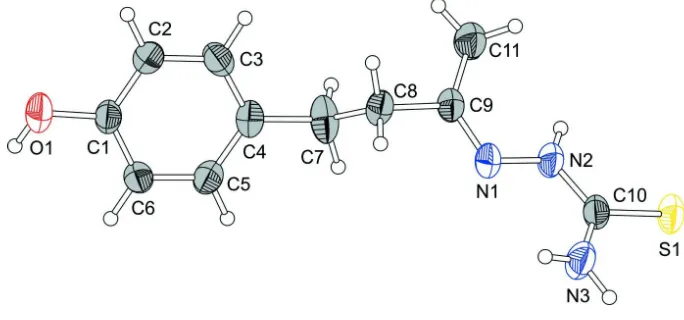
In the title molecule, Fig. 1, the thiosemicarbazone unit is nearly planar showing a torsion angle for the N1—N2—C10
—N3 entity of -3.1 (3)°. The maximum deviation from the mean plane of the non-H atoms of the C9/C10/N1/N2/N3/S1
fragment amounts to 0.085 (5)°. The angle between this mean plane and the aromatic ring is 50.31 (8)°. This strong tilting
is possiblly due to free rotation around the sp3-hybridized C7 and C8 atoms (Fig. 1).
In the crystal, molecules are connected by N—H···O, N—H···S and O—H···S hydrogen bonds, with bridging sulfur
atoms, into a three-dimensional H-bonded network (Figs. 2 and 3, and Table 1). The molecules are arranged along the
[011] direction, but the hydrogen bonding interactions are present along all three directions (Fig. 3).
S2. Synthesis and crystallization
The synthesis of the title compound was adapted from a procedure reported previously (Freund & Schander, 1902). In a
hydrochloric acid catalyzed reaction, a mixture of 4-(4-hydroxyphenyl)-2-butanone (raspberry ketone) (10 mmol) and
thiosemicarbazide (10 mmol) in ethanol (80 mL) was refluxed for 5 h. After cooling and filtering, the title compound was
obtained. Yellow crystals suitable for X-ray diffraction were obtained by slow evaporation of asolution in methanol.
S3. Refinement
Crystal data, data collection and structure refinement details are summarized in Table 1. All the hydrogen atoms were
Figure 1
The molecular structure of the title compound, showing the atom labelling. Displacement ellipsoids are drawn at the 40%
probability level.
Figure 2
A view of the intermolecular hydrogen bonding (dashed lines) in the crystal of the title compound (see Table 1 for details
[image:4.610.133.474.287.391.2]supporting information
[image:5.610.132.477.74.327.2]sup-3 Acta Cryst. (2015). E71, o33–o34
Figure 3
Crystal packing of the title compound viewed along the c axis, with the molecules stacking along the [011] direction.
Hydrogen bonds are shown as dashed lines (see Table 1 for details).
(E)-2-[4-(4-Hydroxyphenyl)butan-2-ylidene]hydrazine-1-carbothioamide
Crystal data
C11H15N3OS Mr = 237.32
Monoclinic, P21/c
Hall symbol: -P 2ybc
a = 13.5604 (7) Å
b = 9.7578 (6) Å
c = 9.3079 (4) Å
β = 95.194 (3)°
V = 1226.56 (11) Å3 Z = 4
F(000) = 504
Dx = 1.285 Mg m−3
Mo Kα radiation, λ = 0.71073 Å Cell parameters from 4899 reflections
θ = 2.9–27.5°
µ = 0.25 mm−1 T = 293 K Rod, yellow
0.17 × 0.13 × 0.09 mm
Data collection
Nonius KappaCCD diffractometer
Radiation source: fine-focus sealed tube, Nonius KappaCCD
Graphite monochromator
Detector resolution: 9 pixels mm-1
CCD rotation images, thick slices scans Absorption correction: multi-scan
(Blessing, 1995)
Tmin = 0.929, Tmax = 0.994 12737 measured reflections 2806 independent reflections 1587 reflections with I > 2σ(I)
Rint = 0.058
θmax = 27.5°, θmin = 3.0°
h = −15→17
k = −11→12
Refinement on F2
Least-squares matrix: full
R[F2 > 2σ(F2)] = 0.043 wR(F2) = 0.116 S = 0.98 2806 reflections 205 parameters 0 restraints
Primary atom site location: structure-invariant direct methods
Secondary atom site location: difference Fourier map
Hydrogen site location: difference Fourier map All H-atom parameters refined
w = 1/[σ2(Fo2) + (0.0576P)2]
where P = (Fo2 + 2Fc2)/3
(Δ/σ)max = 0.001
Δρmax = 0.16 e Å−3
Δρmin = −0.21 e Å−3
Special details
Geometry. All e.s.d.'s (except the e.s.d. in the dihedral angle between two l.s. planes) are estimated using the full covariance matrix. The cell e.s.d.'s are taken into account individually in the estimation of e.s.d.'s in distances, angles and torsion angles; correlations between e.s.d.'s in cell parameters are only used when they are defined by crystal symmetry. An approximate (isotropic) treatment of cell e.s.d.'s is used for estimating e.s.d.'s involving l.s. planes.
Refinement. Refinement of F2 against ALL reflections. The weighted R-factor wR and goodness of fit S are based on F2,
conventional R-factors R are based on F, with F set to zero for negative F2. The threshold expression of F2 > σ(F2) is used
only for calculating R-factors(gt) etc. and is not relevant to the choice of reflections for refinement. R-factors based on F2
are statistically about twice as large as those based on F, and R- factors based on ALL data will be even larger.
Fractional atomic coordinates and isotropic or equivalent isotropic displacement parameters (Å2)
x y z Uiso*/Ueq
S1 0.53385 (4) −0.71017 (6) 0.78232 (6) 0.0603 (2) O1 −0.29137 (12) −0.88281 (19) 0.09365 (18) 0.0714 (5) N1 0.30776 (11) −0.76195 (18) 0.49369 (17) 0.0513 (4) N2 0.38871 (12) −0.7859 (2) 0.59241 (18) 0.0522 (5) N3 0.41152 (14) −0.5570 (2) 0.6114 (2) 0.0635 (5) C1 −0.19991 (14) −0.8803 (2) 0.1707 (2) 0.0480 (5) C2 −0.12722 (15) −0.9634 (2) 0.1242 (2) 0.0538 (5) C3 −0.03396 (16) −0.9638 (2) 0.1973 (2) 0.0550 (5) C4 −0.01049 (14) −0.8834 (2) 0.3178 (2) 0.0493 (5) C5 −0.08500 (15) −0.8014 (2) 0.3617 (2) 0.0515 (5) C6 −0.17858 (16) −0.7981 (2) 0.2904 (2) 0.0496 (5) C7 0.08871 (17) −0.8918 (4) 0.4046 (3) 0.0701 (7) C8 0.17714 (15) −0.8434 (3) 0.3288 (2) 0.0501 (5) C9 0.27054 (13) −0.8668 (2) 0.42605 (19) 0.0467 (5) C10 0.43922 (14) −0.6808 (2) 0.6538 (2) 0.0493 (5)
C11 0.3082 (2) −1.0096 (3) 0.4435 (3) 0.0671 (7)
H1 −0.337 (3) −0.853 (4) 0.148 (4) 0.145 (15)*
H2 −0.1415 (17) −1.020 (2) 0.035 (2) 0.077 (7)*
H3 0.0147 (16) −1.022 (2) 0.1646 (19) 0.054 (6)*
H5 −0.0730 (17) −0.750 (2) 0.449 (2) 0.069 (6)*
H6 −0.2290 (15) −0.741 (2) 0.3238 (19) 0.051 (6)*
H7A 0.101 (2) −0.985 (3) 0.439 (3) 0.114 (11)*
supporting information
sup-5 Acta Cryst. (2015). E71, o33–o34
H9 0.4068 (16) −0.864 (2) 0.614 (2) 0.054 (7)*
H10A 0.3683 (18) −0.544 (2) 0.541 (3) 0.077 (8)*
H10B 0.4429 (17) −0.481 (3) 0.650 (2) 0.078 (7)*
H11A 0.377 (2) −1.011 (3) 0.431 (3) 0.101 (9)*
H11B 0.275 (2) −1.069 (3) 0.380 (3) 0.121 (11)*
H11C 0.302 (2) −1.039 (3) 0.539 (3) 0.119 (11)*
Atomic displacement parameters (Å2)
U11 U22 U33 U12 U13 U23
S1 0.0393 (3) 0.0773 (4) 0.0609 (3) −0.0021 (3) −0.0135 (2) −0.0007 (3) O1 0.0395 (9) 0.0927 (13) 0.0788 (10) −0.0011 (8) −0.0123 (8) −0.0169 (9) N1 0.0360 (9) 0.0618 (11) 0.0537 (9) 0.0004 (8) −0.0091 (7) −0.0022 (8) N2 0.0378 (9) 0.0564 (13) 0.0593 (10) 0.0007 (9) −0.0129 (7) −0.0022 (9) N3 0.0545 (12) 0.0599 (14) 0.0713 (13) −0.0051 (10) −0.0212 (9) −0.0006 (10) C1 0.0352 (10) 0.0496 (13) 0.0583 (11) −0.0043 (10) −0.0016 (8) 0.0011 (9) C2 0.0484 (13) 0.0553 (14) 0.0575 (12) −0.0053 (11) 0.0028 (10) −0.0094 (10) C3 0.0425 (12) 0.0553 (14) 0.0681 (14) 0.0078 (11) 0.0096 (10) 0.0020 (11) C4 0.0388 (11) 0.0577 (14) 0.0504 (11) −0.0057 (10) −0.0006 (8) 0.0100 (9) C5 0.0470 (12) 0.0546 (13) 0.0522 (12) −0.0092 (11) 0.0007 (9) −0.0043 (10) C6 0.0403 (11) 0.0470 (13) 0.0618 (12) 0.0007 (10) 0.0052 (9) −0.0023 (10) C7 0.0400 (13) 0.107 (2) 0.0618 (14) −0.0023 (13) −0.0043 (10) 0.0199 (15) C8 0.0413 (12) 0.0546 (14) 0.0525 (12) −0.0065 (10) −0.0067 (9) 0.0027 (10) C9 0.0360 (11) 0.0555 (14) 0.0476 (11) −0.0003 (10) −0.0015 (8) 0.0010 (9) C10 0.0328 (10) 0.0634 (15) 0.0513 (11) −0.0021 (10) 0.0016 (8) −0.0045 (10) C11 0.0553 (16) 0.0638 (16) 0.0787 (18) 0.0031 (13) −0.0125 (13) −0.0031 (13)
Geometric parameters (Å, º)
S1—C10 1.6973 (19) C4—C5 1.379 (3)
O1—C1 1.376 (2) C4—C7 1.507 (3)
O1—H1 0.89 (4) C5—C6 1.378 (3)
N1—C9 1.281 (2) C5—H5 0.95 (2)
N1—N2 1.386 (2) C6—H6 0.95 (2)
N2—C10 1.333 (3) C7—C8 1.520 (3)
N2—H9 0.82 (2) C7—H7A 0.98 (3)
N3—C10 1.315 (3) C7—H7B 1.05 (3)
N3—H10A 0.85 (2) C8—C9 1.506 (2)
N3—H10B 0.91 (3) C8—H8A 1.01 (2)
C1—C2 1.376 (3) C8—H8B 0.92 (2)
C1—C6 1.382 (3) C9—C11 1.488 (3)
C2—C3 1.381 (3) C11—H11A 0.95 (3)
C2—H2 1.00 (2) C11—H11B 0.91 (3)
C3—C4 1.383 (3) C11—H11C 0.95 (3)
C3—H3 0.94 (2)
C1—O1—H1 110 (2) C1—C6—H6 119.7 (11)
C10—N2—H9 118.6 (14) C8—C7—H7A 109.0 (18)
N1—N2—H9 121.4 (14) C4—C7—H7B 112.1 (15)
C10—N3—H10A 121.7 (16) C8—C7—H7B 104.6 (15)
C10—N3—H10B 121.1 (15) H7A—C7—H7B 104 (2)
H10A—N3—H10B 117 (2) C9—C8—C7 109.24 (17)
O1—C1—C2 117.58 (18) C9—C8—H8A 108.1 (12)
O1—C1—C6 122.95 (19) C7—C8—H8A 112.5 (12)
C2—C1—C6 119.47 (18) C9—C8—H8B 107.2 (12)
C1—C2—C3 119.8 (2) C7—C8—H8B 111.8 (12)
C1—C2—H2 119.8 (13) H8A—C8—H8B 107.9 (17)
C3—C2—H2 120.4 (13) N1—C9—C11 125.35 (18)
C2—C3—C4 122.1 (2) N1—C9—C8 116.50 (19)
C2—C3—H3 118.7 (11) C11—C9—C8 118.01 (19)
C4—C3—H3 119.2 (11) N3—C10—N2 117.12 (19)
C5—C4—C3 116.66 (18) N3—C10—S1 122.95 (16)
C5—C4—C7 121.0 (2) N2—C10—S1 119.93 (17)
C3—C4—C7 122.2 (2) C9—C11—H11A 109.3 (18)
C6—C5—C4 122.55 (19) C9—C11—H11B 112.3 (19)
C6—C5—H5 118.4 (14) H11A—C11—H11B 110 (3)
C4—C5—H5 118.8 (14) C9—C11—H11C 108.9 (19)
C5—C6—C1 119.4 (2) H11A—C11—H11C 106 (2)
C5—C6—H6 120.8 (11) H11B—C11—H11C 110 (3)
C9—N1—N2—C10 171.64 (19) C2—C1—C6—C5 −0.4 (3)
O1—C1—C2—C3 179.56 (19) C5—C4—C7—C8 −118.0 (3)
C6—C1—C2—C3 0.0 (3) C3—C4—C7—C8 66.8 (3)
C1—C2—C3—C4 0.4 (3) C4—C7—C8—C9 −176.4 (2)
C2—C3—C4—C5 −0.3 (3) N2—N1—C9—C11 −1.1 (3)
C2—C3—C4—C7 175.1 (2) N2—N1—C9—C8 174.63 (17)
C3—C4—C5—C6 −0.1 (3) C7—C8—C9—N1 −101.3 (2)
C7—C4—C5—C6 −175.6 (2) C7—C8—C9—C11 74.7 (3)
C4—C5—C6—C1 0.5 (3) N1—N2—C10—N3 −3.1 (3)
O1—C1—C6—C5 −179.97 (19) N1—N2—C10—S1 176.21 (14)
Hydrogen-bond geometry (Å, º)
D—H···A D—H H···A D···A D—H···A
O1—H1···S1i 0.89 (4) 2.32 (4) 3.206 (2) 175 (3)
N3—H10A···O1ii 0.85 (2) 2.22 (2) 2.936 (2) 143 (2)
N3—H10B···S1iii 0.91 (3) 2.73 (3) 3.585 (2) 156.6 (19)