research communications
Acta Cryst.(2014).E70, 587–589 doi:10.1107/S1600536814025604
587
Received 4 November 2014 Accepted 23 November 2014
Edited by M. Weil, Vienna University of Technology, Austria
†
Keywords:Crystal structure; rhenium carbonyl complex; 5-phenyl-3-(pyridin-2-yl)-1H-1,2,4-triazole; hydrogen bonding; slipped– stacking interactions
CCDC reference:1031232
Supporting information:this article has supporting information at journals.iucr.org/e
Crystal structure of bromido-
fac
-tricarbonyl-[5-phenyl-3-(pyridin-2-yl)-1
H
-1,2,4-triazole-j
2N
,
N
000]rhenium(I)
Kseniia Piletska,a* Konstantin V. Domasevitchband Alexander V. Shtemenkoa
a
Department of Inorganic Chemistry, Ukrainian State University of Chemical Technology, Gagarin Ave. 8, Dnipropetrovsk 49005, Ukraine, andbInorganic Chemistry Department, National Taras Shevchenko University of Kyiv, Volodymyrska Street 64/13, Kyiv 01601, Ukraine. *Correspondence e-mail: ksenijapiletska@gmail.com
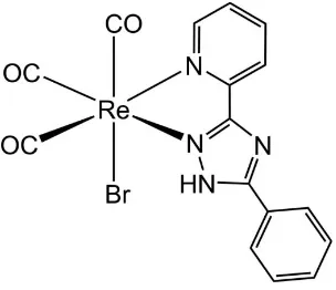
In the title compound, [ReBr(C13H10N4)(CO)3], the Re I
atom has a distorted octahedral coordination environment. Two N atoms of the 5-phenyl-3-(pyridin-2-yl)-1H-1,2,4-triazole ligand and two of the three carbonyl groups occupy the equatorial plane of the complex, with the third carbonyl ligand and the bromide ligand in the axial positions. The three carbonyl ligands are arranged in a fac
configuration around the ReIatom. Mutual N—H Br hydrogen bonds arrange molecules into centrosymmetric dimers. Additional stabilization within the crystal structure is provided by C—H O and C—H Br hydrogen bonds, as well as by slipped – stacking interactions [centroid-to-centroid distance = 3.785 (5) A˚ ], defining a three-dimensional network.
1. Chemical context
The coordination chemistry of rhenium and technetium has been well studied over the last half century, particularly in view of the potential applications of their186/188Re and99mTc isotopes in therapeutic and diagnostic agents in nuclear medicine (Volkert & Hoffman, 1999; Alberto et al., 1999). Complexes of the type [M(CO)3(NN)X] (M= Tc, Re;NN=
bidentate nitrogen donor; X = anionic ligand) have been shown to possess interesting photophysical, photochemical and excited-state redox properties (Striplin & Crosby, 2001; Stufkens & Vlceˇk, 1998), making this class of complexes applicable as fluorescent probes, in addition to their potential usage as radio-imaging and therapeutic agents. Moreover, metal carbonyls display intense infrared absorptions in the range 1800 to 2200 cm1, which is the IR transparency window for biological media (Hildebrandt, 2010). In addition to their luminescent properties, the vibrational signature of fac -[Re(CO)3(NN)] is appropriate for IR imaging (Policaret al.,
2011; Cle`de et al., 2012). They are thus valuable as small molecular units enabling multimodal imaging involving vibrational-based detections (IR, Raman) and fluorescence (Cle`de et al., 2012). In [Re(CO)3(NN)X] compounds, the
photophysical properties of the complexes are closely dependent on the ligand. When NNis a ligand with low* orbitals, the corresponding [Re(CO)3(NN)] unit is
lumines-cent (Wrighton & Morse, 1974) and this property has often been used in subcellular bio-imaging (Loet al., 2012; Baggaley
et al., 2012; Xianget al., 2013; Coogan & Fernandez-Moreira, 2014).
In this communication, we report the synthesis and crystal structure analysis of a novel ReIcomplex which contains the
triazole ligand 5-phenyl-3-(pyridin-2-yl)-1H-1,2,4-triazole, [Re(CO)3(C13H10N4)Br]. Its luminescent properties will be
reported in a forthcoming article.
2. Structural commentary
In the title compound, the ReIatom is in a slightly distorted octahedral coordination environment (Fig. 1). The three carbonyl ligands bonded to the ReIatom are arranged in afac -configuration. The distances of C1, C2, and C3 to the ReIatom are 1.905 (4), 1.915 (4), and 1.922 (6) A˚ , respectively, and the Re—N bonds lengths are 2.201 (3) and 2.164 (3) A˚ . The CO ligands are almost linearly coordinated with O—C—Re bond angles of 178.4 (4), 175.6 (3) and 179.0 (4). The C—Re—C bond angles between CO carbon atoms are 87.78 (17), 90.4 (2) and 89.18 (19), close to ideal values, whereas thecis equa-torial bite angle [N1—Re1—N2] is 74.33 (11). All other bond lengths and angles are comparable to those found for related ReIcomplexes (Rajendranet al., 2000).
3. Supramolecular features
The title compound adopts a typical molecular structure. There is only one relatively strong donor (N—H) and one
acceptor (Br) site for hydrogen-bonding interactions, which arrange molecules into dimers (Table 1, Fig. 1). Weak hydrogen bonds of the type C—H O with carbonyl O atoms as acceptor groups play a supporting role in the crystal packing. Nevertheless, these interactions demonstrate a clear discrimination of the C—H binding sites that follow a common pattern. The C—H O hydrogen bonds present are provided by the 2- and 4-C—H protons of the pyridine ring, which are the most polarized and acidic. Besides C—H Br inter-actions, weak slipped – stacking interactions between pyridine and phenyl rings (symmetry code: 1 x, y, z) [with a shortest separation of C6 C11(1 x, y, z) = 3.265 (6) A˚ , a centroid-to-centroid distance of 3.785 (5) A˚ and an interplanar angle of 7.1 (3)] also appear to be involved in the stabilization of the crystal structure (Fig. 2).
4. Synthesis and crystallization
Pentacarbonylrhenium(I) bromide (0.1 g, 0.246 mmol) was reacted with 5-phenyl-3-(pyridin-2-yl)-1H-1,2,4-triazole (0.1 g, 0.492 mmol) in benzene at 353 K, with stirring, under a steady stream of argon for five h. The dark-yellow solution was removed from the heat and allowed to cool overnight. The yellow product was collected by suction filtration, washed with a 50 ml portion of petroleum ether and dried. Yield = 0.107g,
588
Piletskaet al. [ReBr(C13H10N4)(CO)3] Acta Cryst.(2014).E70, 587–589
[image:2.610.95.247.124.255.2]research communications
Figure 1
The structure of the title complex, showing the association of molecules into a centrosymmetric dimer by means of mutual hydrogen bonds of the N—H Br and C—H Br types. Displacement ellipsoids are drawn at the 40% probability level. [Symmetry code: (i)x+1
2,y+ 1 2,z.]
Table 1
Hydrogen-bond geometry (A˚ ,).
D—H A D—H H A D A D—H A
N3—H3 Br1i 0.87 2.51 3.360 (3) 168
C16—H16 Br1i 0.94 2.87 3.784 (4) 165
C6—H6 O2ii 0.94 2.38 3.194 (5) 145
C8—H8 O1iii 0.94 2.56 3.285 (5) 134
Symmetry codes: (i)xþ1 2;yþ
1
2;z; (ii)xþ 1 2;yþ
1
[image:2.610.319.565.504.693.2]2;z; (iii)xþ1;y;zþ 1 2.
Figure 2
The crystal structure of the title complex, showing weak hydrogen-bonding interactions (indicated by dotted lines) of the type C—H O between carbonyl O atoms and pyridyl C—H groups of the organic ligands. [Symmetry codes: (i)x+1
2,y+ 1
[image:2.610.47.295.515.700.2](76.4%). Crystals suitable for X-ray diffraction were obtained by slow diffusion of hexane into a methanol solution of the complex. IR (KBr, cm1):as(CO) 2028 (s),s(CO) 1912 (s).
5. Refinement
Crystal data, data collection and structure refinement details are summarized in Table 2. H atoms were positioned with
idealized geometry and were refined with C—H = 0.94, N—H = 0.87 A˚ andUiso(H) = 1.2Ueq(C,N).
Acknowledgements
This work was supported by the fund ‘Grant for Science Research’ (No. 0111U000111) from the Ministry of Education and Science of Ukraine. We thank the Taurida National V. I. Vernadsky University, Crimea, Ukraine, for providing the ligand.
References
Alberto, R., Schibli, R., Waibel, R., Abram, U. & Schubiger, A. P. (1999).Coord. Chem. Rev.901, 190–192.
Baggaley, E., Weinstein, J. A. & Williams, J. A. G. (2012).Coord. Chem. Rev.256, 1762–1785.
Brandenburg, K. (1999).DIAMOND. Crystal Impact GbR, Bonn, Germany.
Cle`de, S., Lambert, F., Sandt, C., Gueroui, Z., Re´fre´giers, M., Plamont, M.-A., Dumas, P., Vessie`res, A. & Policar, C. (2012). Chem. Commun.48, 7729–7731.
Coogan, M. P. & Fernandez-Moreira, V. (2014).Chem. Commun.50, 384–399.
Farrugia, L. J. (2012).J. Appl. Cryst.45, 849–854.
Hildebrandt, P. (2010).Angew. Chem. Int. Ed.49, 4540–4541. Lo, K. K.-W., Choi, A. W.-T. & Law, W. H.-T. (2012).Dalton Trans.41,
6021–6047.
Policar, C., Waern, J. B., Plamont, M. A., Cle`de, S., Mayet, C., Prazeres, R., Ortega, J.-M., Vessie`res, A. & Dazzi, A. (2011). Angew. Chem. Int. Ed.50, 860–864.
Rajendran, T., Manimaran, B., Lee, F.-Y., Lee, G.-H., Peng, S.-M., Wang, C.-C. & Lu, K.-L. (2000).Inorg. Chem.39, 2016–2017. Sheldrick, G. M. (2008).Acta Cryst.A64, 112–122.
Stoe & Cie (2001).X-SHAPE,X-REDandIPDS. Stoe & Cie GmbH, Darmstadt, Germany.
Striplin, D. R. & Crosby, G. A. (2001).Coord. Chem. Rev.211, 163– 175.
Stufkens, D. J. & Vlceˇk, A. Jr (1998).Coord. Chem. Rev.177, 127–179. Volkert, W. A. & Hoffman, T. J. (1999).Chem. Rev.99, 2269–2292. Wrighton, M. & Morse, D. L. (1974).J. Am. Chem. Soc.96, 998–1003. Xiang, H., Cheng, J., Ma, X., Zhou, X. & Chruma, J. J. (2013).Chem.
Soc. Rev.42, 6128–6185.
research communications
Acta Cryst.(2014).E70, 587–589 Piletskaet al. [ReBr(C
[image:3.610.43.294.88.364.2]13H10N4)(CO)3]
589
Table 2
Experimental details.
Crystal data
Chemical formula [ReBr(C13H10N4)(CO)3]
Mr 572.39
Crystal system, space group Monoclinic,C2/c
Temperature (K) 213
a,b,c(A˚ ) 20.8082 (15), 7.2521 (4), 24.386 (2)
() 111.599 (7)
V(A˚3) 3421.5 (4)
Z 8
Radiation type MoK
(mm1) 9.46
Crystal size (mm) 0.140.120.11
Data collection
Diffractometer Stoe Imaging plate diffraction
system
Absorption correction Numerical (X-REDand
X-SHAPE; Stoe & Cie, 2001)
Tmin,Tmax 0.319, 0.385
No. of measured, independent and observed [I> 2(I)] reflections
14578, 4092, 2844
Rint 0.057
(sin/ )max(A˚1) 0.661
Refinement
R[F2> 2(F2)],wR(F2),S 0.023, 0.050, 0.84
No. of reflections 4092
No. of parameters 226
H-atom treatment H-atom parameters constrained
max,min(e A˚3) 1.03,1.14
supporting information
sup-1
Acta Cryst. (2014). E70, 587-589
supporting information
Acta Cryst. (2014). E70, 587-589 [doi:10.1107/S1600536814025604]
Crystal structure of bromido-
fac
-tricarbonyl[5-phenyl-3-(pyridin-2-yl)-1
H
-1,2,4-triazole-
κ
2N
,
N
′
]rhenium(I)
Kseniia Piletska, Konstantin V. Domasevitch and Alexander V. Shtemenko
Computing details
Data collection: IPDS (Stoe & Cie, 2001); cell refinement: IPDS (Stoe & Cie, 2001); data reduction: IPDS (Stoe & Cie,
2001); program(s) used to solve structure: SHELXS97 (Sheldrick, 2008); program(s) used to refine structure:
SHELXL2014 (Sheldrick, 2008); molecular graphics: DIAMOND (Brandenburg, 1999); software used to prepare material
for publication: WinGX (Farrugia, 2012).
Bromido-fac-tricarbonyl[5-phenyl-3-(pyridin-2-yl)-1H-1,2,4-triazole-κ2N,N′]rhenium(I)
Crystal data
[ReBr(C13H10N4)(CO)3]
Mr = 572.39 Monoclinic, C2/c a = 20.8082 (15) Å
b = 7.2521 (4) Å
c = 24.386 (2) Å
β = 111.599 (7)°
V = 3421.5 (4) Å3
Z = 8
F(000) = 2144
Dx = 2.222 Mg m−3
Mo Kα radiation, λ = 0.71073 Å Cell parameters from 8000 reflections
θ = 3.0–28.0°
µ = 9.46 mm−1
T = 213 K Prism, yellow
0.14 × 0.12 × 0.11 mm
Data collection
Stoe Imaging plate diffraction system diffractometer
Radiation source: fine-focus sealed tube
φ oscillation scans
Absorption correction: numerical
(X-RED and X-SHAPE; Stoe & Cie, 2001)
Tmin = 0.319, Tmax = 0.385 14578 measured reflections
4092 independent reflections 2844 reflections with I > 2σ(I)
Rint = 0.057
θmax = 28.0°, θmin = 3.0°
h = −27→27
k = −9→8
l = −32→32
Refinement
Refinement on F2 Least-squares matrix: full
R[F2 > 2σ(F2)] = 0.023
wR(F2) = 0.050
S = 0.84 4092 reflections 226 parameters 0 restraints
Primary atom site location: structure-invariant direct methods
Secondary atom site location: difference Fourier map
Hydrogen site location: inferred from neighbouring sites
H-atom parameters constrained
w = 1/[σ2(F
o2) + (0.0225P)2] where P = (Fo2 + 2Fc2)/3 (Δ/σ)max = 0.001
supporting information
sup-2
Acta Cryst. (2014). E70, 587-589
Special details
Geometry. All e.s.d.'s (except the e.s.d. in the dihedral angle between two l.s. planes) are estimated using the full covariance matrix. The cell e.s.d.'s are taken into account individually in the estimation of e.s.d.'s in distances, angles and torsion angles; correlations between e.s.d.'s in cell parameters are only used when they are defined by crystal symmetry. An approximate (isotropic) treatment of cell e.s.d.'s is used for estimating e.s.d.'s involving l.s. planes.
Fractional atomic coordinates and isotropic or equivalent isotropic displacement parameters (Å2)
x y z Uiso*/Ueq
Re1 0.38382 (2) 0.25265 (3) 0.10559 (2) 0.02798 (5) Br1 0.32731 (2) 0.54427 (7) 0.04146 (2) 0.03557 (11) O1 0.40827 (17) 0.4654 (6) 0.21974 (13) 0.0594 (10) O2 0.24980 (15) 0.1146 (5) 0.11477 (13) 0.0516 (10) O3 0.4540 (2) −0.0838 (7) 0.17525 (18) 0.0796 (14) N1 0.48045 (14) 0.3439 (5) 0.09681 (13) 0.0264 (7) N2 0.38108 (14) 0.1420 (5) 0.02237 (12) 0.0244 (7) N3 0.33622 (14) 0.0546 (5) −0.02600 (12) 0.0258 (7)
H3 0.2960 0.0108 −0.0295 0.031*
N4 0.42685 (15) 0.1255 (5) −0.04749 (13) 0.0255 (7) C1 0.3988 (2) 0.3830 (7) 0.17704 (17) 0.0372 (12) C2 0.2983 (2) 0.1674 (7) 0.10904 (15) 0.0361 (11) C3 0.4289 (2) 0.0391 (9) 0.14970 (19) 0.0463 (13) C4 0.49040 (18) 0.2879 (5) 0.04763 (16) 0.0246 (9) C5 0.54965 (18) 0.3314 (7) 0.03688 (17) 0.0297 (9)
H5 0.5554 0.2910 0.0024 0.036*
C6 0.59958 (19) 0.4352 (7) 0.07811 (18) 0.0341 (10)
H6 0.6407 0.4653 0.0725 0.041*
C7 0.58898 (19) 0.4942 (6) 0.12745 (18) 0.0347 (10)
H7 0.6225 0.5668 0.1557 0.042*
C8 0.5289 (2) 0.4467 (7) 0.13551 (17) 0.0340 (10)
H8 0.5220 0.4883 0.1694 0.041*
C9 0.43441 (18) 0.1817 (6) 0.00714 (16) 0.0258 (8) C10 0.36475 (18) 0.0474 (6) −0.06765 (15) 0.0246 (8) C11 0.33235 (18) −0.0329 (6) −0.12595 (15) 0.0268 (9) C12 0.3677 (2) −0.0228 (7) −0.16469 (17) 0.0337 (10)
H12 0.4104 0.0389 −0.1534 0.040*
C13 0.3397 (3) −0.1037 (7) −0.21942 (18) 0.0426 (12)
H13 0.3636 −0.0967 −0.2454 0.051*
C14 0.2775 (3) −0.1946 (7) −0.23681 (19) 0.0448 (12)
H14 0.2592 −0.2506 −0.2741 0.054*
C15 0.2425 (2) −0.2025 (7) −0.19894 (19) 0.0403 (12)
H15 0.1998 −0.2636 −0.2106 0.048*
C16 0.2693 (2) −0.1219 (7) −0.14401 (18) 0.0369 (11)
supporting information
sup-3
Acta Cryst. (2014). E70, 587-589
Atomic displacement parameters (Å2)
U11 U22 U33 U12 U13 U23
Re1 0.02700 (7) 0.03224 (11) 0.02386 (7) −0.00874 (8) 0.00836 (5) 0.00069 (8) Br1 0.03230 (18) 0.0375 (3) 0.03867 (19) −0.0024 (2) 0.01516 (15) 0.0035 (2) O1 0.068 (2) 0.075 (3) 0.0338 (16) −0.021 (2) 0.0179 (15) −0.0196 (19) O2 0.0364 (15) 0.077 (3) 0.0440 (17) −0.0237 (18) 0.0178 (13) −0.0038 (18) O3 0.081 (3) 0.071 (4) 0.079 (3) 0.009 (3) 0.020 (2) 0.034 (3) N1 0.0216 (14) 0.022 (2) 0.0309 (15) −0.0032 (15) 0.0036 (12) 0.0023 (15) N2 0.0224 (14) 0.019 (2) 0.0291 (15) −0.0061 (14) 0.0071 (12) 0.0002 (14) N3 0.0250 (14) 0.023 (2) 0.0270 (14) −0.0053 (15) 0.0074 (11) −0.0012 (15) N4 0.0240 (14) 0.018 (2) 0.0350 (16) −0.0011 (14) 0.0116 (12) 0.0018 (15) C1 0.032 (2) 0.049 (4) 0.0275 (19) −0.009 (2) 0.0074 (15) 0.004 (2) C2 0.036 (2) 0.047 (3) 0.0221 (17) −0.013 (2) 0.0068 (15) −0.0053 (19) C3 0.041 (2) 0.053 (4) 0.043 (2) −0.007 (3) 0.0144 (19) 0.017 (3) C4 0.0250 (16) 0.015 (3) 0.0322 (17) 0.0002 (15) 0.0084 (13) 0.0071 (15) C5 0.0245 (17) 0.023 (3) 0.042 (2) 0.0009 (18) 0.0128 (16) 0.009 (2) C6 0.0243 (18) 0.021 (3) 0.052 (2) −0.0034 (19) 0.0086 (17) 0.014 (2) C7 0.0268 (18) 0.018 (3) 0.047 (2) −0.0065 (18) −0.0005 (16) 0.009 (2) C8 0.038 (2) 0.024 (3) 0.0353 (19) −0.007 (2) 0.0078 (16) −0.003 (2) C9 0.0266 (17) 0.016 (2) 0.0354 (19) 0.0016 (17) 0.0123 (15) 0.0062 (17) C10 0.0298 (18) 0.013 (2) 0.0293 (17) 0.0031 (17) 0.0089 (14) 0.0020 (17) C11 0.0324 (19) 0.018 (3) 0.0287 (17) 0.0041 (18) 0.0099 (15) −0.0024 (17) C12 0.044 (2) 0.022 (3) 0.037 (2) 0.005 (2) 0.0161 (17) 0.0047 (19) C13 0.065 (3) 0.030 (3) 0.039 (2) 0.012 (2) 0.025 (2) 0.002 (2) C14 0.066 (3) 0.030 (3) 0.033 (2) 0.002 (2) 0.012 (2) −0.008 (2) C15 0.048 (2) 0.027 (4) 0.041 (2) −0.006 (2) 0.0107 (19) −0.013 (2) C16 0.037 (2) 0.033 (3) 0.042 (2) 0.001 (2) 0.0155 (18) −0.008 (2)
Geometric parameters (Å, º)
Re1—C1 1.905 (4) C5—C6 1.374 (6)
Re1—C2 1.915 (4) C5—H5 0.9400
Re1—C3 1.922 (6) C6—C7 1.369 (6)
Re1—N2 2.164 (3) C6—H6 0.9400
Re1—N1 2.201 (3) C7—C8 1.380 (5)
Re1—Br1 2.6357 (5) C7—H7 0.9400
O1—C1 1.153 (5) C8—H8 0.9400
O2—C2 1.135 (4) C10—C11 1.453 (5)
O3—C3 1.103 (6) C11—C16 1.380 (6)
N1—C8 1.327 (5) C11—C12 1.397 (5)
N1—C4 1.352 (5) C12—C13 1.376 (6)
N2—C9 1.325 (4) C12—H12 0.9400
N2—N3 1.361 (4) C13—C14 1.373 (7)
N3—C10 1.353 (4) C13—H13 0.9400
N3—H3 0.8700 C14—C15 1.371 (6)
N4—C10 1.328 (5) C14—H14 0.9400
supporting information
sup-4
Acta Cryst. (2014). E70, 587-589
C4—C5 1.388 (5) C15—H15 0.9400
C4—C9 1.442 (5) C16—H16 0.9400
C1—Re1—C2 87.78 (17) C7—C6—C5 119.4 (3)
C1—Re1—C3 90.4 (2) C7—C6—H6 120.3
C2—Re1—C3 89.18 (19) C5—C6—H6 120.3
C1—Re1—N2 168.91 (15) C6—C7—C8 119.7 (4)
C2—Re1—N2 102.56 (14) C6—C7—H7 120.2
C3—Re1—N2 93.71 (18) C8—C7—H7 120.2
C1—Re1—N1 95.31 (14) N1—C8—C7 122.0 (4)
C2—Re1—N1 176.88 (13) N1—C8—H8 119.0
C3—Re1—N1 91.22 (15) C7—C8—H8 119.0
N2—Re1—N1 74.33 (11) N2—C9—N4 114.0 (3)
C1—Re1—Br1 91.81 (14) N2—C9—C4 118.2 (3)
C2—Re1—Br1 93.80 (15) N4—C9—C4 127.6 (3)
C3—Re1—Br1 176.38 (12) N4—C10—N3 110.0 (3)
N2—Re1—Br1 83.63 (9) N4—C10—C11 124.8 (3)
N1—Re1—Br1 85.69 (9) N3—C10—C11 125.2 (3)
C8—N1—C4 118.4 (3) C16—C11—C12 118.9 (4)
C8—N1—Re1 125.5 (3) C16—C11—C10 122.9 (3)
C4—N1—Re1 116.1 (2) C12—C11—C10 118.1 (3)
C9—N2—N3 103.7 (3) C13—C12—C11 119.6 (4)
C9—N2—Re1 116.5 (3) C13—C12—H12 120.2
N3—N2—Re1 139.3 (2) C11—C12—H12 120.2
C10—N3—N2 108.5 (3) C14—C13—C12 121.3 (4)
C10—N3—H3 125.7 C14—C13—H13 119.4
N2—N3—H3 125.7 C12—C13—H13 119.4
C10—N4—C9 103.8 (3) C15—C14—C13 119.0 (4)
O1—C1—Re1 178.4 (4) C15—C14—H14 120.5
O2—C2—Re1 175.6 (3) C13—C14—H14 120.5
O3—C3—Re1 179.0 (4) C14—C15—C16 120.8 (4)
N1—C4—C5 122.4 (4) C14—C15—H15 119.6
N1—C4—C9 114.8 (3) C16—C15—H15 119.6
C5—C4—C9 122.8 (3) C15—C16—C11 120.4 (4)
C6—C5—C4 118.1 (4) C15—C16—H16 119.8
C6—C5—H5 120.9 C11—C16—H16 119.8
C4—C5—H5 120.9
C9—N2—N3—C10 −0.3 (4) C5—C4—C9—N2 177.7 (4)
Re1—N2—N3—C10 170.7 (3) N1—C4—C9—N4 172.3 (4)
C8—N1—C4—C5 1.1 (6) C5—C4—C9—N4 −7.0 (7)
Re1—N1—C4—C5 −178.5 (3) C9—N4—C10—N3 −0.7 (5)
C8—N1—C4—C9 −178.2 (4) C9—N4—C10—C11 179.0 (4)
Re1—N1—C4—C9 2.2 (4) N2—N3—C10—N4 0.7 (5)
N1—C4—C5—C6 0.0 (6) N2—N3—C10—C11 −179.1 (4)
C9—C4—C5—C6 179.3 (4) N4—C10—C11—C16 176.3 (4)
C4—C5—C6—C7 −1.0 (6) N3—C10—C11—C16 −4.0 (7)
supporting information
sup-5
Acta Cryst. (2014). E70, 587-589
C4—N1—C8—C7 −1.3 (6) N3—C10—C11—C12 177.5 (4)
Re1—N1—C8—C7 178.3 (3) C16—C11—C12—C13 −1.0 (7)
C6—C7—C8—N1 0.3 (7) C10—C11—C12—C13 177.5 (4)
N3—N2—C9—N4 −0.1 (5) C11—C12—C13—C14 0.0 (7)
Re1—N2—C9—N4 −173.6 (3) C12—C13—C14—C15 0.8 (7)
N3—N2—C9—C4 175.8 (3) C13—C14—C15—C16 −0.5 (7)
Re1—N2—C9—C4 2.3 (5) C14—C15—C16—C11 −0.5 (7)
C10—N4—C9—N2 0.5 (5) C12—C11—C16—C15 1.2 (7)
C10—N4—C9—C4 −174.9 (4) C10—C11—C16—C15 −177.1 (4)
N1—C4—C9—N2 −3.0 (5)
Hydrogen-bond geometry (Å, º)
D—H···A D—H H···A D···A D—H···A
N3—H3···Br1i 0.87 2.51 3.360 (3) 168
C16—H16···Br1i 0.94 2.87 3.784 (4) 165
C6—H6···O2ii 0.94 2.38 3.194 (5) 145
C8—H8···O1iii 0.94 2.56 3.285 (5) 134
![Crystal structure of diethyl {2,2,2 trichloro 1 [2 (1,3 dioxo 2,3 dihydro 1H isoindol 2 yl) 4 methylpentanamido]ethyl}phosphonate](data:image/gif;base64,R0lGODlhAQABAIAAAP///wAAACH5BAEAAAAALAAAAAABAAEAAAICRAEAOw==)