Original Article
Diagnostic value of high-frequency ultrasound
for submandibular gland sialolithiasis
Hong-Yan Chen1*, Bo-Ji Liu2*, Dan-Dan Li2, Chun-Yun Wu3, Hui Zhu1, Wei-Ping Xu1, Dong-Hua Wang1, Hui-Xiong Xu2
1Department of Ultrasound, Minhang Central Hospital, Fudan University, Xinsong Road, Shanghai 201199, China; 2Department of Medical Ultrasound, Shanghai Tenth People’s Hospital, Ultrasound Research and Education
Institute, Tongji University School of Medicine, Yanchangzhong Road, Shanghai 200072, China; 3Department of
Stomatology, Minhang Central Hospital, Fudan University, Xinsong Road, Shanghai 201199, China. *Equal
con-tributors.
Received September 15, 2015; Accepted January 9, 2016; Epub February 15, 2016; Published February 29, 2016
Abstract: The purpose of the study was to evaluate the diagnostic value of combined use of linear-array and intra-cavity high-frequency transducers in submandibular gland sialolithiasis. This retrospective study involved 77 cases with surgically confirmed submandibular gland sialolithiasis. All patients underwent high-frequency ultrasound with linear-array and intracavity transducers before surgery. Submandibular gland size, shape, echogenicity, blood sup-ply, duct dilation, and other sonographic characteristics were carefully analyzed. Using linear-array high-frequency transducer, among the 77 cases with submandibular gland sialolithiasis, 63 cases were correctly diagnosed before operation, 13 cases had missed diagnoses and 1 case was wrongly diagnosed, resulting in an accurate diagnosis rate of 81.8% (63/77). However, using linear-array together with intracavity transducer, 72 cases were correctly diagnosed before operation, 4 cases had missed diagnoses and 1 case was wrongly diagnosed, resulting in an ac-curate diagnosis rate of 93.5% (72/77). According to location, submandibular gland sialolithiasis was divided into four types: type I, intra-gland stones only; type II, hilum or gland-duct junction stones, with or without intra-gland stones; type III, extra-gland stones (stones in the main duct), with or without gland stones; and type IV, intra-ductal stones at mouth floor close to the intra-ductal opening. Among 17 cases of type III sialolithiasis, 9 were detected by linear transducer and another 7 were detected by additional use of intracavitary transducer under the chin. Two cases of type IV sialolithiasis were detected by putting intracavity transducer under the chin, whereas they were not detected using linear transducer. In conclusion, combined use of linear-array and intracavity high-frequency transducers enables scanning from multiple directions, which leads to improved visualization and high accuracy in detecting submandibular gland sialolithiasis.
Keywords: High frequency ultrasound, linear-array transducer, intracavity transducer, submandibular gland, sialo-lithiasis
Introduction
Submandibular gland sialolithiasis is a relative-ly common form of sialolithiasis, accounting for 80%-90% of all the cases; it is a major cause of acute or chronic submandibular gland inflam-mation [1, 2]. Primary clinical manifestations of submandibular gland sialolithiasis include sub-mandibular pain and swelling. Previously, the diagnosis of submandibular gland sialolithiasis was mainly achieved by palpation, X-ray inspec-tion. However, X-ray examination has difficulty in visualizing soft stones, thus limiting its diag-nostic accuracy. Cone beam computed
that a stone within the duct could be missed. To further improve the detection rate of subman-dibular gland sialolithiasis, US in combination with X-ray examination is generally used. This increases the sensitivity to 80-96% in visualiz-ing soft stones [8, 9], but the radiation associ-ated with X-ray examination cannot be avoided. The intracavity transducer, although designed primarily for gynecology & obstetrics or pros-tate studies, has better contact between the transducer and skin compared to the linear transducer. Therefore, the utilization of the intracavity transducer might provide better visualization of the submandibular duct, lead-ing to an improved diagnostic accuracy for sub-mandibular gland sialolithiasis.
In this retrospective study, the US features of 77 cases of surgically confirmed submandibu-lar gland sialolithiasis over a 9 year period were analyzed. All cases underwent preoperative high frequency US examination with a combina-tion of linear transducer and intracavitary transducer. The aim of the study was to evalu-ate the diagnostic value of the combined use of linear-array and intracavity transducers for sub-mandibular gland sialolithiasis.
Methods and materials
Patients
The study included 77 patients with subman-dibular gland sialolithiasis that were confirmed by surgery between October 2004 and August 2013. There were 51 men and 26 women, rang-ing in age from 12 to 75 years old, with a mean age of 44±12 years. All the cases with sialoli-thiasis were unilateral and all the patients underwent unilateral submandibular gland excision. Forty-five cases were on the right side and 32 cases were on the left side.
Methods
A Philips IU22 color Doppler ultrasound imaging unit (Philips Ultrasound, Bothell, WA, USA) with an intracavity transducer (frequency range, 5-17 MHz) and an Esaote DU-6 color Doppler ultrasound imaging unit (Esaote, Genoa, Italy) with a linear array transducer (frequency range, 7.5-10 MHz) were used. In order to fully expose the submandibular area, patients were scanned in a supine position, with a pillow located under the shoulder when necessary. The
submandib-ular glands were scanned thoroughly to observe the gland size, morphology, internal echogenici-ty, and duct diameter. Color Doppler flow imag-ing was performed to evaluate the vascularity in the gland and was used to make a distinction between blood vessels and submandibular gland duct dilation.
The findings in the suspected submandibular gland were referenced with the contralateral submandibular gland, and the whole course of the submandibular gland duct was traced on the oblique coronal plane parallel to the man-dibular body. Once duct dilation was found, its course was tracked until the stone obstruction location was detected. When the stone was located in the main duct rather than intraglan-dular, an intracavity transducer was applied. A sterile membrane was placed over the intracav-ity transducer, which was placed under the chin or directly placed under the tongue near the lingual frenulum.
Statistical analysis
Statistical analysis was performed using SPSS software (version 18.0, Chicago, IL, USA). Continuous variables were expressed as mean ± standard deviation (SD) and range. The num-bers of four types of submandibular gland sialo-lithiasis on ultrasound characteristics were counted using descriptive statistics. Differ- ences in accuracy were compared using the χ2 test. A two-tailed P<0.05 was defined as statis-tically significant difference.
Results
Using linear-array high-frequency transducer, among the 77 cases with submandibular gland sialolithiasis, 63 cases were correctly diag-nosed before surgery, 13 cases had missed diagnoses and 1 case was wrongly diagnosed, resulting in an accurate diagnosis rate of 81.8% (63/77). However, using linear-array in combi-nation with intracavity transducer, 72 cases were correctly diagnosed before surgery, 4 cases had missed diagnoses and 1 case was wrongly diagnosed, resulting in an statistically significant increased accurate diagnosis rate of 93.5% (72/77) (P=0.048) (Table 1).
1) Type I, intra-gland stones only (n=46): All the lesions appeared hyperechoic, among which 38 cases showed posterior acoustic shadowing and 8 cases did not. Fifteen cases had single stone and 31 cases had multiple stones. Twenty-eight cases showed tree-like duct dila-tion and 18 cases showed thin strip-like duct dilation (Figure 1A).
[image:3.612.89.526.86.138.2]2) Type II, gland hilum (gland-duct junction) stones, with or without intra-gland stones (n=12): All were hyperechoic with acoustic shadowing on US, of which 11 cases had single stone and 1 case had multiple stones. In the 11 cases with single stone at the gland hilum, 4 cases were accompanied by intraglandular stones. Ten cases showed tree-like dilation of
Table 1. The accuracy of two different methods
Methods diagnosedCorrectly diagnosedMissed diagnosedWrongly Accuracy
Linear-array high-frequency transducer 63 13 1 81.8% (63/77)
Together with intracavity transducer 72 4 1 93.5%* (72/77)
[image:3.612.93.524.170.338.2]Note: *P=0.048, compared with accuracy of linear-array high frequency transducer (93.5% vs 81.8%).
Figure 1. Intra-gland stone (Type I), gland hilum stone (Type II) and extra-gland stone (Type III) of submandibular gland sialolithiasis. A: Intra-gland stones and intra-glandular duct dilation (arrows); B: Gland hilum stone is shown in the gland-duct junction (arrows); C: Extra-gland stone is present in the main duct (arrows).
[image:3.612.92.526.401.572.2]the duct and 2 cases showed cystic duct dila-tion (Figures 1B and 2).
3) Type III, extra-gland stones (stones in the main duct), with or without intra-gland stones (n=17): All the lesions were hyperechoic, among which 10 cases showed posterior acoustic shadowing and 7 cases did not. Fifteen cases had single stone and 2 cases had multiple stones (multiple segmental duct obstructions), including 11 cases accompanied by intraglan-dular stones. The duct showed tree-like dilation in 10 cases, thin strip-like dilation in 5 cases, and cystic dilation in 2 cases. Among 17 cases of type III stones, 10 were detected by linear transducer and additional 7 were detected by intracavity transducer under the chin. All the type III stones were located in the middle-lower of the main duct (from gland hilum to the mouth floor close to ductal opening) (Figure 1C).
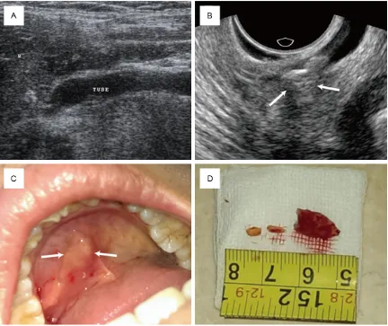
4) Type IV, intraductal stones at the mouth floor close to ductal opening (n=2): Both cases were detected by putting the intracavity transducer under the chin. Neither was detected by the lin-ear transducer (Figure 3).
The ultrasound features of the submandibular glands for the 77 cases were shown in Table 2. Among the 77 cases with submandibular gland sialolithiasis, 17 cases had simple submandib-ular gland duct calculi, 30 cases had coexisting chronic submandibular gland inflammation, and 31 cases had acute episode of chronic inflammation.
[image:4.612.91.523.71.434.2]There were four false-negative cases in this series. Among these cases, three appeared to have submandibular gland enlargement and uneven echo on US, which were speculated to be submandibular gland inflammation without
any signs of duct dilation and stones. Surgical results showed that two cases had muddy intraglandular duct stones and one case had small loose stones in the main duct. Another case was negative on US and surgical result revealed muddy intraglandular duct stones. On the other hand, there was one false-positive case in the study. US images indicated glandu-lar duct dilation with muddy stones and bilat-eral main duct dilation was found. Surgery con-firmed duct dilation due to stricture caused by a long-term chronic inflammatory adhesion; there were inflammatory sediments in the glan-dular duct.
Discussion
The submandibular gland is located within the submandibular triangle. After leaving the gland, the salivary duct runs irregularly, opening lat-eral to the lingual frenulum in the anterior floor of the mouth. The total length of the salivary duct is approximately 6-7 cm and its diameter is about 2 mm. Submandibular duct is slender and long, with a predilection as the site of cal-culi and inflammation [10]. Typical US features for submandibular gland stones consist of round or oval hyperechoic structures with pos-terior acoustic shadowing. Proximal ductal dila-tion can be seen if the stone causes complete
Submandibular duct dilation in combination with inflammatory change of the gland on US always suggests ductal obstruction. As such, the duct should be tracked along its entire course from the gland hilum to determine the nature and location of the obstruction. The diagnosis of submandibular sialolithiasis can be achieved when there is a hyperechoic object with posterior acoustic shadowing. When an extraglandular stone is far from the gland hilum (especially if near the ductal papilla), the con-spicuity of the US will be diminished due to gas interference in the mouth floor or the mandibu-lar tissue blockage. In such a case, we used an intracavity transducer under the chin at a loca-tion near the lingual frenulum to avoid the man-dibular bone interference. If needed, the intra-cavity transducer can also be placed inside the mouth after the transducer is completely cleaned and disinfected, which further improved the diagnostic accuracy for stones located near the papilla. Our accurate diagno-sis rate was 93.5%, condiagno-sistent with previously reported accuracy of about 90% [13].
[image:5.612.91.329.96.294.2]Several factors may have contributed to the 4 false-negative results in our study. In three cases, the intraglandular duct muddy stones were isoechoic and spread in the slightly dilat-ed ducts, making it difficult to be differentiatdilat-ed
Table 2. Ultrasound findings of the submandibular glands in the 77 cases
Characteristics Type I Type II Type III Type IV
Number of cases 46 12 17 2
Submandibular morphology
Swelling 34 10 12 2
Normal 12 2 5 0
Increased volume
Obvious 29 8 7 2
Mild 5 2 5 0
None 12 2 5 0
Uneven echogenicity
Decreased 27 8 10 2
Increased 19 4 7 0
Blood supply
Rich 25 10 12 2
Dotted 21 2 5 0
Note-Types of submandibular gland sialolithiasis: Type I, intra-gland stones only; Type II, gland hilum stones (gland-duct junction), with or without intra-gland stones; Type III, extra-gland stones (stones in the main duct), with or without intra-gland stones; Type IV, intraductal stones at the mouth floor close to ductal opening.
from inflammatory changes of the gland or the duct within normal glands. In the fourth case, small loose stones in the main duct were found. Obstruction was incomplete and thus ductal dilation was not significant. Terraz [4] reported that stone size is an important factor that can influence diagnostic accuracy. Stones <3 mm often do not have strong echo and obvious pos-terior acoustic shadowing; the duct may not be dilated, thus resulting in false-negative results. In addition, the loose stones appeared isoecho-ic, which also contributed to the misdiagnosis. Several useful tips to minimize false negative results are as follows: 1) If there is intraglandu-lar duct dilation, the stones may be present either in the glandular duct or the main duct. If it is located in the main duct, the main duct is usually dilated. 2) The diagnosis of sialolithiasis should be considered if there is a history of alternate submandibular swelling and degrada-tion. 3) A patient who is suspected to have sali-vary obstruction can be given stimulating food in order to make ductal dilation more obvious. Koischwit [9] has injected agents to facilitate salivary secretion and duct dilation and improved display of intraductal stones is avail-able. Other authors reported that vitamin C administration prior to US examination can pro-mote duct dilation and thereby improve the detection rate of duct stones [14, 15]. 4) Missed diagnoses may occur when there are intraductal stones in parenchyma which fails to cause dilation [9, 12].
There was one false-positive case in this study. Although the patient had bilateral main duct dilation, it was caused by the stricture of the main duct due to previous surgery. The debris in the duct mimicked intraductal stones. Therefore, it is necessary to exclude duct obstruction caused by duct wall fibrosis or scar-ring [4, 12], as well as echogenic gas in the duct [11]. In addition, some authors used finger compression from oral cavity towards the trans-ducer, which can also improve the display of stones within the main duct [11, 12]. Patel et al [12] found that the diagnostic sensitivity and specificity of US alone were 91% and 80%, which were improved to 96.9% and 90% using routine US plus finger compression from oral cavity.
Currently, there are several diagnostic imaging methods for submandibular gland sialolithiasis,
including US, X-ray, CT, MRI, iodinated sialogra-phy and salivary endoscopy. Of them, X-ray and CT are associated with radiation exposure. They also have difficulties in visualizing soft stones, thus up to 20% of stones are invisible by these modalities [8, 16], which affects the accuracy of these imaging modalities [17, 18]. Although iodinated sialography is considered as the clinical gold standard, it is invasive and highly dependent on the operator experience. Additionally, the intra- or post-operative pain and possible allergic reactions cannot be neglected. Furthermore, it is contraindicated for submandibular gland inflammation since contrast is apt to increase the extent of inflam-mation [3, 4, 19, 20]. MRI, as a non-invasive method, has a sensitivity and specificity of 80-100% and 90-100% [19, 21, 22]; however, it is not suitable for daily practice due to its high cost. Salivary endoscopy is highly accurate for diagnosis of sialolithiasis and treatment by endoscopic removal of stones is available; nev-ertheless, it is invasive and expensive, acute submandibular gland inflammation/large sto- nes (>10 mm)/intra-gland stones were unsuit-able for this examination [23, 24].
In summary, high-frequency US examination is convenient and noninvasive; it can determine the location the submandibular gland duct cal-culi intuitively and accurately if combined with the use of intracavity transducer, which is of great value in choosing appropriate treatment method and is an important modality in postop-erative follow-up.
Acknowledgements
This work was supported in part by grant 2012045 of Shanghai Talent Development Project from Shanghai Human Resource and Social Security Bureau and grant of 2013SY- 066 from Shanghai Municipal Commission of Health and Family Planning.
Disclosure of conflict of interest
None.
References
[1] Drage NA, Brown JE, Escudier MP, McGurk M. Interventional radiology in the removal of sali-vary calculi. Radiology 2000; 214: 139-142. [2] Su JZ, Yang NY, Liu XJ, Cai ZG, Lv L, Zhang L,
Wu LL, Liu DG, Ren WG, Gao Y, Yu GY. Obstructive sialadenitis of a transplanted sub-mandibular gland: chronic inflammation sec -ondary to ductal obstruction. Br J Ophthalmol 2014; 98: 1672-1677.
[3] Diederich S, Wernecke K, Peters PE. Sialographic and sonographic diagnosis of salivary gland diseases. Radiologe 1987; 27: 255-261.
[4] Terraz S, Poletti PA, Dulguerov P, Dfouni N, Becker CD, Marchal F, Becker M. How reliable is sonography in the assessment of sialolithia-sis. AJR Am J Roentgenol 2013; 201: W104-109.
[5] Koch M, Zenk J, Iro H. Algorithms for treatment of salivary gland obstructions. Otolaryngol Clin North Am 2009; 42: 1173-1192, Table of Contents.
[6] Gritzmann N. Sonography of the salivary glands. AJR Am J Roentgenol 1989; 153: 161-166.
[7] Avrahami E, Englender M, Chen E, Shabtay D, Katz R, Harell M. CT of submandibular gland sialolithiasis. Neuroradiology 1996; 38: 287-290.
[8] Alyas F, Lewis K, Williams M, Moody AB, Wong KT, Ahuja AT, Howlett DC. Diseases of the sub-mandibular gland as demonstrated using high resolution ultrasound. Br J Radiol 2005; 78: 362-369.
[9] Koischwitz D, Gritzmann N. Ultrasound of the neck. Radiol Clin North Am 2000; 38: 1029-1045.
[10] Ellies M, Laskawi R, Arglebe C, Schott A. Surgical management of nonneoplastic dis-eases of the submandibular gland. A follow-up study. Int J Oral Maxillofac Surg 1996; 25: 285-289.
[11] Bialek EJ, Jakubowski W, Zajkowski P, Szopinski KT, Osmolski A. US of the major salivary glands: anatomy and spatial relationships, pathologic conditions, and pitfalls. Radiographics 2006; 26: 745-763.
[12] Patel NJ, Hashemi S, Joshi AS. Sonopalpation: a novel application of ultrasound for detection of submandibular calculi. Otolaryngol Head Neck Surg 2014; 151: 770-775.
[13] Marchal F, Dulguerov P. Sialolithiasis manage-ment: the state of the art. Arch Otolaryngol Head Neck Surg 2003; 129: 951-956. [14] Bozzato A, Hertel V, Bumm K, Iro H, Zenk J.
Salivary simulation with ascorbic acid enhanc-es sonographic diagnosis of obstructive sialad-enitis. J Clin Ultrasound 2009; 37: 329-332. [15] Bozzato A, Hertel V, Koch M, Zenk J, Iro H.
Vitamin C as contrast agent in diagnosis of salivary duct obstruction. Laryngorhinootologie 2009; 88: 290-292.
[16] Steiner E. [Ultrasound imaging of the salivary glands]. Radiologe 1994; 34: 254-263. [17] Drage NA, Brown JE. Cone beam computed
sia-lography of sialoliths. Dentomaxillofac Radiol 2009; 38: 301-305.
[18] Dreiseidler T, Ritter L, Rothamel D, Neugebauer J, Scheer M, Mischkowski RA. Salivary calculus diagnosis with 3-dimensional cone-beam com-puted tomography. Oral Surg Oral Med Oral Pathol Oral Radiol Endod 2010; 110: 94-100. [19] Jager L, Menauer F, Holzknecht N, Scholz V,
Grevers G, Reiser M. Sialolithiasis: MR sialog-raphy of the submandibular duct--an alterna-tive to conventional sialography and US. Radiology 2000; 216: 665-671.
[20] Ngu RK, Brown JE, Whaites EJ, Drage NA, Ng SY, Makdissi J. Salivary duct strictures: nature and incidence in benign salivary obstruction. Dentomaxillofac Radiol 2007; 36: 63-67. [21] Becker M, Marchal F, Becker CD, Dulguerov P,
Georgakopoulos G, Lehmann W, Terrier F. Sialolithiasis and salivary ductal stenosis: di-agnostic accuracy of MR sialography with a three-dimensional extended-phase conjugate-symmetry rapid spin-echo sequence. Radiology 2000; 217: 347-358.
[22] Kalinowski M, Heverhagen JT, Rehberg E, Klose KJ, Wagner HJ. Comparative study of MR sialography and digital subtraction sialography for benign salivary gland disorders. AJNR Am J Neuroradiol 2002; 23: 1485-1492.
[23] Hasan W, Curran A. Sialoendoscopy in the management of salivary gland disorders--4 years experience. Ir Med J 2014; 107: 120-121.