Original Article
The role of efalizumab in protecting ventilator-induced
lung injury in anesthesia rats and related mechanisms
Jianmeng Li1, Lili Xia2, Haibo Wang3, Yingui Sun4, Juntao Pang3
Departments of 1ICU, 2Neonatus ICU, People’s Hospital of Zhucheng, Zhucheng 262200, Shandong, China; 3
De-partment of ICU, Weifang People’s Hospital, Weifang 261053, Shandong, China; 4Department of Anesthesiology,
Weifang Medical University, Weifang 261041, Shandong, China
Received June 24, 2015; Accepted November 21, 2015; Epub February 15, 2016; Published February 29, 2016
Abstract: Although important for advanced life support and critical care, mechanical ventilation frequently caused ventilator-induced lung injury (VILI), manifesting a severe impact on survival rate. As the new generation of immu-nosuppressant, efalizumab was studied on a VILI model in rats to elucidate the protective function against damage and possible mechanisms. SD rats were randomly enrolled into control, VILI model, IgG control, glucocorticoid and efalizumab treated groups (N=15 each). The VILI model was generated by mechanical ventilation. In the experi-mental group, 2.5 mg/kg efalizumab was applied before mechanical ventilation. The total number of nuclear cells and neutrophils in bronchoalveolar lavage fluid (BALF) were counted, and the expressions of tumor necrosis factor (TNF)-α and interleukin (IL)-8 were also determined. Real-time PCR and Western blotting were also employed to detect the expression levels of SP-A gene and protein. Both nuclear cell and neutrophil numbers were significantly increased in VILI model group (P<0.05). The intervention by efalizumab decreased inflammatory cell number, as well as impeding the levels of cytokines such as TNF-α and IL-8 (P<0.05 in all cases). In VILI and IgGgroups, mRNA levels of SP-A gene were significantly decreased (P<0.05) but were potentiated by the addition of efalizumab or glucocorticoid. SP-A proteins had consistent distribution patterns as those of mRNA did. Efalizumab protects lung tissues from VILI via decreasing the activation and infiltration of inflammatory cells, inhibiting inflammatory factor release and facilitating expression of surfactant proteins.
Keywords: Efalizumab, mechanical ventilation, ventilator-induced lung injury
Introduction
As the most important life-saving intervention in patients, mechanical ventilation (MV) me- chanically assists or replaces spontaneous br- eathing, especially helping support people with acute pulmonary injury or acute respiratory dis-tress syndrome (ARDS) [1, 2]. MV can improve functions of critical organs including heart, brain, liver and kidney, and maintain body ho- meostasis, thereby providing opportunities for functional recovery. It may, however, cause structural and functional injuries on the pulmo-nary tissue, leading to ventilator-induced lung injury (VILI). As one severe complication, VILI may aggravate patient’s condition, or even ca- use death as the result of multi-organ failures, thus severely compromising patient’s progno-sis [3-5]. Currently the incidence of VILI in long-term MV patients is as high as 15%, with thou-sands of death even more than the death rate
caused by hypoxic toxicity. Therefore the under-standings of pathogenesis of VILI as well as development of potential drugs remain chal-lenging and unrecognized [6, 7].
Efalizumab is a humanized monoclonal
anti-body (IgG1к) produced by recombinant DNA
technology, which belongs to human CD11a monoclonal antibody. With the approximate mo- lecular size of ~150 KD, efalizumab is a novel immunosuppressive agent and has been certi-
ficated by FDA for treating chronic psoriasis
(moderate or severe) [8-10]. During the usage of respiratory machine, on the other hand, VILI was mainly caused by the hyperextension of pulmonary tissues at the end of respiration cycle as a result of high pressure and/or high volume ventilation. The consequent periodic
re-tension of pulmonary alveoli recruits pro-inflam -matory cells and increases the production of
the alveolar epithelial cells followed by the occurrence and progression of VILI [11, 12]. The inhibition of immune response and further
down-regulation of pro-inflammatory cytokines
may effectively suppress the activation and recruitment of neutrophils, thereby reversing the progression of VILI [13, 14]. This study
aimed to evaluate the treatment efficacy of
efalizumab in the therapy of VILI, via the func-tional study of efalizumab on VILI model of anesthesia rats.
Materials and methods
Animal model
A total of 75 healthy SD rats (SPF grade, fema- le, 8 weeks old, body weight=250 g±30 g) we- re purchased from Laboratory Animal Unit of Weifang Medical University and were kept in a SPF-grade facility. The protocol has been pre-approved by the ethical committee of Weifang Medical University.
All animals were randomly divided into three groups (N=15 each): control group; VILI model group, IgG control, glucocorticoide and efali-zumab treated group. Except control group, ani-mals in other 4 groups were anaesthetized by 10% chloral hydrate (in-house prepared) fol-lowed by tracheal intubation. MV was then per-formed on an animal respiratory machine (Ha- rvard, US) using the parameters (tidal volume: 20 mL/kg; oxygen concentration: 21%; respira-tory rate: 70 per minute; H2O PEEP: 0 cm). The MV lasted for four hours. In IgG, glucocorticoid andefalizumab groups, animals received drug injection of IgG, glucocorticoid andefalizumab
(final concentrations were 2.5 mg/kg in PBS,
Life technologies, US), respectively, 72 hours before the surgery as previously reported [15]. Control group received the same surgical
pro-cedure without connecting respiratory machine. Four hours after surgery, all animals were
sacri-ficed for sample collection.
Analysis of BALF
1×PBS (2 mL per reaction) was added for rins -ing left pulmonary tissues. The rins-ing buffers were collected and centrifuged for 10 min at 4°C. The supernatant was frozen at -80°C. Cell
pellets were re-suspended in 1×PBS (0.5 mL).
Total cell number was deduced from a hemato-cytometer. Other suspensions were prepared using slide centrifugation for 15 min. A clear pattern of evenly distributed cell was stained
for further classification.
Enzyme-linked immunosorbent assay (ELISA)
Both TNF-α and IL-8 levels in BALF were deter -mined by ELISA using relevant test kits (Abcam, US) following manual instruction. In brief, serial diluted standard samples were added into 96-well plate. Test samples were also put into pre-designated wells, which were added with enzyme-linked reagents for 30-min incubation at 37°C. After gentle washing, chromogenic
substrate A and B were sequentially added
in-to each well, followed by 10-mn incubation at 37°C. The reaction was stopped by adding st- opping buffer. A microplate reader was used to measure the optical density (OD) value at 450 nm in each well. A linear regression was per-formed to plot a standard curve, by which we could evaluate the sample concentration.
Real-time PCR
Animals were sacrificed for collecting pulmo -nary tissues, which were grinded by tissue ho- mogenizer on ice. The lysis mixture was inten-sively rinsed by buffer, and was centrifuged for 2 min with its supernatants being transferred into a new tube. Total RNA was extracted by Trizol (Invitrogen, US) and RNA extraction kit (Invitrogen, US). cDNA was then synthesized
using specific primers (SP-A-F: 5’-GATCG CTCGT
AGCAC TAGGA C-3’; SP-A-R: 5’-TATCC CGGCC GGTCA GCCT-3’; GAPDH-F: 5’-CAGTA GTACC TG- TTG CTGG-3’; GAPDH-R: 5’-TAATA GACCC GGTCT GGTAT G-3) with reverse transcription kit (In- vitrogen, US). The real-time PCR was carried
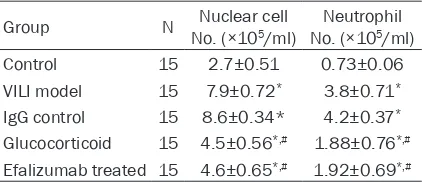
[image:2.612.91.302.96.187.2]out using pre-mixture (Invitrogen, US) on a fluo -rescent PCR machine. PCR conditions were set as: 90°C denature for 30 sec, 58°C annealing Table 1. Number of nuclear cells and neutrophils
in BALF
Group N No. (×10Nuclear cell 5/ml) No. (×10Neutrophil 5/ml)
Control 15 2.7±0.51 0.73±0.06 VILI model 15 7.9±0.72* 3.8±0.71*
IgG control 15 8.6±0.34* 4.2±0.37*
Glucocorticoid 15 4.5±0.56*,# 1.88±0.76*,#
for 50 sec, plus 72°C extension (35 cycles in total). The relative level of mRNA was deter-mined using 2-ΔCt method, and GAPDH gene
was used as the reference gene. Western blot
Pulmonary tissues were lysed in lysis buffer on ice for 15~30 min. After sonic rupture of cells for 5 sec (4 times), lysate was centrifuged at 10 000 g for 15 min, with the supernatant being transferred to a new tube. Proteins extracted were separated by 10% SDS-PAGE, and were transferred to PVDF membrane (Pall Life, US).
Non-specific binding sites were blocked by
blocking buffer contains 5% skimmed milk pow-der for 2 hours at room temperature. Rabbit anti-human SP-A monoclonal antibody (1:1000, Cell signaling, US) was used to incubate the membrane at 4°C overnight. After gentle
wash-ing in PBST, the membrane was further incu -bated in goat anti-rabbit IgG conjugated with horseradish peroxidase (HRP) (1:2000, Cell sig-naling, US) for 30 minutes. The results were
visualized by ECL reagents (AmershamBiosci.,
US) and were exposed under X-ray. Images were captured and analyzed for relative density by Quantity One software.
Statistical analysis
SPSS 20.0 software (SPSS, Chicago, IL, USA) was used for data analyses. Enumeration data were compared by using chi-square test, and measurement data were presented as mean ± standard deviation (_x±s). Multiple group
com-parison was finished by one-way analysis of
variance (ANOVA). Differences P<0.05
indicat-ed statistically significant.
Results
Effect of efalizumab on the cell count of nuclear cells and neutrophils
BALF collected from all groups were quantified
for the numbers of nuclear cells and
neutro-phils. Results showed significantly elevated numbers of both cells in BALF of VILI model
rats, compared to controlled animals (P<0.05). The intervention by efalizumabor glucocorticoid
significantly decreased the number of inflam -matory cells, although still higher than those in the control and IgGgroups (Table 1). These
results suggest the up-regulation of inflamma -tory cells in VILI, and the protective role of
efali-zumab by decreasing inflammatory cell
numbers.
TNF-α and IL-8 expression levels in BALF
The contents of TNF-α and IL-8 in BALF of all
animals were determined by ELISA. Results
illustrated significantly rise of TNF-α and IL-8 levels in BALF in VILI model rats in comparison
to controlled animals (Figure 1A and 1B, P<
0.05). The application of efalizumab significant
-ly decreased levels of both cytokines in BALF,
although still higher than those in the control and IgGgroup (Figure 1A and 1B, P<0.05). The expressions of cytokines in glucocorticoid and
efalizumab groups showed no significant
di-fference.
SP-A gene expression in pulmonary tissues
We firstly used real-time PCR to detect mRNA
levels of SP-A gene in rat pulmonary tissues. It
[image:3.612.99.518.73.200.2]has been demonstrated that there were signifi -cantly depressed level of SP-A mRNA in VILI
Figure 1. TNF-α and IL-8 levels in BALF. A. TNF-α concentrations; B. IL-8 concentrations. *P<0.05 compared to the
model rats, compared to the control group (Fi- gure 2A, P<0.05). The application of efalizum-ab restored SP-A mRNA levels, although still lower than the control and IgGgroups. These results indicated that the potential injury of MV on pulmonary tissues was via decreasing SP-A expression. Efalizumab, on the other hand, may provide protection of pulmonary tissues via facilitating SP-A gene expression.
Western blotting assay consistently confirmed that VILI rats had significantly suppressed SP-A
protein expression compared to those in the control group (Figure 2B, 2C, P<0.05). Similar to the result in the glucocorticoid group, the intervention by efalizumab elevated SP-A pro-tein levels but still lower than the control and IgGgroups. These results further suggest the role of efalizumab in protecting pulmonary tis-sues via potentiating SP-A protein expression levels.
Discussion
MV plus primary pulmonary disease may aggra-vate the biological injury of the lung tissue, leading to other organ failure or even multip- le organ dysfunction syndromes. It has been shown that the hyperextension of lung tissue by
MV may elicit biochemical signals transducing
into the cell, thus activating local inflammatory
cells and inducing release of large amounts of
inflammatory cytokines or factors [16, 17]. Our
study demonstrated the aggravated pulmonary damages during VILI by the increased secretion
of TNF-α and IL-8. Efalizumab, as a humanized
monoclonal antibody (IgG1к), a novel immuno
-suppressive agent, inhibits the release of in-
flammatory cytokines. Our data demonstrated
thatefalizumab decreased expressions of TNF-
α and IL-8by interfering the function of IgG1к.
Nonetheless, the presence of IgG showed
ele-vated expressions of TNF-α and IL-8 during VILI
that even caused severer injury, suggesting that efalizumabexerts an alleviated effecton VILI treatment.
Mainly targeting T cell for immune suppression, efalizumab interferes with primary T cell
activa-tion, thus mediating the release of inflammato -ry cytokines in addition to the regulation of memory T cell production. It also inhibits body’s immune system via impeding the migration of T cells, and hindering the binding between lym-phocyte adhesion molecule LFA-1 and endothe-lial ICAM-1 in a rate-limit step of early migration phase [18, 19]. Efalizumab therefore exerts its
[image:4.612.91.518.73.320.2]anti-inflammatory roles in inhibiting the release
Figure 2. SP-A expression level in pulmonary tissues. A. SP-A mRNA relative level (against GADPH); B. Western blot -ting bands of SP-A protein in pulmonary tissues from all rats (1. Control; 2. VILI model; 3. IgG control; 4. Glucocor-ticoid; 5. efalizumab); C. Quantitative results of SP-A protein levels against β-actin levels. *P<0.05 compared to the
of pro-inflammatory cytokines. During the oc-currence and progression of VILI, inflammatory
cells including neutrophils and polymorphic nu-
clear cells may lead to inflammatory response.
The mechanical stretch may also facilitate the recruitment of neutrophils in pulmonary alveoli
and release of inflammatory factors. For exam -ples, TNF induces endothelial cells in micro vessels to expression E-selectin, initiating an interaction with its partners to activate adhe-sion, migration, penetration and chemotactic
aggregation of neutrophils into the inflamma -tory sites, further aggravating pulmonary
dam-age [20, 21]. Our study showed significantly
elevated numbers of both nuclear cells and
neutrophils in BALF of VILI model rats. The
intervention of efalizumab suppressed the total number of those two types of cells, suggesting
the potentiation of inflammatory cells in VILI
and protection by efalizumab by decreasing
inflammatory cell number.
Pulmonary surfactant (PS) is a mixture contain-ing lipoproteins and sugars, and is synthesized and secreted by type II epithelial cells of pulmo-nary alveoli. It plays an important role in main-taining normal cell structure and function via decreasing surface tension of pulmonary alveo-li, keeping alveoli dry, preventing pulmonary edema, relaxing smooth muscle, participating local defense and improving mucosa
proper-ties. SP-A belongs to one of specific markers of
PS [22] and was shown to be down regulated in VILI pulmonary tissues via both mRNA and pro-tein assays. The application of efalizumab, be- sides the abovementioned functions including
inhibiting chemotactic recruitment of inflamma
-tory cells and suppressing inflamma-tory fac -tors, also facilitate the expression of mRNA and protein of SP-A in rat lung tissues. Glucocor-
ticoid, on the other hand,exerts anti-inflamma -tory function that reduces alveolar tension and also plays a favorable role in the treatment of VILI. However, long-term use of glucocorticoids causesa variety of side effects including the aggravation of infection, induction of cardio-vascular disease [23]. Therefore in this study,
we compared theefficacy of damage alleviation
between the treatment of glucocorticoid and efalizumab. The results unraveleda similar role of both treatments in reducingon the cell count of nuclear cells and neutrophils, inhibiting the
secretion of inflammatory factors, and prevent -ing further injuries in lung tissue.
Our study thus suggests the contribution of efalizumabin facilitating the therapy of VILI without potential side effects of glucocorticoid. In summary, efalizumab may provide protec-tions against the damage of VILI, via multiple pathways including inhibiting recruitment of
inflammatory cells, suppressing release of pro-inflammatory cytokines, and potentiating SP-A
expression in lung tissues. This study eluci-dates the protection of efalizumab on VILI and possible mechanisms, and provides novel drug targets for future clinical treatment of VILI. Disclosure of conflict of interest
None.
Address correspondence to: Dr. Juntao Pang, De- partment of ICU, Weifang People’s Hospital, 151 Guangwen Street, Kuiwen District, Weifang 261041, Shandong, China. Tel: 536-8192599; Fax: +86-536-8192599; E-mail: llihing789@163.com
References
[1] Herter JM, Kraft F, Van Aken H, Meersch M, Zarbock A, Rossaint J. GDF-15 prevents venti-lator-induced lung injury by inhibiting the for-mation of platelet-neutrophil aggregates. Thro- mb Haemost 2015; 114: 434-7.
[2] Tsushima K and Tatsumi K. Noninvasive me-chanical ventilation and neutrophil elastase inhibitor: a new potential approaching to acute hypoxemic failure. J Crit Care 2014; 29: 1124-5.
[3] Ghanbarpour R, Saghafinia M, Ramezani Bi-nabaj M, Madani SJ, Tadressi D, Forozanmehr MJ. Pulmonary infections in ICU patients with-out underlying disease on ventilators. Trauma Mon 2014; 19: e15958.
[4] Gao S, Guan S, Li H, Su A, Wang Y. Ameliorat-ing effects of low tidal volume ventilation with associated hypercapnia on pneumoperitone-um-induced lung injury by inhibition of Toll-like receptor 4. Int J Clin Exp Med 2015; 8: 1814-23.
[5] Retamal J, Bergamini BC, Carvalho AR, Bozza FA, Borzone G, Borges JB, Larsson A, Heden -stierna G, Bugedo G, Bruhn A. Non-lobar atel -ectasis generates inflammation and structural alveolar injury in the surrounding healthy tis-sue during mechanical ventilation. Crit Care 2014; 18: 505.
me-chanical ventilation aggravates ventilator-in-duced lung injury in mice. Crit Care 2015; 19: 23.
[7] Carvalho NC, Güldner A, Beda A, Rentzsch I, Uhlig C, Dittrich S, Spieth PM, Wiedemann B, Kasper M, Koch T, Richter T, Rocco PR, Pelosi P, de Abreu MG. Higher levels of spontaneous breathing reduce lung injury in experimental moderate acute respiratory distress syndrome. Crit Care Med 2014; 42: e702-15.
[8] Chetty M, Li L, Rose R, Machavaram K, Jamei M, Rostami-Hodjegan A, Gardner I. Prediction of the Pharmacokinetics, Pharmacodynamics, and Efficacy of a Monoclonal Antibody, Using a Physiologically Based Pharmacokinetic FcRn Model. Front Immunol 2014; 5: 670.
[9] Belinchón I, Arribas MP, Soro P, Betlloch I. Re -covery of the response to biological treatmen- ts using narrow band ultraviolet-B in patients with moderate to severe psoriasis: a retrospec-tive study of 17 patients. Photodermatol Photo-immunol Photomed 2014; 30: 316-22. [10] Mitroulis I, Alexaki VI, Kourtzelis I, Ziogas A,
Ha-jishengallis G, Chavakis T. Leukocyte integrins: role in leukocyte recruitment and as therapeu-tic targets in inflammatory disease. Pharmacol Ther 2015; 147: 123-35.
[11] Futier E, Godet T, Millot A, Constantin JM, Jaber S. Mechanical ventilation in abdominal sur-gery. Ann Fr Anesth Reanim 2014; 33: 472-5. [12] Moraes L, Santos CL, Santos RS, Cruz FF,
Sad-dy F, Morales MM, Capelozzi VL, Silva PL, de Abreu MG, Garcia CS, Pelosi P, Rocco PR. Ef-fects of sigh during pressure control and pres-sure support ventilation in pulmonary and ex-trapulmonary mild acute lung injury. Crit Care 2014; 18: 474.
[13] Saddy F, Sutherasan Y, Rocco PR, Pelosi P. Ventilator-associated lung injury during assist-ed mechanical ventilation. Semin Respir Crit Care Med 2014; 35: 409-17.
[14] Park M, Pires-Neto RC and Nassar Junior AP. Awaking, exercising, sitting, walking and extu-bating: moving on the paradigms for mechani-cally ventilated patients. Rev Bras Ter Intensiva 2014; 26: 203-4.
[15] Coffey GP, Fox JA, Pippig S, Palmieri S, Reitz B, Gonzales M, Bakshi A, Padilla-Eagar J, Fielder PJ. Tissue distribution and receptor-mediated clearance of anti-CD11a antibody in mice. Drug Metab Dispos 2005; 33: 623-9.
[16] Yang S, Stepien D, Hanseman D, Robinson B, Goodman MD, Pritts TA, Caldwell CC, Remick DG, Lentsch AB. Substance P mediates redu-ced pneumonia rates after traumatic brain in-jury. Crit Care Med 2014; 42: 2092-100. [17] Cross LJ, O’Kane CM, McDowell C, Elborn JJ,
Matthay MA, McAuley DF. Keratinocyte growth factor in acute lung injury to reduce pulmonary dysfunction--a randomised placebo-controlled trial (KARE): study protocol. Trials 2013; 14: 51.
[18] Vilarrasa E and Puig L. Monoclonal gammopa-thy of undetermined significance (MGUS) in patients with psoriasis may be associated with long-term treatment with efalizumab, but not with anti-TNF-alpha treatments or ustekinum-ab. J Eur Acad Dermatol Venereol 2015; 29: 609-11.
[19] Mosli MH, Rivera-Nieves J and Feagan BG. T-cell trafficking and anti-adhesion strategies in inflammatory bowel disease: current and fu -ture prospects. Drugs 2014; 74: 297-311. [20] Shu YS, Tao W, Miao QB, Zhu YB, Yang YF. Im
-provement of ventilation-induced lung injury in a rodent model by inhibition of inhibitory kap-paB kinase. J Trauma Acute Care Surg 2014; 76: 1417-24.
[21] Mao P, Wu S, Li J, Fu W, He W, Liu X, Slutsky AS, Zhang H, Li Y. Human alveolar epithelial type II cells in primary culture. Physiol Rep 2015; 3. [22] Hallman M. The surfactant system protects
both fetus and newborn. Neonatology 2013; 103: 320-6.