Original Article
Comparison between transforaminal lumbar interbody
fusion and posterior lumbar interbody fusion in
treatment of lumbar spondylolisthesis
Shao-Yu Han1*, Quan Xiao2*, Guo-Tai Zhu3, Jian Dai3, Xiao-Ming Tang3, Hai-Lang Sun3
1Third Department of Orthopedics, Huaiyin Hospital, Huai’an 223300, Jiangsu, China; 2Department of
Orthopedics, Lianshui People’s Hospital, Huai’an 223400, Jiangsu, China; 3Department of Orthopedics, Huai’an
First People’s Hospital, Huai’an 223001, Jiangsu, China. *Co-first authors.
Received September 11, 2015; Accepted January 30, 2016; Epub February 15, 2016; Published February 29, 2016
Abstract: The aim of this study was to compare the clinical efficacy between transforaminal lumbar interbody fusion
(TLIF) and posterior lumbar interbody fusion (PLIF) in treatment of lumbar spondylolisthesis. A total of 62 patients with lumbar spondylolisthesis were treated at our hospital from 2010 to 2013. These patients were divided into a TLIF group that included 36 patients (mean age: 60 years) and a PLIF group that included 26 patients (mean
age: 57 years) according to different surgical methods. For example, the pedicle screws were fixed first and then
transforaminal lumbar interbody fusion or posterior lumbar interbody fusion was performed, and the pedicle screws or intralaminar spreading device were used to distract. Next, the surgical durations, the volume of bleeding during surgery, postoperative drainage, and complications were compared between the two groups. All 62 patients were well operated, 2 patients developed a dural sac tear, and 1 patient had injury to the nerve roots in the PLIF group. The average follow-up duration was 20 months and 18 months in the TLIF group and PLIF group respectively. Both
TLIF and PLIF technologies could effectively decompress the interbody fusion and fix posterior endplates steadied
centrum, which alleviated clinical symptoms, but TLIF had some advantages over PLIF, such as smaller trauma, low incidence rate of nervous injury or dural sac injury, and better protection of end structures.
Keywords: Lumbar spondylolisthesis, pedicle screws fixation, transforaminal lumbar interbody fusion, posterior
lumbar interbody fusion
Introduction
Lumbar spondylolisthesis (LSL) is a common disease and causes lumbosacral pain [1-5]. The pathological mechanisms of LSL are very complicated; furthermore, the duration is long
and the efficacy of treatment of LSL is uncer -tain. Conservative treatment is not effective and patients develop progressive neuropathic dysfunction. Spondylolisthetic fusion is the gold standard treatment to be performed [2, 3].
Pedicle screw fixation combined with grating
fusion has been acknowledged widely by many experts, but the type of grating fusion used is still controversial. This study was a
retrospec-tive analysis to compare the clinical efficacy of
treatment of LSL through transforaminal bar interbody fusion (TLIF) and posterior lum-bar interbody fusion (PLIF).
Materials and methods
General data
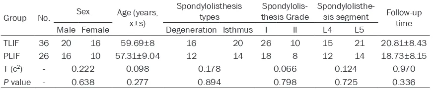
A total of 62 patients with LSL, including 36 men and 26 women, aged 45 to 76 years, with an average age of 50 years, were enrolled in this study from March 2010 to March 2013. The duration ranged from 9 months to 20 years, with an average of 48 months. All patients were divided into the TLIF group, which included 20 men and 16 women, aged 48 to 78 years with an average of 60 years; and the PLIF group that included 16 men and 10 women, aged 45 to 72 years with an average of 57 years.
Table 1. The general data of two groups
Group No. Sex Age (years, x±s)
Spondylolisthesis
types thesis GradeSpondylolis- Spondylolisthe-sis segment Follow-up time Male Female Degeneration Isthmus I II L4 L5
TLIF 36 20 16 59.69±8 16 20 26 10 15 21 20.81±8.43 PLIF 26 16 10 57.31±9.04 12 14 18 8 12 14 18.73±8.15 T (c2) - 0.222 0.098 0.178 0.066 0.124 0.970
P value - 0.638 0.277 0.894 0.798 0.725 0.336
12 patients with lumbar degenerative spondy-lolisthesis and 14 with isthmic spondylolisthe-sis. Moreover, according to Meyerding’s Cla-
ssification, the TLIF group had 26 patients with
grade I and 10 with grade II injuries, whereas the PLIF group had 18 patients with grade I and 8 with grade II injuries.
According to spondylolisthesis segment
classi-fication, the TLIF group had 15 patients with L4
and L5 spondylolisthesis and 21 patients with L5-S1 spondylolisthesis, whereas the PLIF group had 12 patients with L4 and L5 lolisthesis and 14 patients with L5-S1 spondy-lolisthesis. All patients manifested single lum-bar spondylolisthesis and had differing degrees of lumbar pain plus low limb symptoms; more-over, all patients were treated for at least six months by conservative therapy that had resulted in a poor outcome. Furthermore, rou-tine examinations such as radiography, com-puted tomography (CT), and magnetic reso-nance imaging (MRI) were performed before the surgery. Patients with a history of a lumber operation, spondylolisthesis over grade II, tumor or malformation were excluded from this study. This study was conducted in accordance with the declaration of Helsinki. This study was conducted with approval from the Ethics Committee of the Huai’an First People’s Hospital. Written informed consent was obtained from all participants.
Operation
Patients underwent general anesthesia and
the surgeon began by making a vertical incision to expose the facet joints and the lordotic posi-tion of the lumbar spine. Spondylolisthetic lum-bar loosening was seen in patients with isthmic spondylolisthesis and distinguished by rising spinous processes; additionally, zygopophysis hypertrophy was obviously seen in patients with lumbar degenerative spondylolisthesis.
Under the assistance of fluoroscopy, the posi -tions of upper and lower pedicle screws were
confirmed and tracing pins were places. Pedicle
screws were then placed in the standard fashion.
In the TLIF group, we adhered to the following procedure: The zygopophysis was removed with the use of an osteotome, and the skin, mus-cles, and soft tissues were gently retracted to expose the lateral aspect of the spinous pro-cess, the lamina, and the facet joint. The thecal sac and traversing nerve roots were mobilized and retracted to the midline, with care taken to protect the dural sac and neural contents with a retractor. Then the interbody disc was exposed.
In the PLIF group, we adhered to the following procedure: The corresponding lumbar
seg-ments and ligamentum flavum were removed to
expose the thecal sac and traversing nerve roots, which were slightly pulled with a nerve hook to expose the interbody disc. Next, the interbody disc was incised in the two groups, and the vertebral endplates were removed with
Table 2. Comparisons of postoperation parameters between two groups
Group n Surgical Duration (min) Amount of bleeding during operation (ml) Volume of postoperative drainage (ml) Complications (n) TLIF 36 134.17±27.40 246.94±48.33 151.67±46.93 1 PLIF 26 130.38±30 271.92±42.43 181.15±60.29 3 T (c2) - 0.515 2.111 2.165 1.919
[image:2.612.91.524.211.288.2]were confirmed and broken bones were added
into the interbody space as trial bone grating.
Finally, screws were fixed and a titanium rod was placed. After they were identified to be
without active bleeding, drainage was per-formed, followed by closing of the incisions. The Pedicle screw system used was CD Horizon Legacy system (Medtronic), and the interbody fusion instrument used was Capstone lumbar interbody fusion instrument (Medtronic) (Minneapolis, Minnesota, USA).
Postoperative treatment
Patients were nursed and drainage tubes were placed for 24-48 h after the surgery. Meanwhile, antibiotics were administrated for 1 day, ste-roids and dehydrating agents were used for 3 days after the surgery. Sutures were removed 12-14 days after the surgery. Furthermore, patients needed to exercise the lumbar and back muscles. According to the patients’ condi-tion, they tried to walk with a waistband sup-port about 1 week after the surgery. Additionally, postoperative follow-up was performed. Assessment
The operative duration, amount of bleeding during the surgery, and postoperative drainage volume were observed and recorded. The fol-lowing Imaging evaluations were performed: Interbody fusion was evaluated according to the Simmons Method. There was no bright area around cage on the radiogram, and the cage did not move above six months after the sur-gery. The angle of the fused segment on the
flexion-extension radiograph was less than 5°.
Clinical effect was assessed by visual analogue score (VAS) and oswestry disability index (ODI). The clinical symptoms, physical sign, and sphincter function were assessed before and after the surgery, and during the last follow-up. The improvement rate was calculated accord-ing to ODI scores, (preoperative
scores-postop-erative scores)/preopscores-postop-erative scores × 100%. “Excellent” meant that the improvement rate was more than 75%; “good” meant that the improvement rate ranged from 50% to 74%; “medium” meant that the improvement rate ranged from 25% to 49%, and “poor” meant that the improvement rate was less than 24%. Statistical analysis
Statistical analysis was performed using the SPSS10.0 software. Measured data was pre-sented as means ± SD. Student t test and chi-square test were used to compare data between the two groups. P < 0.05 denoted a
significant statistical difference.
Results
A total of 62 patients underwent the proce-dures successfully. Two patients developed dural sac tears, and 1 patient had an injury to the nerve roots in the PLIF group. There was no obvious difference in the general data between the two groups (Table 1). The surgical duration
time was of statistical significance between the
TLIF group (134.17±27.40 min) and the PLIF group (130.38±30 min) (t = 2.111, P = 0.039). The volume of postoperative drainage was of
statistical significance between the TLIF group
(151.67±46.93 ml) and the PLIF group (181.15±60.29 ml) (t = 2.165, P = 0.034, Table 2). The follow-up time ranged from 12 to 48 months, with an average of 20 months in the TLIF group and 9-42 months (mean age: 18 months) in the PLIF group. The VAS and ODI
scores were not statistically significant at any
time between the TLIF group and the PLIF group (P > 0.05, Table 3). The improvement rate and acceptance rate were 75±17% and 88.9%, respectively, in the TLIF group and 70±16% and 84.5%, respectively, in the PLIF group. One fusion device moved without nervous symp-toms in the PLIF group, and 1 patient devel-oped fat liquefaction at the site of the incision in the TLIF group. Both were treated
according-Table 3. The results of VAS and ODI scores in two groups
Group n VAS scores ODI scores
Pre-operation Post-operation Last follow-up Pre-operation Post-operation Last follow-up TLIF 36 7.11±0.95 2.44±1.42* 2.25±1.59* 67.50±13.43 24.50±12.19* 20.56±10.33*
[image:3.612.92.523.85.140.2]Figure 1. Female with the age of 59 years, had lumbar pain for over 10 years, and the lumbar pain worsened and right low limb was numbness and pain six months
ago. A-D showed the lumbar anterio-posterior position and flexion-extension X-ray, which indicated the lumbar degeneration and L4 spondylolisthesis forward. E, F
showed the lumbar CT, which also noted the lumbar degeneration and L4 spondylolisthesis forward. G, H showed the lumbar MRI, which implied the lumbar degen-eration and bulged interbody disc of L1-2, L2-3, and L4 spondylolisthesis forward. I, J postoperative lumbar anterio-posterior position X-ray showed the posterior
pedicle screws fixation at L4-5, and L5/L5 right side by interbody rod decompression plus cage grafting, meanwhile, L4 spondylolisthesis reduction was satisfaction and symptoms such as lumbar pain and low limbs numbness were improved. K, L showed the lumbar fixation still existed through X-ray of lumbar anterio-posterior
ly. The pedicle screw system had no loosening or fractures in the two groups, and the inter-body fusion rate was 94.4% (34/36) and 92.3% (24/26) in the TLIF group and PLIF group respectively. The average duration of bone grat-ing fusion was 6.5 months. In addition, Figure 1
showed the typical cases.
Discussion
LSL is a common disease. About 5% patients with lumbosacral pain have LSL, but the cause of LSL remains unclear. Most studies indicate that congenital dysplasia and chronic strain may be the most important factors that cause LSL [4]. Congenital isthmus diastasis or isth-mus cracking caused by chronic strain, degen-erative intervertebral disc, and unstable inter-vertebral facet joints cause and increase in lumbar instability and occur in spondylolisthe-sis between the upper and lower centrum. Usually, lumbar degenerative spondylolisthesis and isthmic lumbar spondylolisthesis are com-mon cases in clinical practice [3]. Lumbar spon-dylolisthesis aggravates spinal canal stenosis and compresses nerves so as to present cor-responding symptoms. In addition, durative vertebral instability or increase of stress makes corresponding smaller joints wear down and cause hyperplasia. Moreover, many prolifera-tive scars were formed in the area of isthmus diastasis, which aggravates spinal canal and nerve roots stenoses. Furthermore, lumbar spondylolisthesis is generally accompanied by a herniated disk and spinal stenosis, which
increases the difficulties of diagnosis and
treatment.
Degenerative lumbar vertebrae, discontinuous pedicles, and lumbar spondylolisthesis affect stability of three lumbar spines. Meanwhile, spi-nal mechanics are changed and the erected body increased stress on the lower lumbar spines, which further causes intervertebral labilization and compensatory hypertrophy that leads to vicious circle [5, 6]. Therefore, better decompression, reduced distraction of lumbar spondylolisthesis and interbody fusions are the basis to treat LSL.
LSL can be treated by surgery or nonsurgical treatment. Generally, surgical treatment is for patients who have found conservative treat-ment ineffective and have progressive nervous
dysfunction [7]. Moreover lumbar instability and spinal canal stenosis cause degenerative spondylolisthesis and isthmic lumbar spondylo-listhesis; thus, the treatment principle of LSL presented is that injured spines are reduced,
fixated, and a bone graft fusion is performed.
Furthermore, the spinal canal and nerve roots should be completely decompressed which makes the bone graft fusion the most important.
The three spines fusion is the key measure that makes up spinal stabilization for spondylolis-thesis; meanwhile, spine fusion could prevent breakage of pedicle screws [8, 9]. Spine fusion approaches contained PLF and LIF that includ-ed ALIF, PLIF, and TLIF according to bone graft parts [10]. The bone graft fusion parts of PLIF contained the basal part of transverse process and outside of small joints, which is easy to operate and has a big bony mattress with plen-ty of vessels. However, the exposed wide range of the two sides might cause more bleeding; furthermore, the joint sac and muscles covered the transverse process and zygopophysis so the bone graft fusion bed is not established. Simple periosteal bone graft fusion might lead to posterior loosening and breakage of pedicle screws [11, 12]. Interbody fusion not only avoids increasing bleeding problems after strip-ing muscles and establishstrip-ing a bone graft bed, but also provides plenty of blood support between the maximum distraction and recov-ery of the intervertebral space height. Moreover, interbody fusion indirectly expands the nerve roots and recovers the spinal physiologic curve. Most of researchers considered that the fusion rate of interbody fusion was higher than poste-rior fusion [13]. Cheng et al. [14] compared the effect of PLIF and PLF in treatment of LSL and the results indicated that the fusion rate of PLIF was higher than that of PLF, but there was no
obvious difference, which was confirmed in a
study by Barbanti Brodano et al. [15].
affects the dural sac and nerve roots. PLIF also acts as a fulcrum for a dural retractor and to preserve a tension band posteriorly. Dural sac formation leads to iatrogenic spinal canal ste-nosis [13, 16]. In order to reach the aim of dis-traction, infusion and increase spine stability to preserve more posteriors structures, TLIF tech-nology was termed on the basis of PLIF by Harms in 1982. Harms and Rolinger reported use of a bone graft packed in a titanium mesh that was inserted via a transforaminal route into the disc space, which had similar charac-teristics, such as small surgical trauma and lit-tle bleeding. Additionally, TLIF effectively reduced compression and achieved the better effect of 360 degree fusion for patients with LSL [17].
Xu et al. [18] also concluded that the TLIF pro-cedure was a safe and effective treatment for 60 the Hans with LSL. Furthermore, Kleinstueck et al. and his team [19] found that decompres-sion with fudecompres-sion better improved lumbar pain in patients with LDS than with decompression alone, which corroborated with the study of Ha et al. [20]. A study suggested that the TLIF tech-nique had a better treatment for patients with LSL than the PLIF technique [13], in which the results, such as less bleeding and a lower nerve injury rate were also similar with this study. Here, 2 out of 26 patients had dural sac fraction in the PLIF group, which then required additional treatment. Fortunately, no severe
leakage of the cerebrospinal fluid occurred
after the surgery, and 1 case occurred of nerve root injury, which was then treated with ste-roids. After a six month follow-up, the symp-toms of this patient improved. Our experience was that zygapophyseal joints were properly knocked out during PLIF, which avoided exces-sive tension on the nerve roots. Also, TLIF was tried in order to reduce the destruction of tis-sue and structures as much as possible. In summary, both the TLIF and PLIF techniques were effective methods to treat LSL and achieve interbody fusion with nerve
decom-pression. Posterior fixation and unfold inter -body fusion could achieve distraction, stability, and fusion of the spine. Compared with PLIF, TLIF had some advantages, such as less blood loss, reducing potential nerve injury, and prop-erly preserving the posterior structure of spine.
Disclosure of conflict of interest
None.
Address correspondence to: Hai-Lang Sun, De- partment of Orthopedics, Huai’an First People’s Hospital, No. 6 West Beijing Road Huaiyin District, Huai’an 223001, Jiangsu, China. Tel: +86 517 80872310; Fax: +86 517 80872310; E-mail: hai-langsuncn@163.com
References
[1] Alfieri A, Gazzeri R, Prell J and Rollinghoff M.
The current management of lumbar spondylo-listhesis. J Neurosurg Sci 2013; 57: 103-113. [2] Matsudaira K, Yamazaki T, Seichi A, Takeshita
K, Hoshi K, Kishimoto J and Nakamura K. Spinal stenosis in grade I degenerative lumbar spondylolisthesis: a comparative study of out-comes following laminoplasty and laminecto-my with instrumented spinal fusion. J Orthop Sci 2005; 10: 270-276.
[3] Martin CR, Gruszczynski AT, Braunsfurth HA, Fallatah SM, O’Neil J and Wai EK. The surgical management of degenerative lumbar spondy-lolisthesis: a systematic review. Spine (Phila Pa 1976) 2007; 32: 1791-1798.
[4] Garet M, Reiman MP, Mathers J and Sylvain J. Nonoperative treatment in lumbar spondyloly-sis and spondylolisthespondyloly-sis: a systematic review. Sports Health 2013; 5: 225-232.
[5] Majid K and Fischgrund JS. Degenerative lum-bar spondylolisthesis: trends in management. J Am Acad Orthop Surg 2008; 16: 208-215. [6] Toyone T, Tanaka T, Kato D, Kaneyama R and
Otsuka M. Anatomic changes in lateral spondy-lolisthesis associated with adult lumbar scolio-sis. Spine (Phila Pa 1976) 2005; 30: E671-675.
[7] Steiger F, Becker HJ, Standaert CJ, Balague F, Vader JP, Porchet F and Mannion AF. Surgery in lumbar degenerative spondylolisthesis: indica-tions, outcomes and complications. A system-atic review. Eur Spine J 2014; 23: 945-973. [8] Sivaraman A, Altaf F, Jalgaonkar A, Kakkar R,
Sirigiri PB, Howieson A and Crawford RJ. Prospective study of Posterior Lumbar Interbody Fusion with either interbody graft or interbody cage in the treatment of degenera-tive spondylolisthesis. J Spinal Disord Tech 2015; 28: E467-471.
[9] Resnick DK, Watters WC 3rd, Sharan A, Mummaneni PV, Dailey AT, Wang JC, Choudhri TF, Eck J, Ghogawala Z, Groff MW, Dhall SS and Kaiser MG. Guideline update for the perfor-mance of fusion procedures for degenerative disease of the lumbar spine. Part 9: lumbar fu-sion for stenosis with spondylolisthesis. J Neurosurg Spine 2014; 21: 54-61.
fluence Outcome? Four-Year Results of the
Spine Patient Outcomes Research Trial. Spine (Phila Pa 1976) 2009; 34: 2351-2360. [11] Goto K, Tajima N, Chosa E, Totoribe K, Kubo S,
Kuroki H and Arai T. Effects of lumbar spinal fusion on the other lumbar intervertebral
lev-els (three-dimensional finite element analysis).
J Orthop Sci 2003; 8: 577-584.
[12] Liu X, Wang Y, Qiu G, Weng X and Yu B. A sys-tematic review with meta-analysis of posterior interbody fusion versus posterolateral fusion in lumbar spondylolisthesis. Eur Spine J 2014; 23: 43-56.
[13] Cole CD, McCall TD, Schmidt MH and Dailey AT. Comparison of low back fusion techniques: transforaminal lumbar interbody fusion (TLIF) or posterior lumbar interbody fusion (PLIF) ap-proaches. Curr Rev Musculoskelet Med 2009; 2: 118-126.
[14] Cheng L, Nie L and Zhang L. Posterior lumbar interbody fusion versus posterolateral fusion in spondylolisthesis: a prospective controlled study in the Han nationality. Int Orthop 2009; 33: 1043-1047.
[15] Barbanti Brodano G, Lolli F, Martikos K, Gasbarrini A, Bandiera S, Greggi T, Parisini P and Boriani S. Fueling the debate: Are out-comes better after posterior lumbar interbody fusion (PLIF) or after posterolateral fusion (PLF) in adult patients with low-grade adult
isthmic spondylolisthesis? Evid Based Spine
Care J 2010; 1: 29-34.
[16] Kunze B, Drasseck T and Kluba T. [Posterior and transforaminal lumbar interbody fusion (PLIF/TLIF) for the treatment of localised seg-ment degeneration of lumbar spine]. Z Orthop Unfall 2011; 149: 312-316.
[17] Quante M, Kesten H, Richter A and Halm H. [Transforaminal lumbar interbody fusion for the treatment of degenerative spondylolisthe-sis]. Orthopade 2012; 41: 153-162.
[18] Xu H, Tang H and Li Z. Surgical treatment of adult degenerative spondylolisthesis by instru-mented transforaminal lumbar interbody fu-sion in the Han nationality. J Neurosurg Spine 2009; 10: 496-499.
[19] Kleinstueck FS, Fekete TF, Mannion AF, Grob D, Porchet F, Mutter U and Jeszenszky D. To fuse or not to fuse in lumbar degenerative spondylolisthesis: do baseline symptoms help
provide the answer? European spine journal: official publication of the European Spine
Society, the European Spinal Deformity Society, and the European Section of the Cervical Spine Research Society 2012; 21: 268-275. [20] Ha KY, Na KH, Shin JH and Kim KW. Comparison