Original Article
The effects of BML-111 and MAPK signaling pathway on
the expression of AQP1 in rat lung tissue of acute
lung injury caused by endotoxic shock
Ning Luo1, Xiu-Hua Wang2, Yin Li1, Bing Wang1, Yong-Qiang Wang1*, Hong-Mei Gao1*
1Intensive Care Unit, Tianjin First Center Hospital, Tianjin, China; 2First People’s Hospital of Jinan, Shandong,
China. *Equal contributors.
Received January 14, 2016; Accepted March 27, 2016; Epub May 15, 2016; Published May 30, 2016
Abstract: Objective: This study aims to explore the effects of BML-111 and MAPK signaling pathway on the expres-sion of AQP1 in rat lung tissue of acute lung injury caused by endotoxic shock. Methods: Acute lung injury caused by LPS mouse model was established. They were divided into normal group, ALI model group, BML group, SB group and SP group. Wet and dry weight of the right lung was detected, number of neutrophils and pulmonary permeability index were determined. Pathological morphology of lung tissues was determined by HE staining. The levels of TNF-α, IL-1β and IL-6 were determined by ELISA. The levels of AQP1, p38 and JNK were determined by western-blotting method. Results: There was pulmonary interstitial edema and thickening alveolar wall in ALI model group, the ra-tion of W/D, PPI, the number of neutrophils and the levels of TNF-α, IL-1β, IL-6, p38 MAPK and JNK phosphorylara-tion increased, while the levels of AQP1 decreased when compared with normal group (P<0.01). However, W/D, PPI, the levels of TNF-α, IL-1β, IL-6 and the phosphorylation levels of p38 MAPK and JNK in BML group, SB group and SP group were significantly lower than that of model group (P<0.01), while the levels of AQP1 increased in BML group, SB group and SP group (P<0.01). Conclusions: BML-111, P38 MAPK inhibitor and JNK inhibitor could significantly inhibit the lung injury in ALI rats induced by LPS and increased the levels of AQP1, which may be through the inacti-vation of MAPK p38 and JNK signaling pathway.
Keywords: BML-111, mitogen activated protein kinase (MAPK), signaling pathway, lipopolysaccharide (LPS), acute lung injury (ALI), aquaporin 1 (AQP1)
Introduction
Endotoxin shock is a common complication in clinical burn, trauma and surgery and easily leads to multiple organ dysfunction or even fail-ure. Lung is one of the most vulnerable organ and acute lung injury (ALI) easily occurs in the process of endotoxin shock. The mechanism of
ALI is complex, the imbalance between inflam -matory response and compensatory anti in-
flammatory response make the lung environ -ment abnormal and pulmonary edema play an important role [1, 2]. Therefore, it will have a
promoting role in treating ALI if the inflamma -tory response of ALI can be inhibited and make the alveolar dry.
Lipoxin is a kind of endogenous anti-inflamma -tory and pro-remission lipid medium. BML-111 is an agonist of the A4 lipoxin receptor, which
can inhibit neutrophil chemotaxis and alleviate tissue damage. It was reported that BML-111 could reduce the hemorrhagic lung injury and the lung injury caused by mechanical
ventila-tion, and reduce the levels of IL-1β, IL-6, TNF-α
and BALF [3, 4].
Many kinds of signaling pathways are involved in the process of ALI. Mitogen activated protein kinase (MAPK) signaling pathway has the func-tion of the sensor and regulates the expression
of various inflammatory factors in ALI [5].
Pre-vious study suggested that the development
of ALI was significantly limited by making the
MAPK signaling pathway inactivation or treat-ment of BML-111 [6].
to water permeability, and 6 kinds of AQPs have been found in the lung. AQP1 is mainly distrib-uted in the pulmonary microvascular
endothe-lial cells. It was confirmed that the expression of AQP1 significantly decreased in the process
of ALI [7-10]. The pulmonary edema could be improved if the expression of AQP1 increased in the process of ALI [11]. The expression of AQP1 could be increased in animal models when they were treated by p38 MAPK inhibitor SB203580 [12], which suggested that MAPK signaling pathway was closely related to AQP1. BML-111 could reduce the lung injury in hemor-rhagic shock by MAPK/AP-1 signaling pathway [13]. Therefore, we explored the effects of BML-111 and MAPK signaling pathway on the ex- pression of AQP1 in ALI rats induced by LPS. Materials and methods
Experimental animals
A total of 60 male SD rats weight 200±20 g were obtained from Shanghai Slac Laboratory Animal Co. Ltd. These rats were pre-feeding for 7 days with free access to food and water to adapt to the environment. Feeding room venti-lation is good with natural lighting. The ALI model was induced by LPS. Animals were anes-thetized with 10% chloral hydrate (400 mg/kg)
and fixed onto the platform in a prone position.
A longitudinal incision in the neck was per-formed. The internal carotid artery was exposed and ligated at the distal end, detecting instru-ment of the artery was connected to observe the changes of blood pressure in rats. The changes of blood pressure were recorded after the treatment of LPS (10 mg/kg) for 30 min, 60 min, 120 min and 240 min. The model was believed to be successfully established if the
Rats were executed after inducing by LPS for 6 h. Aortic blood and middle lobe of right lung were taken out. The lung was washed with PBS and the surface was dried. Its wet weight was determined (W) and dry weight was determined (D) after drying at 80°C for 48 h. The ratio of W/D was calculated. The number of neutrophils was counted under microscope. PPI was calcu-lated by Lowry method. PPI = protein content
of bronchoalveolar lavage fluid (BALF)/protein
content of serum.
H&E staining
The lung tissues were fixed in 10% formalin and embedded in paraffin routinely. The paraffin
blocks of specimen were cut into continuous
sections with 5 μm respectively. The sections
were dewaxed with xylene and washed with ethanol and water. They were stained with He- matoxylin after that and then differentiated, washed and stained with eosin, then
dehydrat-ed, hyalinized and finally mounted on slides and
observed under microscope, pictures were taken.
Detection of IL-1β, IL-6 and TNF-α with ELISA
20-50 mg middle lobe of right lung was taken and homogenized and the supernatant were
collected. The levels of IL-1β, IL-6 and TNF-α in
lung tissues were detected with ELISA kit according to the manual. OD values at 450 nm were determined by ELISA detector.
RNA extraction and real-time PCR
Total RNA was extracted from lung tissue using trizol Kit according to the manufacturer’s proto-col. Their concentration and purity were
detect-ed with Qubit Fluorometer. 1 μg RNA was sub -jected to reverse transcription using reverse
[image:2.612.92.289.98.200.2]transcription kit (Promega). Real-time PCR were performed using SYNBR Green PCR Master Mix (Qigen). The primers used in this study were as follows: AQP1 forward: 5’-CTGGCCTTTGGTTT- GAGCAT-3’; reverse: 5’-CCACACACTGGGCGATG- AT-3’. GAPDH gene was used as an internal con-trol for normalization of RNA quantity and qu- ality differences in all samples. GAPDH for- ward: 5’-AGCCACATCGCTCAGACA-3’; reverse: 5’-TGGACTCCACGACGTACT-3’. Reaction param-eters were degeneration at 95°C for 30 sec, annealing at 59°C for 45 sec and extension at 72°C for 60 sec with 40 cycles.
Western blotting detection
The lung tissues were lysed with RIPA lysis buf-fer and total proteins were extracted and ana-lyzed with SDS-PAGE electrophoresis. Then it was electrotransferred to the PVDF membrane. After the transmembrane, PVDF membrane was rinsed with TBS for 10 to 15 min, placed in TBS/T blocking buffer containing 5% (w/v) skimmed milk powder and shook at room tem-perature for one hour. It was incubated at room temperature for two hours after added with appropriate dilution degree of primary antibody (diluted with TBST containing 1% (w/v) skimmed milk powder). Then the membrane was rinsed with TBST for three times (5 to 10 minutes one time). The membrane was incubated at room temperature for one hour with HRP labeled sec-ondary antibody (1:10000) diluted with TBST
containing 0.05% (w/v) skimmed milk powder and rinsed for three times with TBST (5 to 10 minutes at a time). The protein bands were scanned and gray values was determined using “Quantity One” software.
Statistical analysis
The results are expressed as mean ± SD and analyzed with SPSS 17.0 software. Student t-test were used to evaluate the differences between groups. A value of P<0.05 and P<0.01
were taken to denote statistical significance.
Results
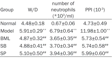
The results of W/D, number of neutrophils and PPI in different groups
The results of W/D, number of neutrophils and PPI in different groups were shown in Table 1. It showed that the ratio of W/D, the number of
neutrophils and PPI significantly increased in
model group when compared with normal group (P<0.01); while they significantly
de-creased in BML group, SB group and SP group when compared with model group (P<0.01).
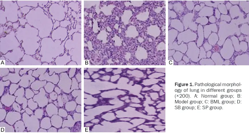
Pathological morphology of lung in different groups
As shown in Figure 1, the lung tissue in normal
[image:3.612.90.522.71.303.2]group was complete without infiltration of neu -trophils. There was pulmonary interstitial ede-
ma, thickening alveolar wall and infiltration of
neutrophils in model group. There was mild
pul-monary edema and infiltration of neutrophils in
BML, SB and SP groups.
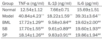
The levels of TNF-α, IL-1β and IL-6 in different groups
The levels of TNF-α, IL-1β and IL-6 in different
groups were shown in Table 2. It showed that
the levels of TNF-α, IL-1β and IL-6 in model group were significantly higher than that of nor -mal group (P<0.01). However, the levels of
TNF-α, IL-1β and IL-6 in BML group, SB group and SP group were significantly lower than that of
model group (P<0.01).
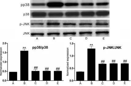
Phosphorylation levels of p38 MAPK and JNK in different groups
Western blotting results of p38 MAPK, JNK and their phosphorylation levels were shown in Figure 2. It showed that the phosphorylation levels of p38 MAPK and JNK in model group
were significantly higher than that of normal
group (P<0.01). However, the phosphorylation levels of p38 MAPK and JNK in BML group, SB
group and SP group were significantly lower
than that of model group (P<0.01).
The level of AQP1 in different groups
Western blotting results of AQP1 levels were shown in Figure 3. It showed that the level of
AQP1 in model group was significantly lower
than that of normal group (P<0.01). However, it
was significantly higher in BML group, SB group
and SP group than that of model group (P<0.01). Discussion
ALI is a clinical syndrome with characteristic pathological changes in the lung, it could cause
SP600125 group, the ratio of W/D, the number
of neutrophils and the levels of TNF-α, IL-1β and IL-6 decreased, degree of infiltration was
reduced. These results suggested that BML-111, SB203580 and SP600125 could
attenu-ate the ALI of rat by inhibiting the inflammatory
response. We also found that in BML-111 group, SB203580 group and SP600125 group, the phosphorylation levels of p38 MAPK and JNK decreased, which suggested that BML-111
could reduce the expression of inflammatory
factors through inactivating the MAPK p38 and JNK signaling pathway, and then to resist in-
flammation of ALI rats.
ALI could be inhibited by anti-inflammatory
reaction through p38 MAPK signaling pathway, pulmonary edema induced by ALI also could be alleviated by regulation of AQP1 through p38 MAPK signaling pathway [13]. AQP1 could pro-mote the migration of non arterial smooth mus-cle cells through p38-MAPK signaling pathway [14]. AQP1 could be down-regulated by peptido-glycan via p38 MAPK pathways and could be down-regulated by LPS via p38 MAPK, JNK and ERK pathways [15, 16]. These results suggest-ed that p38 MAPK and JNK pathways could also play an important role in pulmonary edema induced by ALI and were closely related to the expression of AQP1.
AQP1 and AQP5 participated in the abnormal transport of liquid in the pulmonary edema. Previous study found that water permeability decreased 10 times in AQP-5 knockout mice, while it decreased 25-30 times in AQP-1 and AQP-5 knockout mice [17]. ALI induced by LPS in rats could get the best treatment effect with up regulation of AQP1 and AQP5 [18]. In this
[image:4.612.88.299.97.178.2]sug-gested that BML-111, SB203580 and SP- 600125 could alleviate pulmonary edema by up-regulation of AQP1.
In a word, BML-111 could significantly inhibit
the lung injury in ALI rats induced by LPS and increased the levels of AQP1, which may be through the inactivation of MAPK p38 and JNK
signaling pathway. It has important theoreti-
cal significance for the clinical application of
BML-111.
Acknowledgements
[image:5.612.95.522.75.356.2]This work was supported by the National Clinical Key Specialty Project Foundation of the
Figure 2.Western blotting results of Phosphorylation levels of p38 MAPK and JNK in different groups. A: Normal group; B: Model group; C: BML group; D: SB group; E: SP group. Compared with normal group, **P<0.01; Compared
with model group, ##P<0.01.
Figure 3.The levels of AQP1 in different groups. A: Normal group; B: Model group; C: BML group; D: SB group; E: SP group; A: Western-blotting results; B: RT-PCR results. Compared with normal group, **P<0.01; Compared with model
[image:5.612.99.521.419.574.2]References
[1] Peters AL, Van Stein D and Vlaar AP. Antibody-mediated transfusion-related acute lung inju-ry; from discovery to prevention. Br J Haematol 2015; 170: 597-614.
[2] Ortiz-Diaz E, Festic E, Gajic O and Levitt JE. Emerging pharmacological therapies for pre-vention and early treatment of acute lung inju-ry. Semin Respir Crit Care Med 2013; 34: 448-458.
[3] Li H, Wu Z, Feng D, Gong J, Yao C, Wang Y, Yuan S, Yao S and Shang Y. BML-111, a lipoxin re-ceptor agonist, attenuates ventilator-induced lung injury in rats. Shock 2014; 41: 311-316. [4] Gong J, Li HB, Guo S, Shang Y and Yao SL.
Lipoxin receptor agonist, may be a potential treatment for hemorrhagic shock-induced ac- ute lung injury. Med Hypotheses 2012; 79: 92-94.
[5] He DK, Shao YR, Zhang L, Shen J, Zhong ZY, Wang J and Xu G. Adenovirus-delivered an- giopoietin-1 suppresses NF-kappaB and p38 MAPK and attenuates inflammatory responses in phosgene-induced acute lung injury. Inhal Toxicol 2014; 26: 185-192.
[6] Wei D, Huang ZH, Zhang RH, Wang CL, Xu MJ, Liu BJ, Wang GH and Xu T. Roles of p38 MAPK in the regulation of the inflammatory response to swine influenza virus-induced acute lung in -jury in mice. Acta Virol 2014; 58: 374-379. [7] Hasan B, Li FS, Siyit A, Tuyghun E, Luo JH, Upur
H and Ablimit A. Expression of aquaporins in the lungs of mice with acute injury caused by LPS treatment. Respir Physiol Neurobiol 2014; 200: 40-45.
[8] Li Z, Gao C, Wang Y, Liu F, Ma L, Deng C, Niu KC, Lin MT and Wang C. Reducing pulmonary injury by hyperbaric oxygen preconditioning during simulated high altitude exposure in rats. J Trauma 2011; 71: 673-679.
Invest 1999; 103: 555-561.
[12] Yao C, Purwanti N, Karabasil MR, Azlina A, Javkhlan P, Hasegawa T, Akamatsu T, Hosoi T, Ozawa K and Hosoi K. Potential down-regula-tion of salivary gland AQP5 by LPS via cross-coupling of NF-kappaB and p-c-Jun/c-Fos. Am J Pathol 2010; 177: 724-734.
[13] Li HB, Wang GZ, Gong J, Wu ZY, Guo S, Li B, Liu M, Ji YD, Tang M, Yuan SY, Shang Y and Yao SL. BML-111 attenuates hemorrhagic shock-in-duced acute lung injury through inhibiting ac- tivation of mitogen-activated protein kinase pathway in rats. J Surg Res 2013; 183: 710-719.
[14] Gao J, Chen L, Zeng J, Cui J, Ning JL, Wang GS, Belguise K, Wang X, Qian GS, Lu KZ and Yi B. The involvement of aquaporin 1 in the hepato-pulmonary syndrome rat serum-induced mi-gration of pulmonary arterial smooth muscle cells via the p38-MAPK pathway. Mol Biosyst 2015; 11: 3040-3047.
[15] Liu L, Du L, Chen Y, Qin S, Liang Q, Zou X, Liang X, Jiang J, Chen Q, Wang K and Xie C. Down-regulation of Aquaporin1 (AQP1) by peptidogly-can via p38 MAPK pathways in primary rat pleural mesothelial cells. Exp Lung Res 2014; 40: 145-153.
[16] Liu L and Xie C. Effects of downregulation of aquaporin1 by peptidoglycan and lipopolysac-charide via MAPK pathways in MeT-5A cells. Lung 2011; 189: 331-340.
[17] Ma T, Fukuda N, Song Y, Matthay MA and Verkman AS. Lung fluid transport in aquapo -rin-5 knockout mice. J Clin Invest 2000; 105: 93-100.