Original Article
Acute and subchronic toxicity as well as evaluation of
safety pharmacology of
Coptis chinensis Franch
solution
Xin Lai1*, Qingsheng Pei2*, Chunjuan Lei3*, Lianci Peng1*, Zhongqiong Yin1*, Renyong Jia1, Xu Song1, Yuanfeng Zou1, Lixia Li1, Lizi Yin1, Xiaoxia Liang1, Cheng Lv1
1Natural Medicine Research Center, College of Veterinary Medicine, Sichuan Agricultural University, Chengdu
611130, China; 2Qinghai Academy of Animal Science and Veterinary Medicine, Xining 810016, Qinghai, China; 3China Animal Disease Control Center, Beijing 100125, China. *Equal contributors and co-first authors.
Received November 8, 2015; Accepted April 12, 2016; Epub May 15, 2016; Published May 30, 2016
Abstract:Coptis chinensis Franch is a medicinal plant that is used as a traditional treatment for clearing heat, detoxicating, and treating dysentery and cholera. However, Coptis chinensis Franch has not previously been evalu-ated for safety through systematic toxicological studies. The aim of the present study was to evaluate the acute and subchronic toxicity of CoptischinensisFranch Solution (CCFS) in specific pathogen-free (SPF) Sprague-Dawley (SD) rats. In addition, we conducted a safety pharmacological evaluation of CCFS to supplement the toxicity tests. The acute administration of CCFS was performed at top dose of 5000 mg per kg/bodyweight (bw) and Groups of male and female SPF SD rats received 3000, 4000 and 5000 mg/kg doses of CCFS orally for 30 days. The acute toxicity study showed that the LD50 of CCFS was greater than 5000 mg/kg, whereas the subchronic oral toxicity study sug-gested that the growth of rats could be related to different doses of CCFS. The clinical pathology showed that a CCFS
dose of 5000 mg/kg could cause reversible damage to the liver, kidney, spleen, testis and ovary lesions. The NOAEL of CCFS is greater than 3000 mg/kg body weight but less than 4000 mg/kg body weight. Safety pharmacological findings indicated that CCFS has no side effects on the central nervous system, cardiovascular system and respira-tory system. These results suggest that CCFS is a safe veterinary medicine for external use.
Keywords:Coptis chinensis Franch solution, acute toxicity, subchronic toxicity, NOAEL, safety pharmacology
Introduction
In recent years, the risk of infections caused by pathogenic microorganisms has increased among humans and animals. Natural products, including those from plants, animals and miner-als, have been used as basic treatments for human disease [1]. The underlying principles of these products are different from those of the current popular western medical approaches [2]. Therefore, safe medicinal plants must be identified to replace these synthetic chemicals.
Coptis chinensis Franch has been officially list-ed in the Chinese Pharmacopoeia in the past few years [3]. However, the safety, efficacy, and quality of Coptis chinensis Franch solution have not been evaluated by the FDA as of this writing.
Coptis chinensis Franch (C. chinensis Franch), which is found in many parts of China
(espe-cially in Sichuan Province), mainly consists of alkaloids, such as berberine, epiberberine, and jatrorrhizine. C. chinensis Franch is traditionally used for heat clearing, detoxicating, and treat-ing dysentery and cholera [4]. This plant also possesses anti-microbial [5, 6], anti- inflamma-tory [7], anti-cancer [8, 9], analgesic, anti
-hypertension, and anti-oxidant properties [10]. It is worth highlighting that Coptis chinensis Franch can be widely used in daily life, espe-cially as an anti-bacterial agent.
Streptococcus agalactiae, also called Group B streptococcus, remains a major cause of sub-clinical mastitis in dairy cattle, and this disease has caused economic losses in industry [11].
Coptis chinensis Franch showed the best anti
-bacterial effects on Streptococcus agalactiae
modern scientific evidence on the efficacy and safety of herbal products remains lacking [13, 14]. This study estimates a no-observed
-adverse-effect level (NOAEL) and conducts toxi-cological and safety pharmatoxi-cological evalua-tions of CCFS to develop a safe formulation of
CCFS.
Materials and methods
Coptis chinensis Franch solution
Coptis chinensis Franch was supplied by the Xinran Biotechnology Co., Ltd (Shanghai, PR China). The water extracts of C. chinensis Franch and its solution (The Effective compo-nents of C. chinensis Franch was more than 70%) were prepared in a laboratory. Different concentrations of CCFS, including 30%, 40%, and 50%, were prepared by mixing appropriate doses of stabilizer and antioxidant.
Animals
Young adult male (average weight 100±5 g) and female (average weight 100±5 g) SPF Sprague-Dawley (SD) rats were purchased from Chengdu Dossy Experimental Animals Co., Ltd. [License No. SCXK (Sichuan) 2009-26]. Animal experiments were conducted under the princi-ples of proper laboratory animal care, and were approved by the ethical committee of the Laboratory Animals Care and Use of Sichuan Agriculture University (Chengdu, China). Each cage contained five rats of the same sex. Based on the guidelines of the International Committee on Laboratory Animals, the rats were main-tained at a controlled temperature of 20-25°C and relative humidity of 55±5% and 12 h light/ dark cycle with the lights off at 8 p.m. The experiments were conducted after acclimating the rats for 1 week. The animals were treated with a starter diet from Nuvital Nutrients (Colombol, PR, Brazil) and were given access to distilled water ad arbitrium.
Oral acute toxicity
An oral study for calculating LD50 was per-formed according to the guidelines of the Organization for Economic Cooperation and Development (OECD) [15-17]. Animals were dosed one at a time. If the first animal survived, after treatment, the next animal received a
higher dose. If the first animal died, the dose given to the next animal was reduced. Before the formal experiments, a pre-test study on five experimental groups with 2 rats each was per-formed. In the pre-test, CCFS treatment was conducted at a top-dose of 5000 mg/kg. If the animal survived, the surviving animals were divided into 5 experimental groups with 10 rats each. If no mortality, occurred after the top
-dose treatment was repeated twice, the test was stopped. In each case, the product volume administered by gavage was 1 mL/100 g body weight (b.w). The animals were observed for gross behavioral neurologic, autonomic, and toxic effects for 24 h and then daily for 14 d [18]. The toxicological effect was assessed based on mortality, which was expressed as an LD50 value.
30-day subchronic oral toxicity Treatments
According to the OECD guideline 407, four groups of 10 rats (each comprising 5 males and 5 males), were given CCFS doses of 3000 (Group II), 4000 (Group III), and 5000 (Group IV) mg/kg for 30 d consecutively [15]. The ani-mals were monitored for clinical and behavioral symptoms such as diarrhea, immobility, and mortality over this period. Each rat was marked with a unique identification number by using tri-nitrophenol. Rat body weight was measured once a week.
Clinical examination
All animals were observed once daily for clinical signs of toxicity. The changes in their hair, eyes, mucous membrane, respiratory system, ner-vous system, physical activity, and behavior were recorded. To reduce the residual interac-tion between the animals and postmortem tis-sue autolysis, the dead animals and those that are at risk of dying were dissected in a timely manner [19].
Bodyweight and food consumption
lung, kidney, testis, uterus, and ovary were recorded and expressed in relation to the final body weight of each animal.
Clinical pathology
Clinical pathology was performed to analyze the blood chemistry and hematology of the ter-minal sacrifice animals that were approaching the end of their in-life phase. For the hematol-ogy assessment, 0.5 mL of blood was collected from each animal in a pre-calibrated tube that contained EDTA. For the clinical chemistry assessments, approximately blood sample (1 mL) was collected in a tube that contained no preservatives. These blood samples were cen-trifuged and refrigerated, and the serum was transferred to a labeled tube.
Hematology: The hematological parameters included white blood count (WBC), red blood cell count (RBC), hemoglobin concentration
lung, testis and ovaries were measured imme-diately after dissection to avoid drying. The tis-sues and organs were obtained and preserved in 10% neutral buffered formalin, and pro-cessed for histopathological assessment.
[image:3.612.91.380.73.182.2]Histopathological examination: The preserved organs and tissues of rats from the control group (Group I) and treated groups (from Group II to Group V) were subjected to histological examination. They were pressed in a fixation medium of 10% buffered formalin (pH 7.4) and enclosed in paraffin for subsequent histopatho-logical examination. A 5 μm section of each organ tissue was stained with hematoxylin and eosin. Each section was examined under an optical microscope. Three slides from different part of each tissue (3 rats per group) were ana-lyzed. The whole lesions for each tissue were scored by multiplying the degree of severity (0 = no lesions, 1 = mild lesions, 2 = moderate Figure 1. Effect of subchronic administration of CCFS on body weight of male
rats. Average body weights for male rats during the 30-day oral (gavage) toxic-ity study. The values are presented as means ± standard deviation (5 rats/ sex/group).
Figure 2. Effect of subchronic administration of CCFS on body weight of female rats. Average body weights for male rats during the 30-day oral (gavage) toxic-ity study. The values are presented as means ± standard deviation (5 rats/ sex/group).
(HGB), hematocrit (HCT), mean corpuscular volume (MCV), mean corpuscular hemoglobin (MCH), MCH concentration (MCHC), pla- telet count (PLT), and leuko-cyte differential count (lym-phocytes, neutrophils, and monocytes).
Clinical chemistry: The mea-sured clinical chemistry pa- rameters included Albumin (ALB), total protein (TP), ala-nine aminotransferase (ALT), aspartate aminotransferase (AST), urea nitrogen (BU), creatinine (CRE), glucose (GLU), triglycerides (TG), to- tal cholesterol (TCH), creati-nine (CRE) potassium (K), sodium (Na), and chlorine (Cl).
Pathology
[image:3.612.90.381.255.393.2]lesions, and 3 = severe lesions) with the extent of lesions (1 = low extent, 2 = intermediate extent, and 3 = large extent) [20]. For each organ, the maximal lesional score was 9 and the minimal score was 0.
Safety pharmacology assay Treatment
Five groups of 10 rats, each comprising 5 males and 5 females, consumed saline control group (Group I), solvent control group (Group II), 3000 mg/kg (Group III), 4000 mg/kg (Group IV), 5000 mg/kg (Group V) doses of CCFS. The back of each rat was smeared with a 0.3 mL for 5 d.
Central nervous system assay
[image:4.612.91.523.84.190.2]During the last day of the experiment, the changes in the behavior, posture, gait and pupils of each rat were closely observed at 4 h intervals [21]. The rats with or without saliva-tion or muscle trembling were also recoded. The situations of these animals were observed for 7 d after the last administration of CCFS. Their independent activities were observed and recorded using a versatile recorder of locomo-tor activity at 0.5 h after the last administration of CCFS. Each rat was also subjected to a pole test at 0.5 h after the last administration of CCFS. During the test, the rats were placed on the tip of a rod that was fixed on the base. Each Table 1. Effect of subchronic administration of CCFS on hematological parameters
Parameters Group I Group II Group III Group IV
HGB, g/L 152.50±3.83 149.50±3.51 156.67±5.15 152.00±3.58
RBC, 1012/L 7.36±0.25 7.13±0.41 7.38±0.23 7.77±0.39
WBC, 109/L 9.78±3.89 11.70±2.34 10.23±2.99 10.03±1.79
GRA, % 2.25±0.95 2.18±0.46 2.62±0.89 2.70±0.49
LYM, % 7.28±2.86 9.25±2.11 7.33±1.99 7.02±1.28
MON, % 0.25±0.84 0.27±0.05 0.28±0.15 0.32±0.12
PLT, 109/L 1139.50±82.05 1089.67±154.46 945.17±129.17 888.17±158.83
The values are presented as means ± standard deviation (10 rats/group). HGB: hemoglobin; RBC: red Blood Cell; WBC: white
Blood Cell;GRA: neutrophils; LYM: lymphocytes; MON: monocytes; PLT: blood platelet. There was no significant difference in test groups and the control (P>0.05).
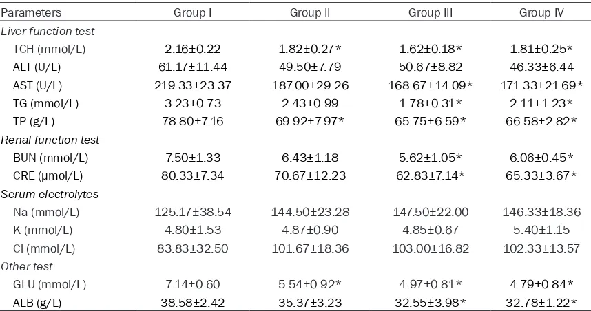
Table 2. Effect of subchronic administration of CCFS on serum biochemistry parameters
Parameters Group I Group II Group III Group IV
Liver function test
TCH (mmol/L) 2.16±0.22 1.82±0.27* 1.62±0.18* 1.81±0.25*
ALT (U/L) 61.17±11.44 49.50±7.79 50.67±8.82 46.33±6.44
AST (U/L) 219.33±23.37 187.00±29.26 168.67±14.09* 171.33±21.69*
TG (mmol/L) 3.23±0.73 2.43±0.99 1.78±0.31* 2.11±1.23*
TP (g/L) 78.80±7.16 69.92±7.97* 65.75±6.59* 66.58±2.82*
Renal function test
BUN (mmol/L) 7.50±1.33 6.43±1.18 5.62±1.05* 6.06±0.45*
CRE (µmol/L) 80.33±7.34 70.67±12.23 62.83±7.14* 65.33±3.67*
Serum electrolytes
Na (mmol/L) 125.17±38.54 144.50±23.28 147.50±22.00 146.33±18.36
K (mmol/L) 4.80±1.53 4.87±0.90 4.85±0.67 5.40±1.15
Cl (mmol/L) 83.83±32.50 101.67±18.36 103.00±16.82 102.33±13.57
Other test
GLU (mmol/L) 7.14±0.60 5.54±0.92* 4.97±0.81* 4.79±0.84*
ALB (g/L) 38.58±2.42 35.37±3.23 32.55±3.98* 32.78±1.22*
The values are presented as means ± standard deviation (10 rats/sex/group). ALB: albumin; ALT: alanine aminotransferase;
[image:4.612.92.521.256.482.2]rat crawled down the rod and rated based on the following criteria:
0: Step by step to climb down; 1: Downward side; 2: Unable to grasp the stick; 3: Loss of righting reflex.
Heart rate assay and respiratory rate assay
The heart rate and respiratory rate of each group of rats was measured by using BL-420F to record their electrocardiogram after they were anesthetized by ether before administra-tion, after the second administration of CCFS and a week after the last administration.
Statistical analysis
The means and standard deviations of the measured data from each group, including body weight, food consumption, clinical patho-logical data, organ weights, heart rate and respiratory rate, were calculated. The statisti-cal significance of the data from control and experimental groups was compared by one way analysis of variance (ANOVA) and the Student-Newman-Keuls test.
Results
Acute toxicity study
During the pre-experiment, the rats that were given an optimum CCFS dose showed no
mor-and 2. Compared with the control group (Group I), the female rats in Group II had significantly lower body weight (P<0.05), whereas the male rats in Group IV had significantly higher body weight (P<0.05).
Clinical pathology
Hematology parameters: The changes in the hematology parameters including hemoglobin (HGB), red blood cell (RBC), white blood cell (WBC), neutrophils (GRA), lymphocytes (LYM), monocytes (MON), blood platelet (PLT), are shown in Table 1. No significant differences were observed between the control group and experimental groups (P>0.05). The values are presented as means ± standard deviation (10 rats/group).
[image:5.612.91.372.107.306.2]Serum biochemical parameters: The changes in the serum biochemical parameters including albumin (ALB); amine aminotransferase (ALT); aspartate transaminase (AST); total protein (TP); blood urea nitrogen (BUN); glucose (GLU); Creatinine (CRE); total cholesterol (TCH); triglyc-erides (TG); sodium (Na); potassium (K); chlo-rine (Cl), are shown in Table 2. No significant differences were observed in the ALT, TCH, TP, BUN, K, and Alb of the control group and the experimental groups (P>0.05). The TP, CH, and GLU of the rats in low dose Group were signifi-cantly different from those of the rats in Group Table 3. Effect of subchronic administration of CCFS on terminal
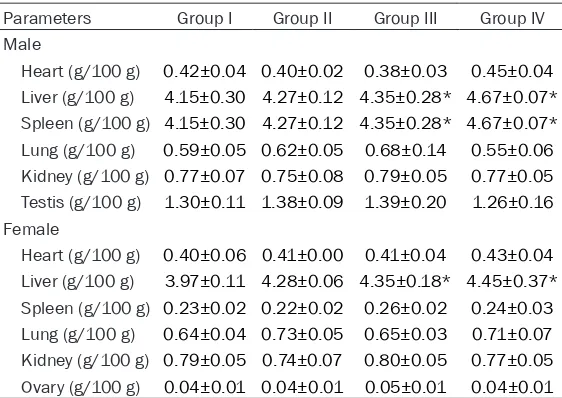
body weight and organic coefficient (g/100 g) in grams of male and female rats
Parameters Group I Group II Group III Group IV
Male
Heart (g/100 g) 0.42±0.04 0.40±0.02 0.38±0.03 0.45±0.04
Liver (g/100 g) 4.15±0.30 4.27±0.12 4.35±0.28* 4.67±0.07*
Spleen (g/100 g) 4.15±0.30 4.27±0.12 4.35±0.28* 4.67±0.07*
Lung (g/100 g) 0.59±0.05 0.62±0.05 0.68±0.14 0.55±0.06
Kidney (g/100 g) 0.77±0.07 0.75±0.08 0.79±0.05 0.77±0.05
Testis (g/100 g) 1.30±0.11 1.38±0.09 1.39±0.20 1.26±0.16
Female
Heart (g/100 g) 0.40±0.06 0.41±0.00 0.41±0.04 0.43±0.04
Liver (g/100 g) 3.97±0.11 4.28±0.06 4.35±0.18* 4.45±0.37*
Spleen (g/100 g) 0.23±0.02 0.22±0.02 0.26±0.02 0.24±0.03
Lung (g/100 g) 0.64±0.04 0.73±0.05 0.65±0.03 0.71±0.07
Kidney (g/100 g) 0.79±0.05 0.74±0.07 0.80±0.05 0.77±0.05
Ovary (g/100 g) 0.04±0.01 0.04±0.01 0.05±0.01 0.04±0.01
The values are presented as means ± standard deviation (10 rats/group). *P<0.05 shown there was significantly difference from control.
tality. A zero mortality rate was also achieved during the re- peated experiments.
Subchronic toxicity study
Clinical observations
The behavior of rats was not adversely affected by CCFS
doses of 3000, 4000, and 5000 mg/kg (Group II-Group IV). The rats remained healthy and demonstrated no signs of toxicity during the experimen-tal period.
Body weight and feed con-sumption
I (P<0.05). The ALB, GLU, CRE, BUN, TP, TG, AST, and TCH of the rats in the middle and high dose group were significantly different from those of the rats in the control group (P<0.05).
Organ coefficient
The organ coefficient results are shown in Table 3. The organ coefficient of heart, liver, spleen, kidney, testis (in male), and ovary (in female) in the test groups were not statistically different from those in the control group (P>0.05). How- ever, the organ coefficient of the lung in the medium and high dose groups was significantly different from that of the control group (P<0.05).
Histopathological analysis
The cross-section in the liver of the rats in the control group showed a normal and conserved appearance of liver, sinusoids, and hepato-cytes (Figure 3A). Central venous extended with hyperemia and varying degrees of
vacuo-lar degeneration of hepatocytes were found in the liver of the rats in the experimental groups (Figure 3B).
The cross-section in the spleen of the rats in the control group showed a normal appearance of spleen, white pulp, red pulp, and spleen tra-becula (Figure 4A). Red pulp extended with hyperemia and a large number of macrophages and Langhans cells (LC) infiltration were ob- served in the liver of the rats in the experimen-tal groups (Figure 4B).
[image:6.612.90.288.70.386.2]The cross-section in the kidney of the rats in the control group showed a normal appearance of kidney, glomerulus, renal capsule, and renal tubular epithelial cell (RTEC) (Figure 5A). Glo- merulus with varying degrees of hyperemia, RETC with varying degrees of granular degen-eration, and a narrowed renal tubular were found in the liver of the rats in the experimental groups (Figure 5B).
[image:6.612.325.523.70.384.2]Figure 3. A. The liver of rats in the control group (HE, 200×). B. The liver of rats in the experiment group (3000 mg/kg, 4000 mg/kg, 5000 mg/kg doses of CCFS), vesicular degeneration (↑↓) and central ve-nous hyperemia (←) (HE, 400×).
The heart cross section of the rats in the con-trol group had normal appearance, striations, nucleus, and interstitial cells (Figure 6A). In rats in the experimental group, myocardial interstitium extended with hyperemia, and a small amount of inflammatory cell in filtration was observed (Figure 6B).
The cross-section in the lung of the rats in the control, middle dose, and high dose groups showed a normal appearance of the lung, bron-chia, and arterioles (Figure 7A). The capillaries in the alveolar walls extended with hyperemia and thickened alveoli septum were found in the heart of the rats in the high dose group (Figure 7B).
The cross-section in the ovaries of the rats in the control group showed that the ovary and fol-licles have a normal appearance (Figure 8A). Granular degeneration and inflammatory cell
infiltration were observed in the heart of the rats in the experimental groups (Figure 8B). In the testicles of the control group, the cross
-section showed the normal appearance of testicles, convoluted seminiferous tubule and Leydig cell (Figure 9A). A wide gap among Leydig cells was observed in the testicles of the rats in the experimental groups (Figure 9B). In the stomach and intestines, the cross-sec-tion showed no significant lesions among the control groups and the experiment groups. These observations were also proved by the lesional score (Table 4).
Safety pharmacology study The central nervous system
No abnormalities were observed on the behav-ior, posture, gait, and pupils of the rats. Bizarre Figure 5. A. The kidney of rats in the control group,
glomerulus, renal capsule and RTEC (↑) (HE, 200×). B. The kidney of rats in the experiment group (3000 mg/kg, 4000 mg/kg, 5000 mg/kg doses of CCFS), glomerulus hyperemia (→), RETC with granular de-generation (↑) (HE, 400×).
behaviors, such as salivation and muscle trem-bling, were not observed. No changes were also observed in both the control and experimental groups after the climbing pole test. The level of all the groups was “0”.
Cardiovascular and respiratory system
The safety pharmacology results for the cardio-vascular system are shown in Table 5. After measuring the heart rate and respiratory rate of all rats, no significant changes were found in the cardiovascular system and respiratory sys-tem of the rats in both the experimental and control groups (P>0.05).
Respiratory system
The result was shown in the Table 6. After mea-suring the respiratory rate of all rats, there were no significant changes were observed in the respiratory system of the rats in both the exper-imental and control group (P>0.05).
Discussion
Not much is known regarding the toxicity of
CCFS. Given the potential health risk of using this drug, the efficacy and safety of CCFS re- quires further evaluation because the demand for this drug is growing. Currently, we conduct-ed a comprehensive toxicological evaluation and a necessary safety pharmacology evalua-tion on CCFS by performing acute and 30 d sub-chronic oral toxicity studies and a safety ex- periment.
[image:8.612.324.523.70.385.2]In this study, the acute toxicity results showed that the LD50 value of CCFS was more than 5000 mg/kg by oral route. The data are of high-ly reliable because the value is in the 95% con-fidence interval. According to the acute toxicity grading standards [21], if LD50 is in the range 5000-15000 mg/kg, the drug is non-toxic. Therefore, CCFS is non-toxic.
[image:8.612.89.289.70.388.2]Figure 7. A. The lung in the control group bronchia and arterioles (HE, 200×). B. The lung of rats in the high dose group (5000 mg/kg dose of CCFS), capil-laries in the alveolar walls hyperemia and thickened alveoli septum (→) (HE, 400×).
The LD50 of Coptis chinensis Franch to mice is 2950 mg/kg body weight [22] but data on rats are not available. These results can be a foun-dation for further study.
To assess the long term hazard of using CCFS, subchronic toxicity studies are always valuable in evaluating the safety of xenobiotics [23]. Changes in body weight have been used as an indicator of adverse effects of drugs and chem-icals [24]. In this study, significant changes (P<0.05) were found with the low dose (3000 mg/kg) on female rats and the high dose (5000 mg/kg) of male rats compared with the control group. A feasible dose of CCFS may be able to speed up the weight gain of male rats. Moreover, no significant changes in the general behavior were observed. Therefore, low-dose (3000 mg/ kg) CCFS has no significant differences in the growth and functions of rats.
The hematopoietic system is one of the most sensitive parameters that can be used to assess toxicity of drugs in humans and animals [25]. This study indicated that no significant dif-ferences were observed in HGB, RBC, WBC, GRA, LYM, MON and PLT between the control and the treated groups, and CCFS showed no effects on the circulating blood cells.
Liver is the main site of the synthesis of plasma proteins, and any damage to the liver results in elevations of both ALT and AST in the blood [25]. Moreover, ALT found in the serum consid-ered as a first sign of cell and liver damage [26, 27]. GLU, TG, and TCH are also important bio-chemical indicators related to the liver. The changes in these three indicators suggest inflammation, necrosis, poisoning, and biliary disease of the liver. The present study indicat-ed that ALT and AST in the treatindicat-ed group decreased slightly compared with the control group. In addition, TG and TCH of high-dose (5000 mg/kg) and middle-dose groups (4000 mg/kg) were also decreased compared with the control group. These results indicated that the liver was the target organ of CCFS toxicity. Creatinine is known as a good indicator for renal function. Increased creatinine levels indi-cate obvious damage to kidney [25, 28]. A decline in creatinine levels does not indicate abnormality, but rather, malnutrition. The pres-ent study indicated that CRE and BUN of high-dose and middle-high-dose groups were lower than those of the control groups, thereby indicating that CCFS did not damage the kidney of rats. The characteristic histopathological findings in the kidney were mainly granular degeneration and glomerulus hyperemia in the rats at 5000 mg/kg CCFS. These results suggested that
CCFS at 5000 mg/kg might slightly alter the renal function.
Organ index is the radio of organ weight to body weight and [29] is an important indicator of the functional status of the animals. The increase of organ coefficient indicates organ congestion, edema, or hypertrophy, whereas the decrease of organ coefficient indicates organs atrophy and other degenerative changes [30, 31]. In this study, the results suggested that the index of livers of the high-dose (5000 mg/kg) and middle-dose groups (4000 mg/kg) were higher than those of the control group (Group I), there-by indicating that livers were significantly oncot-Figure 9. A. The testicles of rats in the control group,
[image:9.612.90.289.69.387.2]ic (P<0.05). The indices of spleens of males in the high-dose (5000 mg/kg) and middle-dose groups (4000 mg/kg) were higher than the con-trol group, indicating the spleens were signifi-cantly oncotic (P<0.05).
No observed adverse effect level (NOAEL) was observed for select target organs [32]. To de- termine the NOAEL of CCFS, the pathological examination of principal vital organs in rats at different doses of CCFS is indispensable. The results of oral administration with CCFS for 30 d from the study showed that CCFS at 3000 mg/kg had no recoverable damage on vital organs. The medium (4000 mg/kg) and high (5000 mg/kg) doses of CCFS caused damage on the liver, kidneys, spleen, ovary, and testicle, at varying degrees, and such damage was mainly detected as granular and vacuolar degeneration in cells, focal necrosis, vascular congestion, and damage on Langhans cells. LC derived from bone marrow has Ia antigen, C3, and Fc-IgG receptors and functions in antigen presentation and allogeneic stimulation, si- milar to the function of macrophage [33]. Macrophages and Langhans cells found in the spleen suggest that CCFS stimulated the body’s immune system and boosted immunity. The lesions found in the liver and kidney suggested that the target organs of CCFS are the liver and kidney, which are also consistent with the hematology and biochemical findings. The slight lesions found in the heart, lung, ovaries and testicles suggest that CCFS may have mild effects on organs with middle and high dose. These findings indicated that the NOAEL of
CCFS is greater than 3000 mg/kg body weight
Based on the technical research guidelines of veterinary medicine and natural medicine on safety pharmacology, we measured the effect of CCFS on the cardiovascular system in anes-thetized rats via non-invasive route for the first time. This method effectively reduced the inter-ference of related physiological indicators caused by stress response and surgical inter-ference, and ensured that the various physio-logical indictors could reflect the true state of the rats. The results suggest that CCFS had no effect on the nervous system, respiratory system and cardiovascular system.
Conclusions
In conclusion, the LD50 value of CCFS by oral route was more than 5000 mg/kg, thereby indi-cating that CCFS is a non-toxic drug. Subchronic oral toxicity test results showed that the appro-priate dose of CCFS may not affect the growth of rats. The target organs of the toxic effects of
CCFS might be the liver and kidney. The NOAEL of CCFS is greater than 3000 mg/kg body weight but less than 4000 mg/kg body weight. In the safety pharmacological study, CCFS
administration did not cause produce any side effect on nervous system, cardiovascular sys-tem and respiratory syssys-tem of rats. Thus, CCFS
is a safe veterinary medicine for use in minimiz-ing Streptococcus agalactiae growth.
Acknowledgements
[image:10.612.91.342.84.192.2]This research was financially supported by National Natural Science Foundation of China (Grant No. 31372477); The Sichuan Youth Table 4. Lesional scores for each group
Parameters Group I Group II Group III Group IV
Liver 0.67±0.58 1.33±0.58 4.33±1.73* 2.58±1.98*
Spleen 1.67±0.58 2.00±1.00 3.00±1.00 2.33±0.58
Kidney 1.33±0.58 3.67±2.08 4.67±1.16* 5.33±1.16*
Heart 2.33±0.58 4.00±1.73 4.67±1.15 6.33±2.52*
Lung 2.33±0.58 3.00±1.00 3.33±0.58 4.67±1.15*
Ovary 2.00±1.00 3.67±2.08 3.67±0.58 5.33±1.16*
Testicle 2.33±0.58 3.00±1.00 4.00±1.73 6.33±2.52*
The values are presented as means ± standard deviation (3 rats/group).
*P<0.05 shown there was significantly difference from control (group I).
Lesional scores of each organ were obtained by multiplying the degree of severity (0 = no lesions, 1 = mild lesions, 2 = moderate lesions, and 3 = severe lesions) with the extent of lesions (1 = low extent, 2 = interme-diate extent, and 3 = large extent).
Science and Technology Innovation Research Team for waterfowl disease prevention and control (2013TD0015); the Sichuan Interna- tional Cooperation Projects (2014HH0058). Disclosure of conflict of interest
None.
Address correspondence to: Zhongqiong Yin, Col- lege of Veterinary Medicine, Sichuan Agricultural University, Chengdu, 611130, PR China. Tel: +86 28 86291176; Fax: +86 28 86291176; E-mail: yinzhongq@163.com
References
[1] Sathya M, Kokilavani R and Teepa KSA. Acute and subacute toxicity studies of ethanolic ex-tract of acalypha indica linn in male wistar al-bino rats. Asian Journal of Pharmaceutical & Clinical Research 2012; 97-100.
[2] Poon SK, Goyal S, Cheng A and Poon J. Search-ing for Evidence in Traditional Chinese Medi-cine Research: A Review and New Opportuni-ties. In: editors. Data Analytics for Traditional Chinese Medicine Research. Springer; 2014. pp. 1-16.
[3] Kuang L, Zhang K and Commission CP. Phar-macopoeia of the People’s Republic of China 2005. People’s Medical Publishing House; 2005.
[4] Herbals C. Board, the State Administrative Bu-reau of Chinese Medicine of China. Chinese Herbals 1999; 7: 942-953.
[5] Chen J, Wang F, Liu J, Lee SC, Wang X and Yang H. Analysis of alkaloids in Coptis chinensis
Franch by accelerated solvent extraction com-bined with ultra performance liquid chromato-graphic analysis with photodiode array and tandem mass spectrometry detections. Anal Chim Acta 2008; 613: 184-195.
[6] Bing FH, Liu J, Li Z, Zhang GB, Liao YF, Li J, Dong CY. Anti-influenza-virus activity of total alkaloids from Commelina communis L. Arch Virol 2009; 154: 1837-1840.
[7] Nam KN, Choi YS, Jung HJ, Park GH, Park JM, Moon SK, Cho KH, Kang C, Kang I and Oh MS. Genipin inhibits the inflammatory response of rat brain microglial cells. Int Immunopharma-col 2010; 10: 493-499.
[8] Wang XN, Xu LN, Peng JY, Liu KX, Zhang LH, Zhang YK. In vivo inhibition of S180 tumors by the synergistic effect of the Chinese medicinal herbs Coptis chinensis and Evodia rutaecarpa. Planta Med 2009; 75: 1215-1220.
[9] Deng XK, Wu Y, Li WD, Yin FZ, Lu XY, Zhang XC, Hua ZC and Cai BC. The anti-tumor effects of alkaloids from the seeds of Strychnos nux-vomica on HepG2 cells and its possible mech-anism. J Ethnopharmacol 2006; 106: 179-186.
[10] Xu D and Zhou C. Antioxidative effects of ber-berine pre-treatment on hydrogen peroxide-in-duced PC12 cell toxicity. Neural Regen Res 2010; 5: 1391-1395.
[11] Keefe GP. Streptococcus agalactiae mastitis: a review. Can Vet J 1997; 38: 429.
[image:11.612.87.526.86.150.2][12] LianCi P, ZhongQiong Y, RenYong J, Li L, RuYi D, Jing Q, MingHui L and Ping C. Effects of twenty traditional Chinese medicine extracts against Streptococcus agalactiae in vitro. Journal of South China Agricultural University 2014; 35: 22-25.
Table 5. Effects on the heart rate of rats
The time of measuring (Beats/min)Group I (Beats/min)Group II (Beats/min)Group III (Beats/min)Group IV (Beats/min)Group V
Before dosing 387.12±6.23 387.56±10.09 387.12±6.23 388.56.±8.91 387.00±7.95
The second day after dosing 375.26±3.68 380.53±9.77 375.26±3.68 375.96±14.00 380.00±13.57
One week after dosing 395.76±21.37 390.27±9.98 395.76±21.37 397.83±29.75 395.39±14.00
The values are presented as means ± standard deviation (10 rats/group). There was no significant difference in test groups and the control (P>0.05).
Table 6. Effect on the respiratory rates of rats
The time of measuring (Beats/min)Group I (Beats/min)Group II (Beats/min)Group III (Beats/min)Group IV (Beats/min)Group V
Before dosing 89.49±3.78 91.27±6.38 85.65±2.27 93.18±9.86 90.28±7.27
The second day after dosing 90.27±3.65 90.32±7.21 87.28±4.76 89.29±10.28 90.18±2.98
One week after dosing 86.79±7.28 85.37±6.83 86.38±8.38 85.38±3.98 86.28±9.73
[image:11.612.90.524.207.272.2][13] Seeff LB. Herbal hepatotoxicity. Clin Liver Dis 2007; 11: 577-596.
[14] Tang JL, Liu BY and Ma KW. Traditional chinese medicine. Lancet 2008; 372: 1938-1940. [15] Co-operation OfE and Development. Test No.
425: Acute Oral Toxicity: Up-and-Down Pro- cedure. OECD Publishing, 2008.
[16] Jung H and Choi S. Sequential method of esti-mating the LD50 using a modified up-and-down rule. J Biopharm Stat 1994; 4: 19-30. [17] Bruce RD. An up-and-down procedure for acute
toxicity testing. Fundam Appl Toxicol 1985; 5: 151-157.
[18] Food and Administration D. Single dose acute toxicity testing for pharmaceuticals. Fed Regist 1996; 61: 43934-43935.
[19] Schneeman BO. Dietary fiber and gastrointes-tinal function. Nutr Rev 1987; 45: 129-132. [20] Duan W and Liang X. Technical guidelines
assembly of veterinary medicine research. Bei-jing: Chemical In dustry Press; 2011.
[21] Ma BL, Ma YM, Shi R, Wang TM, Zhang N, Wang CH and Yang Y. Identification of the toxic constituents in Rhizoma Coptidis. J Eth-nopharmacol 2010; 128: 357-364.
[22] Aniagu SO, Nwinyi FC, Akumka DD, Ajoku GA, Dzarma S, Izebe KS, Ditse M, Nwaneri PE, Wambebe C and Gamaniel K. Toxicity studies in rats fed nature cure bitters. Afr J Biotechnol 2005; 4: 72-78.
[23] Tofovic SP and Jackson EK. Effects of long-term caffeine consumption on renal function in spontaneously hypertensive heart failure prone rats. J Cardiovasc Pharmacol 1999; 33: 360-366.
[24] Rahman MF, Siddiqui MK and Jamil K. Effects of Vepacide (Azadirachta indica) on aspartate and alanine aminotransferase profiles in a subchronic study with rats. Hum Exp Toxicol 2001; 20: 243-249.
[25] Jaouad EH, Israili ZH and Badiaa L. Acute and chronic toxicological studies of Ajuga iva in ex-perimental animals. J Ethnopharmacol 2004; 91: 43-50.
[26] Auza NJ, Olson WG, Murphy MJ and Linn JG. Diagnosis and treatment of copper toxicosis in ruminants. J Am Vet Med Assoc 1999; 214: 1624-1628.
[27] Lameire N, Van BW and Vanholder R. The changing epidemiology of acute renal failure. Nat Clin Pract Nephrol 2006; 2: 364-377. [28] Xia JY, Lei PQ, Zeng XL and Li H.
Determina-tions to the weight of main organs and bio-chemical indexes of SPF KM Mice. Sichuan Journal of Physiological Sciences 2009. [29] Wei JZ, Xuan_Guang WU and Lin HH. A further
observation on the serum biochemical values of chickens infected with Eimeriatenella or E.acervulina and the discussion on the patho-logical lesions. Chin J Prev Vet Med 2003. [30] Kristiansen E and Madsen C. Induction of
pro-tein droplet ( α 2 μ -globulin) nephropathy in male rats after short-term dosage with 1,8-cin-eole and l -limonene. Toxicol Lett 1995; 80: 147-152.
[31] Nabae K, Hayashi SM, Kawabe M, Ichihara T, Hagiwara A, Tamano S, Tsushima Y, Uchida K, Koda T and Nakamura M. A 90-day oral toxicity study of purple corn color, a natural food colo-rant, in F344 rats. Food Chem Toxicol 2008; 46: 774-780.
[32] Stingl G, Katz SI, Clement L, Green I and She-vach EM. Immunologic functions of Ia-bearing epidermal Langerhans cells. J Immunol 1978; 121: 2005-2013.