Original Article
Application of a modified cannula insertion technique
for selective endoscopic lumbar discectomy
Lei Ma1,2*, Wei Chen2*, Sidong Yang1,2*, Di Zhang1,2, Hui Wang1,2, Yong Shen1,2, Wenyuan Ding1,2, Wei Zhang1,2
1Department of Spinal Surgery, The Third Hospital of Hebei Medical University, No. 139 Ziqiang Road, Shijiazhuang 050051, China; 2Hebei Provincial Key Laboratory of Orthopedic Biomechanics, Shijiazhuang 050051, China. *Equal contributors.
Received October 18, 2015; Accepted January 6, 2016; Epub February 15, 2016; Published February 29, 2016
Abstract: In this prospective randomized study, we try to find a modified insertion technique in selective endoscopic discectomy (SED) with good outcome but lower incidence of complication. Patients with L4/5 lumbar disc hernia-tion were enrolled from March 2009 and randomly divided into two groups which were tradihernia-tional inserhernia-tion group I and modified insertion group II. Compared with traditional SED technique, the direction of the tube in modified insertion technique was about 20°-25° cephalic inclination and the entry point moved to cranial direction. Surgical outcome and complication were compared in this study. There were 46 patients in group I and 49 patients in group II completed the follow up with 15.39±1.98 months and 15.14±2.06 months respectively. There were no significant differences in ODI and VAS scores in both group pre and post the operation. X-Ray exposure time in Group II (2.59±0.30 minutes) was lower than that in Group I (2.44±0.28 minutes) (P=0.014). Radiating pain during the operation occurred in 8 patients in group I and 2 in group II with significant inter-group difference (P=0.046). Numbness of the lower extremities occurred in five patients in Group I and none patient in Group II (P=0.027). So the modified cannula insertion technique can achieve comparable outcome with lower incidence of complication and less X-Ray exposure.
Keywords: Lumbar disc herniation, selective endoscopic discectomy, modified technique, clinical study
Introduction
Endoscopic spinal surgery is evolving rapidly with the improvement of surgical technique and instrumentation. In comparison with open sur-gery, the endoscopic technique is better to visualize and treat the pathological lumbar disc in a minimally invasive fashion without destabi-lizing the posterior muscles, vertebral column and facets [1-6]. However, a long-term learning curve is required to master endoscopic tech-nique for spinal surgery [7, 8]. In addition, there are potential complications associated with this procedure [9-15].
There are currently two main techniques for endoscopic spinal surgery: the technique devel-oped by Dr. Yeung, which utilizes the Yeung endoscopic spine system, (YESS) [1, 4, 16, 17], and the transforaminal endoscopic technique (TESSYS) [18]. Mastering YESS is the first step for most newcomers to minimally invasive
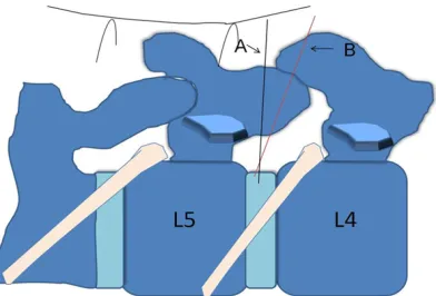
proce-were treated with selective endoscopic discec-tomy (SED) using the traditional Yeung’s inser-tion method (Figure 1, needle A), and patients in Group II underwent SED using the modified cannula insertion technique by a 20-25° cephalic inclination angle (Figure 1, needle B). Two senior surgeons both with more than five years of experience in the application of selec-tive endoscopic lumbar discectomy conducted all the operations in turn.
Surgical techniques
All the procedure was performed under local anesthesia in the prone position on a radiolu-cent table in all patients. Before the surgery, patients were informed with all steps of the pro-cedure. Patients communicated with the sur-geon during the procedure.
Patients in Group I underwent SED using the traditional Yeung’s technique to identify the cannula insertion point (Figure 2, point A) and angle, (Figure 1 needle A) which is based on the foraminal approach procedure developed by Yeung in 2003 [4].
Patients in Group II underwent SED with a mod-ified cannula insertion technique, whose proce-dure was described briefly as follows.
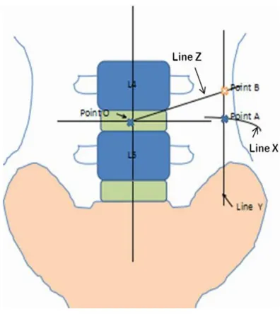
The postero-anterior view of the lumbar spine was obtained with the use of C-arm unit to get the topographical location of the midline, trans-verse plane of the L4/5 disc and the intersec-tion point O (Figure 2).
Intra-operative lateral X-ray imaging was taken to get the location of the lateral disc center (Figure 3).
A line (named as line X) between the lateral center of the disc and the posterior lateral skin was drawn along the inclination of the disc (Figure 3). In the traditional Yeung’s procedure, a line (named Line Y) was drawn which is paral-lel with the midline and the distance between the line Y and midline was equal to the length of Line X. The intersection point A between Line X and Y was the entry point for traditional Yeung’s technique (Point A in Figure 2). In the modified technique, Line Z was drawn from point O with 20-25° cephalic to the end plate and intersect-ed with line Y. The intersection namintersect-ed point B which was the modified entry point (Point B in dure [10], we modified the traditional Yeung’s
insertion technique by a 20-25° cephalic incli-nation angle. We aimed to reduce the irritation of the nerve root during insertion while main-tain the ‘inside out’ concept.
Method
A prospective randomized control study was conducted to assess the effectiveness and safety of the modified technique by comparing surgical outcomes and complications of L4/5 lumbar disc herniation treated using this tech-nique with those treated using the traditional Yeung technique.
[image:2.612.92.288.70.203.2]The study was approved by the local ethics committee and informed consent was obtained from all patients. The patients with L4/5 lum-bar disc herniation, who were admitted to the Department of Spinal surgery from March 2009, were randomly assigned to two groups to treat using either the traditional or modified Yeung technique. The inclusion criteria were as follows: (1) sciatica of one lower extremity; (2) L4/5 disc herniation with evidence on MRI image; and (3) no improvement after at least three-month conservative treatment. The exclusion criteria were: (1) painless motor weakness; (2) previous surgical treatment; (3) the presence of calcified fragments; (4) with disc extrusions, sequestrations, subarticular or foraminal stenosis, or multilevel of herniation. The patients were randomly divided into two groups. After admission to the study, all patients were assessed according to the Oswestry Disability Index (ODI) criteria [21] and Visual analog scales (VAS) [10]. Patients in Group I
wire. After insertion of the obturator a bevel-ended working cannula was inserted into the disc along the obturator (Figures 4 and 5). Then an endoscope was inserted through the work-ing cannula. The blue-stained disc was removed using forceps. After herniated fragment was all removed, the endoscope was removed, and a sterile dressing was applied with a 1-point suture.
All the patients were advised to walk within six hours after the surgery and to do functional practice under the guidance of rehabilitation physician.
The operation time, blood loss and X-ray expo-sure time were recorded. The intra-operative complications, including radiating pain caused by nerve root irritation and dura matter leakage were recorded. The postoperative complica-tions such as: infection, fat liquefaction, hema-toma, numbness and decline in myodynamia of lower extremity were recorded before and after surgery, pain was measured by the 10-point VAS scoring and function was assessed by the ODI in percent (0-100%).
The patients were instructed for follow up at one day, 3 months, 6 months, 12 month after operation. Physical assessment was taken at each follow-up. Antero-posterior and lateral X-rays of the lumbar spine (or CT scan) were taken at the one-year postoperative time point.
Statistical analysis
Based on the data resulted from a preliminary experimental study, a total sample size of not less than ninety patients were required to per-form the statistical analysis. The data were analyzed with SPSS 14.0 k for Windows (SPSS Inc., Chicago, IL, USA). Continuous variables with normality were expressed as mean ± stan-dard deviation (SD). If not, median (interquartile range, IQR) was used. Categorical variables presented as the number of cases. Between-group differences, pre- and post-operative dif-ference in clinical parameters were compared using Student’s t-test. Student’s t test and Fisher’s exact text were used to assess between-group differences in clinical outcomes and incidence of complications. A p-value of less than 0.05 was considered statistically significant.
Figure 2). Then an 18 guage spinal needle was inserted along the direction of line B-O from lat-eral side to the midline with a 20-25° cephalic inclination under the guidance of a fluoroscopic image. After insertion of the spinal needle into the disc the nucleus pulposus was stained blue with 1 ml of indigo carmine. The next steps were as follows: a guide wire was inserted through the spinal needle and then the spinal needle was removed. A small incision was made at the entry point and a tapered cannu-lated obturator was inserted along the guide
[image:3.612.89.287.69.291.2]Figure 2. The intra-operative postero-anterior imag-ing was taken to identify the midline, one line beimag-ing parallel with the L4/5 disc and the intersection point O. The point A which was the intersection of Line X and Line Z was the entry point of traditional Yeung’s technique. The point B was the entry point of modi-fied technique.
[image:3.612.91.287.390.517.2]13.98±4.38 one year after surgery. In Group II, the mean VAS score was 7.43±0.87 before sur-gery, and was improved to 0.59±0.65 one year after surgery. The mean ODI scores were improved from 57.02±7.28 before surgery to 12.78±3.50 one year after surgery. There were no significant between-group difference both in VAS (P=0.368) and ODI scores (P=0.141) one year after operation (Table 2).
During the operation, radiating pain occurred in eight patients in Group I and two patients in Group II. There was significant between group Four patients in group I and one patient in group II drop out during the follow-up peri-od and the follow-up periperi-od was at least 12 months. The mean follow-up periods was 15.39±1.98 months in Group I and15.14±2.06 months in Group II (Table 1).
The mean operation time was 63.52±4.26 minutes in Group I and 60.92±5.37 min-utes in Group II. There was significant difference between two groups (P=0.011). X-Ray exposure time in Group I was 2.59±0.30 minutes and 2.44±0.28 minutes in Group II. There was also significant inter-group difference (P= 0.014). No significant inter group difference was found in blood loss with 21.39±6.26 ml in Group I and 22.94±6.77 ml in Group II respectively (P=0.251). There were no significant between group dif-ference in pre-operative VAS and ODI scores respectively (P=0.055 P=0.207). One year post-operative VAS and ODI scores improved significantly compared with those of pre operation both in Group I (P<0.001) and Group II (P<0.001). In Group I, the mean VAS score declined from 7.75±0.70 pre-operation to 0.72±0.71 one year post-operation. The mean ODI scores were improved from 58.76±5.95 before surgery to Results
From March 2009 to April 2013, 100 patients met the inclusion criteria were enrolled into this study. All patients signed the informed con- sents.
[image:4.612.89.377.72.262.2]In Group I there were 21 women and 29 men and the mean age of the patients was 44.17±7.67 years (range, 25-58 years). There were 26 women and 24 men in Group II and the mean age was 41.37±7.06 years (range, 30-59 years). The demographic profiles of the patients in both groups were similar (Table 1).
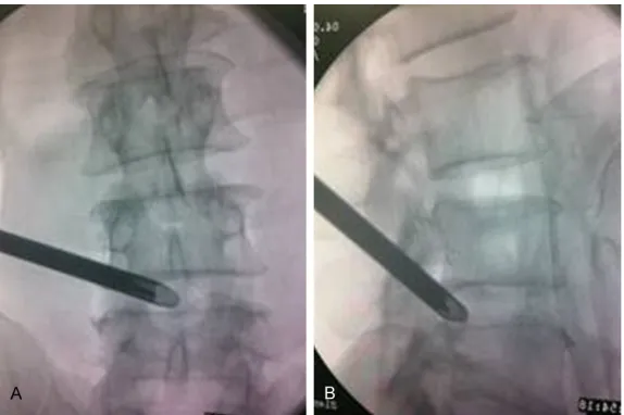
Figure 4. The traditional approach for Yeung’s technique. (A) Intra-operative posteroanterior and (B) lateral fluoroscopic imaging showing the Yeung’s cannula insertion technique.
[image:4.612.90.377.321.512.2]incidence of the nerve irritation in Group I was the same as the data previous reported. That may be related with the feature of the Yeung’s insertion technique. Yeung’s technique is an ‘inside-out’ [4, 16, 19] technique and the work-ing cannula is firstly placed into the disc through the ‘safe triangle’. The ‘safe-triangle’ was also called ‘Kambin triangle’ [16, 20] which is bounded medially by the dural sac, inferiorly by the superior endplate of the caudal vertebra, and its hypotenuse is formed by the exiting nerve root [23, 24]. The exiting dorsal root gan-glion located just cranial to the ‘Kambin trian-gle’. So based on the anatomical character of the ‘safe-triangle’ we hypothesized that the fol-lowing three aspects of the traditional YESS technique may cause nerve irritated symptom: First, the location of cannula tip in the disc near differences (P=0.046) (Table 3). Post-operative
numbness of the lower extremities occurred in seven patients in group I and one patient in group II. There was significant inter-group dif-ference (P=0.027). None dura matter leakage infection and hematoma occurred both in Group I and in Group II. Post-operative fat lique-faction occurred in one patient in Group I and no patient in Group II (P=1.000). The patient with fat liquefaction was treated by physical therapy and was recovered 2 weeks after. Post-operative muscle strength decline in quadri-ceps and hallux extensor muscles occurred in five and three patients respectively in group I but none in group II. There were no significant inter-group differences (P=0.056 P=0.242 respectively). All the patients were recovered three months after the operation (Table 3).
Discussion
In this study, we compared clini-cal outcomes of traditional and modified SED techniques for lum-bar disc herniation. Favorable outcomes could be found in both of the two groups by comparing the pre and post-operative VAS and ODI scores. However, when comparing the complications of the SED techniques, the inci-dence of radiating pain, numb-ness of lower extremity were obviously lower in Group II than those in Group I. The X-ray expo-sure time in Group II was also apparently lower than that in Group I.
[image:5.612.90.364.85.153.2]In SED procedure, approach re- lated irritation of existing nerve root, dorsal ganglia or possibly the furcal nerve in the foraminal area was a thorny issue. Even if the traversing root has been successfully decompressed [1]. Previous studies have reported that the incidence of hyperalge-sia and hypesthehyperalge-sia after the YESS technique was as high as 5%-15% [1, 17]. Ahn reported that the complication of post-operative dysesthesia (POD) was as high as 4.7% [22]. In this study, Table 1. Demographic characteristics of the patients
Gender
Age (year) symptom (month)Duration of Follow-up (month) Male Female
Group I 29 21 44.17±7.67 9.15±3.02 15.39±1.98
Group II 24 26 41.37±7.06 8.51±2.60 15.14±2.06
[image:5.612.92.361.185.319.2]P 0.316 0.066 0.269 0.551
Table 2. Comparison of surgical outcomes between two groups
Group I Group II P
Operating time (min) 63.52±4.26 60.92±5.37 0.011
X-ray exposure (min) 2.59±0.30 2.44±0.28 0.014
Blood loss (ml) 21.39±6.26 22.94 ±6.77 0.251
Pre-opera 7.75± 0.70 7.43± 0.87 0.055
VAS scores
One year post-opera 0.72±0.71 0.59±0.65 0.368
Pre-opera 58.76±5.95 57.02±7.28 0.207
ODI scores
One year post-opera 13.98±4.38 12.78±3.50 0.141
Table 3. Comparison of complication between two groups
Group I Group II P
Radiating pain of the lower extremities 8 2 0.046
Dura matter leakage 0 0 1.000
Numbness of the lower extremities 7 1 0.027
Decline in quadriceps myodynamia 5 0 0.056
Decline in hallux extensor myodynamia 3 0 0.242
Infection 0 0 1.000
Fat liquefaction 1 0 1.000
[image:5.612.92.362.356.473.2]the exiting nerve. In traditional Yeung’s tech-nique, the tip of the cannula was at the point near to the exiting nerve. One study had also showed that the distance between landing point and nerve root was just 3.5±1.4 mm [25]. Another cadaveric study showed that the exit-ing nerve was found to be 2-3 mm (mean: 2.3 mm) from tube with diameter of 2.7 mm. Given a 7 mm cannula is used at the same center, the exiting nerve is 0.15 mm compressed or 0.75 mm away from the cannula. If any tissue con-nected to the exiting nerve is pulled into can-nula, traction injury to the exiting nerve would occur [5]. So it may safer if the tip move toward the median line. In the modified SED technique, we shifted the entry point cephalically. With changed entry point more it is easier for shift-ing the tip of cannula to median. So the change of the entry point may be helpful to reduce the risk of nerve injury.
Second, the obliquity between the cannula and the exiting nerve root (that is parallel to the ver-tebral endplate in traditional insertion tech-nique). Anatomic character of Kambin triangle show that the exiting nerve forms the lateral edge. During the insertion procedure the direc-tion of needle or working cannula is vertical with the exiting nerve in traditional Yeung’s technique. If the superior articular process is hyperplasia the cannula may be pushed later-ally during the insertion procedure and that may raise the risk of nerve injury. So the ideal direction of the cannula may be parallel with the exiting nerve. The angle of the cannula in the coronal plane was changed from 0° to about 20° in the cephalic direction in modified technique. After this adjustment, the angle between the needle and nerve root was signifi-cantly less oblique, thus theoretically reducing the risk of nerve root injury (Figure 1). This is consistent with the theory proposed by Ebraheim that the needle should be as close as possible to the center of the disc to avoid the exiting nerve root [24]. Accordingly this study provided strong evidence for the hypothesis. In the modified insertion group (Group II), the inci-dence of the radiating pain due to the irritation of exiting nerve root during the insertion proce-dure decreased obviously. At mean while the incidence of the numbness of the lower extrem-ity was also decreased. So using this modified insertion technique can effectually reduce the complication of nerve injury.
For the surgeon, X-Ray exposure was another important aspect to be considered [26-28]. In this study we found that X-Ray exposure time could be reduced obviously by using the modi-fied insertion technique. That may because insertion procedure may be repeated if the nerve root irritation occurred. And that may extend operation time and also increase the X-Ray exposure. So the successful insertion can avoid excessive X-Ray exposure. It may be helpful for surgeons especially for newcomers. There were also previous experiments in modi-fying approach for preventing complications. In 2011, Cho, J. Y. reported one technique named by floating retraction technique by which can reduce the incidence of approach related exist-ing dorsal root ganglion injury. The cannula was inserted towards the middle-upper border of the lower pedicle and can be placed by gentle retraction of the root. In this report, 154 patients underwent SED and no patient suf-fered from post-operative dysesthesia [10]. Ahn, Y showed his technical guidelines to pre-vent complications by which changing the initial landing as close as the target as possible. But there were no statistical data for supporting his view [12]. In 2012, Wang reported 50 patients with lumbar disc herniation underwent discec-tomy using unilateral portal full endoscopic inter-laminar approach and 5 patients failed to finish this procedure and converted to open surgery [29, 30]. But this kind of approach was not transforaminal approach.
There were some limitations to our study. Only the segment L4/5 was involved in this study. So further study is needed to identify whether the modified technique is suitable for other seg-ments. Our sample size was limited and the follow-up period was short. Further multi-cen-ter, long term follow up and large sample size study needs to be done to further confirm the efficacy and safety of this modified surgical method.
Disclosure of conflict of interest
None.
zhuang 050051, China. Tel: +86031188602317; E-mail: [email protected]
References
[1] Tzaan WC. Transforaminal percutaneous endo-scopic lumbar discectomy. Chang Gung Med J 2007; 30: 226-234.
[2] Ahn SS, Kim SH, Kim DW, Lee BH. Comparison of outcomes of percutaneous endoscopic lum-bar discectomy and open lumlum-bar microdiscec-tomy for young adults: a retrospective matched cohort study. World Neurosurg 2016; 86: 250-8.
[3] Mayer HM, Brock M. Percutaneous endoscopic discectomy: surgical technique and prelimi-nary results compared to microsurgical discec-tomy. J Neurosurg 1993; 78: 216-225. [4] Yeung AT, Yeung CA. Advances in endoscopic
disc and spine surgery: foraminal approach. Surg Technol Int 2003; 11: 255-263.
[5] Osman SG, Marsolais EB. Posterolateral ar-throscopic discectomies of the thoracic and lumbar spine. Clin Orthop Relat Res 1994; 122-129.
[6] Kambin P. Arthroscopic microdiscectomy. Arthroscopy 1992; 8: 287-295.
[7] Lee DY, Lee SH. Learning curve for percutane-ous endoscopic lumbar discectomy. Neurol Med Chir 2008; 48: 383-389.
[8] Jhala A, Mistry M. Endoscopic lumbar discec-tomy: Experience of first 100 cases. Indian J Orthop 2010; 44: 184-190.
[9] Ahn Y, Lee HY, Lee SH, Lee JH. Dural tears in percutaneous endoscopic lumbar discectomy. Eur Spine J 2011; 20: 58-64.
[10] Cho JY, Lee SH, Lee HY. Prevention of develop-ment of postoperative dysesthesia in transfo-raminal percutaneous endoscopic lumbar dis-cectomy for intracanalicular lumbar disc her-niation: floating retraction technique. Minim Invasive Neurosurg 2011; 54: 214-218. [11] Tenenbaum S, Arzi H, Herman A, Friedlander A,
Levinkopf M, Arnold PM, Caspi I. Percutaneous posterolateral transforaminal endoscopic dis-cectomy: clinical outcome, complications, and learning curve evaluation. Surg Technol Int 2011; 21: 278-283.
[12] Ahn Y. Transforaminal percutaneous endo-scopic lumbar discectomy: technical tips to prevent complications. Expert Rev Med Devices 2012; 9: 361-366.
[13] Ahn Y, Kim JU, Lee BH, Lee SH, Park JD, Hong DH, Lee JH. Postoperative retroperitoneal hematoma following transforaminal percuta-neous endoscopic lumbar discectomy. J Neurosurg Spine 2009; 10: 595-602.
[14] Ahn Y, Lee SH. Postoperative spondylodiscitis following transforaminal percutaneous endo-scopic lumbar discectomy: clinical characteris-tics and preventive strategies. Br J Neurosurg 2012; 26: 482-486.
[15] Wen BT, Zhang XF, Wang Y, Xiao SH, Liu ZS, Liu BW, Zhang YG, Song J, Zhong YX, Sun JH. Complication and treatment of the lumbar in-tervertebral disc herniation using percutane-ous endoscopic lumbar discectomy. Zhonghua Wai Ke Za Zhi 2011; 49: 1091-1095.
[16] Yeung AT, Yeung CA. Minimally invasive tech-niques for the management of lumbar disc herniation. Orthop Clin North Am 2007; 38: 363-372.
[17] Yeung AT, Tsou PM. Posterolateral endoscopic excision for lumbar disc herniation: Surgical technique, outcome, and complications in 307 consecutive cases. Spine 2002; 27: 722-731. [18] Gibson JN, Cowie JG, Iprenburg M. Trans-
foraminal endoscopic spinal surgery: the fu-ture ‘gold standard’ for discectomy? -A review. Surgeon 2012; 10: 290-296.
[19] Yeung AT. Minimally Invasive Disc Surgery with the Yeung Endoscopic Spine System (YESS). Surg Technol Int 1999; 8: 267-277.
[20] Civelek E, Solmaz I, Cansever T, Onal B, Kabatas S, Bolukbasi N, Sirin S, Kahraman S. Radiological analysis of the triangular working zone during transforaminal endoscopic lumbar discectomy. Asian Spine J 2012; 6: 98-104. [21] Djurasovic M, Glassman SD, Dimar JR,
Crawford CH, Bratcher KR, Carreon LY. Changes in the oswestry disability index that predict im-provement after lumbar fusion. J Neurosurg Spine 2012; 17: 486-490.
[22] Ahn Y, Lee SH, Park WM, Lee HY, Shin SW, Kang HY. Percutaneous endoscopic lumbar discectomy for recurrent disc herniation: surgi-cal technique, outcome, and prognostic fac-tors of 43 consecutive cases. Spine 2004; 29: E326-332.
[23] Kambin P, Zhou L. Arthroscopic discectomy of the lumbar spine. Clin Orthop Relat Res 1997; 49-57.
[24] Ebraheim NA, Xu R, Huntoon M, Yeasting RA. Location of the extraforaminal lumbar nerve roots. An anatomic study. Clin Orthop Relat Res 1997; 230-235.
[25] Xin G, Shi-Sheng H, Hai-Long Z. Morphometric analysis of the YESS and TESSYS techniques of percutaneous transforaminal endoscopic lumbar discectomy. Clin Anat 2013; 26: 728-734.
instrumentation. J Neurosurg Spine 2005; 3: 98-105.
[27] Huppertz A, Radmer S, Asbach P, Juran R, Schwenke C, Diederichs G, Hamm B, Sparmann M. Computed tomography for preoperative planning in minimal-invasive total hip arthro-plasty: radiation exposure and cost analysis. Eur J Radiol 2011; 78: 406-413.
[28] Schils F. O-arm guided balloon kyphoplasty: preliminary experience of 16 consecutive pa-tients. Acta Neurochir Suppl 2011; 109: 175-178.
[29] Wang B, Lu G, Liu W, Cheng I, Patel AA. Full-endoscopic interlaminar approach for the sur-gical treatment of lumbar disc herniation: the causes and prophylaxis of conversion to open. Arch Orthop Trauma Surg 2012; 132: 1531-1538.