Original Article
The expression of imprinted genes IGF2 and PHLDA2
in mid-pregnancy have predictive values for the
development of pre-eclampsia
Xueqing Guo1,2, Mei Zhong1, Yiping Luo2, Li Xiao2, Xin Chen1
1Department of Obstetrics and Gynecology, Nanfang Hospital, Southern Medical University, Guangzhou 510515, Guangdong, PR China; 2Department of Gynecology, the First People’s Hospital of Shunde District, Foshan 528300, Guangdong, PR China
Received January 24, 2017; Accepted April 25, 2017; Epub June 15, 2017; Published June 30, 2017
Abstract: Objective: Insulin-like growth factor-2 (IGF-2) and pleckstrin homology-like domain, family A, member 2 (PHLDA2) appear to play an important role in paracrine interactions at the maternal-fetal interface in human preg-nancy. But the potential predictive value of serum IGF-2 and PHLDA2 concentrations in pre-eclampsia remains to be established. The goal of the present study was to explore the expression of IGF2 and PHLDA2 in mid-pregnancy with pre-eclampsia (PE). Methods: Samples of serum were collected from women with normal mid-pregnancy women (control group, n=21), mild pre-eclampsia (M-PE group, n=19) and severe pre-eclampsia women (S-PE, n=19). The expressions of IGF2 and PHLDA2 in the serum were determined by ELISA and western blot. After delivery, the fresh placental tissues were collected. The expression levels of IGF2 and PHLDA2 in placenta were detected by real-time PCR, western blot and immunohistochemistry. Results: It was found that the concentrations of IGF2 and PHLDA2 were remarkably dropped in comparison with control group. And there was significant difference in PHLDA2 expres -sion between mild pre-eclampsia and severe pre-eclampsia in serum. After delivery, the differences in imprinted genes IGF2 and PHLDA2 were found in placental tissues between PE and normal pregnancy. mRNA and protein expression levels were all obviously decreased in PE groups. Conclusion: These data are the first to demonstrate imprinted genes IGF2 and PHLDA2 associated with PE in mid-pregnancy. It illustrates that IGF2 and PHLDA2 may participate in PE pathogenesis.
Keywords: Pre-eclampsia, imprinted genes, IGF2, PHLDA2
Introduction
Preeclampsia (PE), identified by the presence
of hypertension and proteinuria after 20 weeks of pregnancy [1], is a systemic disease that involved many organs such as the brain, eyes, liver and kidneys [2]. It’s one of the most com-mon hypertensive disorders and is a leading cause of morbidity and mortality for pregnant women and perinatal babies [3, 4]. Furthermore, PE is strongly associated with intrauterine growth restriction, iatrogenic prematurity, pla-cental abruption, and stillbirth of the child [5-7]. Despite great efforts, the exact pathophysiolo-gy of PE remains unknown. Because of the seri-ous health consequences of PE, risk
assess-ment and identification of women at risk early
in the pregnancy remain a major challenge in prenatal care.
some 7 in mice and chromosome band 11p15.5 in humans, is controlled by the Kvdmr1 DNA element [13, 14]. Imprinted gene PHLDA2 is upregulated in placentas from intrauterine growth restricted human pregnancies. Elevated expression of the maternally expressed imprint-ed gene PHLDA2, has been reportimprint-ed in the human placenta of growth restricted pregnan-cies. However, the expression of IFG2 and PHLDA2 in mid-pregnancy or pre-eclampsia has not yet been elucidated.
To address this lack of information, this study examined expression of the maternally ex- pressed imprinted genes PHLDA2 and IFG2 in mid-pregnancy or pre-eclampsia.
Materials and methods
Study participants
The study was approved by Nanfang Hospital, Southern Medical University of China Ethics Committee and informed consent was obtained from patients and control women. 19 patients with mild pre-eclampsia (M-PE), 19 patients with severe pre-eclampsia (S-PE) and 21 nor-mal control pregnant women were recruited from Southern Medical University of China between October 2015 and September 2016. Exclusion criteria were cardiac and multi-foetus pregnancies, systemic lupus erythematosus, renal disease and structural or chromosomal anomalies. None of the PE patients and healthy controls had history of smoking or hyperten-sion. The diagnose of PE was based on
modi-fied American Congress of Obstetricians and
Gynaecologists criteria.
Sample collection
Placentas were excised immediately after ery (elective caesarean section or vaginal deliv-ery). Small pieces of placenta tissues (1*1*1 cm3) in the central region avoiding calcification
and infarct areas were obtained. Tissues were immediately frozen and stored at -80°C.
Quantitative RT-PCR was performed using Chromo Four Colour Real Time Detector (MJ Research) in a 20 ml reaction containing 5 ml of cDNA (diluted 1 in 50), 1X Buffer 2 mM MgCl2, 2 mM dNTPs, 0.65 Units Taq (Fermentas (Thermo), Loughborough, UK), 1 mM of each primer (SigmaeAldrich, Dorset, UK) and 0.12X Sybr Green (Invitrogen, Paisley, UK). Conditions
for amplification were 15 min at 94°C, and then
subjected to 30 thermal cycles at 94°C (30 sec), 65°C (1 min) and 72°C (1 min), followed
by a final extension at 72°C (30 sec) using
a thermal sequencer (Zymoreactor, Atto, Tokyo, Japan). The internal control was b-actin. Primer sequences were as follows: IGF2 for-ward: 5’-GGACTTGAGTCCCTGAACCA-3’, reve- rse: 5’-TGAAAATTCCCGTGGAAGG-3’. PHLDA2 forward: 5’-CCATCCTCAAGGTGGACTGC-3’, reve- rse: 5’-TTCCTGGCGGCTGCGAAAGT-3’. GAPDH forward: 5’-CCATCGTCCACCGCAAA T-3’, and reverse: 5’-GCTGTCACCTTCACCGTTC-3’.
Western blotting analysis
NP-40 lysis buffer (Beyotime Institute of Biotechnology, Haimen, China) containing the protease inhibitor (PMSF) was used and total proteins were isolated. Western blots were per-formed as previously described [15]. Samples
containing 60 μg protein were electrophoresed
on 12% (w/v) SDS polyacrylamide gel and tr-
ansferred onto polyvinylidene difluoride (PVDF)
membranes. Then the PVDF membranes were blocked with 5% nonfat milk for 1 h followed by incubation with the following primary antibod-ies: PHLDA2 antibody and IGF2 antibody (1:400 dilution, BIOSS, Beijing, China) at 4°C over-night. After washing with Tween-20 in Tris-buffered saline (TTBS), the membrane was incubated with horseradish peroxidase (HRP)-conjugated goat anti-rabbit secondary antibody (1:5000 dilution, Beyotime Institute of Biote- chnology) at 37°C for 1 hr. The optical density of each protein was normalized to the
corre-sponding β-actin signal using ImageJ software
Immunohistochemistry analysis
Staining of the paraffin-embedded sections
was performed with the CSA II kit (Dako,
Hamburg, Germany). Briefly, antigen retrieval
was performed by microwaving in citrate buffer, pH 6.0, for 15 min. The tissues were then blocked for endogenous peroxidase, incubated with IGF2 or PHLDA2 antibody (1:3000 dilution, BIOSS, Beijing, China), washed in PBS (0.1% Tween 20), and followed by incubation with anti-rabbit HRP polymer (1:200, Envision, Dako) for 30 minutes. Normal healthy pregnant women were used as positive controls in all experi- ments.
Enzyme-linked immunosorbent assay (ELISA)
Concentrations of IGF2 and PHLDA2 in serum were measured using ELISA kits (Boster, Wuhan, China) according to the manufacturer’s instructions.
Statistical analysis
Data with normal distribution were also evalu-ated by one-way ANOVA with the Bonferroni’s post hoc test or repeated measures ANOVA. All results were presented as mean ± SD from a minimum of three replicates. P < 0.05 was
con-sidered statistically significant.
Results
the mRNA level of IGF2 and PHLDA2 in pla-centa
[image:3.612.96.520.73.207.2]The IGF2 and PHLDA2 expression levels in the placenta tissues of the control women and PE patients were analyzed by real-time polymerase chain reaction (PCR). We found that the mRNA expression of IGF2 and PHLDA2 was notably reduced in mild pre-eclampsia (M-PE group), compared with conreol group (Figure 1, P < 0.05). In Severe preeclampsia (S-PE group),
Figure 1. mRNA levels of IGF2 and PHLDA2 in placenta. RT-PCR analysis was used to determine the relative mRNA expression levels of IGF2 and PHLDA2 in placenta. Expression levels were normalized against GAPDH expression. Data values and error bars show the means and standard devations. Each experiment was repeated at least three times and typical results are shown. *P < 0.05 and **P < 0.01 verusus control group, #P < 0.05 versus M-PE group.
[image:3.612.91.521.283.387.2]imprinted genes IGF2 and PHLDA2 were re- duced greatly in comparison with control group or M-PE group (Figure 1, P < 0.05 or P < 0.01).
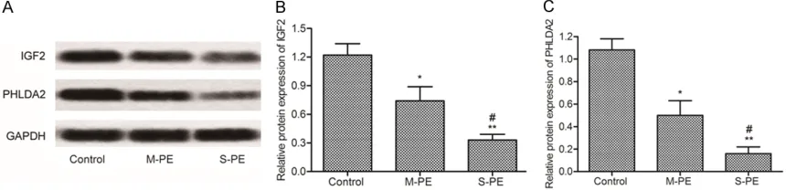
Protein expression level of IGF2 and PHLDA2 in placenta
Using western blotting, we further compared the protein expression of IGF2 and PHLDA2 in placenta. The results showed that the PHLDA2 protein (Figure 2B, P < 0.05) and the IGF2 pro-tein (Figure 2C, P < 0.05) levels in PE group
were significant lower than those in the control
group. As compared with the M-PE group, the expression of two imprinted genes was less in
S-PE group. The results showed that the expres-sion of IGF2 and PHLDA2 was inversely propor-tional to the severity of the PE.
[image:4.612.90.529.73.286.2]In order to further explore, we detected IGF2 and PHLDA2 expression by immunohistochem-istry. Immunohistochemistry was performed on histologic sections. As shown in Figure 3, we could obviously see that those two imprinted genes were all evidently depressed decreased in PE groups. There was almost no protein expression of IGF2 and PHLDA2 in S-PE group, compared with the control/M-PE group. The results were similar to western blotting analysis.
Figure 3. The level of IGF2 and PHLDA2 in placenta was measured by immunohistochemistry analysis. Immuno-histochemical detection shows IGF2 (A) and PHLDA2 (B) protein level in placenta. The staining is both nuclear and cytoplasmic.
[image:4.612.97.516.347.478.2]Level of IGF2 and PHLDA2 in serum
To investigate the expression level of IGF2 and PHLDA2 in serum, ELISA was performed to assess the concentration of those in serum. The M-PE and S-PE patients showed a decrease of concentration levels of IGF2 and PHLDA2 (P
< 0.05), as shown in Figure 4.
the mRNA level of IGF2 and PHLDA2 in serum
The PHLDA2 expression levels in the serum of the control women and PE patients were ana-lyzed by RT-PCR. Clearly, the level of IGF2 and PHLDA2 were obviously down-regulated in PE patients, compared with that in the control group (Figure 5, P < 0.05). Furthermore, the two imprinted genes, IGF2 and PHLDA2, had the least mRNA expression in S-PE group (Figure 5, P < 0.01).
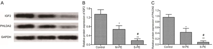
Protein expression level of IGF2 and PHLDA2 in serum
At the same time, protein was extracted from the serum. Similar results were obtained for
protein expression. These results were
con-firmed using western analysis (Figure 6). It showed that proteins of IGF2 and PHLDA2 were
significantly reduced in M-PE and S-PE groups,
compared with the control group (P < 0.05). We found that when compared to M-PE group, pro-tein expression of IGF2 and PHLDA was much fewer in S-PE. Those results were similar with RT-PCR analysis in Figure 5.
Discussion
[image:5.612.93.522.72.179.2]PE is the most common gestational complica-tion and the patients show several typical clini-cal symptoms. It affects approximately 2% of pregnant women worldwide and responsible for more than 50,000 maternal deaths annually [16, 17]. PE is the leading cause of maternal and perinatal morbidity and mortality, particu-larly when it occurs before the 34th week of gestation [18, 19]. In addition, it expressly initi-ates intrauterine placental abruption, iatrogen-ic prematurity, growth restriatrogen-iction, and stillbirth of the child. Because we unable to predict PE by previous obstetric history and risk factors,
Figure 5. mRNA levels of IGF2 and PHLDA2 in serum. A: mRNA Expression of IGF2 and PHLDA2 was analyzed by RT-PCR. B: Graphs showing the mRNA level of IGF2. C: Graphs showing the mRNA level of PHLDA2. Each experiment was repeated at least three times and typical results are shown. *P < 0.05 and **P < 0.01 verusus control group, #P
< 0.05 versus M-PE group.
[image:5.612.91.521.251.351.2]dictive test would allow specific therapeutic
interventions [22].
This study was set up to identify novel potential PE markers, to subsequently be tested in bio-marker discovery approaches. Many research-ers have found some candidate biomarkresearch-ers
[23, 24]. The finding that these markers are also identified by their approach demonstrates
that the approach has the potential to identify other promising biomarkers as well.
Imprinted genes have been demonstrated to regulate the endocrine lineage of the mouse placenta, in particular expression of placental lactogens [25-27]. IGF2, an imprinted gene and peptide hormones, mediates a variety of meta-bolic and mitogenic effects on the surface of target tissues and cells. Several studies have shown that IGF2 is expressed in many tissues and regulates the proliferation and survival of a variety of tissues [28]. In addition, IGF2 stimu-lates various processes that are involved in the
proliferation and apoptosis of first trimester tro -phoblasts. IGF2 appears to play an important role in EVCT proliferation, and facilitates the nutrient and oxygen supply through placental exchange. Compelling evidence in recent years has also suggested that IGF2 contributes to the regulation of placentation and prenatal and postnatal growth. In our experiments, we focus on the detection of IGF2 mRNA and protein expression in serum and placenta, which were collected from PE patients and healthy preg-nant women. As described in the results sec-tion, placental IGF2 expression level was remarkably fell in PE patients, at RNA level and protein level. We subsequently analysed im- printed gene IGF2 in serum. It is obvious that IGF2 from PE women were also inhibited, com-pared with control group. The IGF2 expression was inversely proportional to the severity of PE. PHLDA2 is another maternally expressed imprinted gene that participates in the early growth and development of the placenta [14].
expression of PHLDA2 from PE women also were inhibited, compared with healthy preg-nant women. The imprinted gene PHLDA2 expression was inversely proportional to the severity of PE. A number of studies have dem-onstrated abnormally changed placental PHLDA2 in pregnancies. As reported that it’s upregulated expression inhibits placental growth and increases the risk of low birth
weights in infants [14, 29]. There was a signifi -cant change of PHLDA2 expression and mater-nal serum PHLDA2 level in our study. PHLDA2
expression was significantly associated with
placental weight. This is of clinical interest as RFM is thought to represent a fetal adaptation
to prolonged placental insufficiency which, if
undetected, may result in still birth [30]. So
detecting PHLDA2 expression is significative
for pregnant women.
To summarize, we have used integrative data to identify two changed imprinted genes in serum from PE. It’s just a preliminary investigation for IGF2 and PHLDA2 in serum of PE. These data
are the first to demonstrate imprinted genes
IGF2 and PHLDA2 associated with PE in mid-pregnancy. It illustrates that IGF2 and PHLDA2 may participate in PE pathogenesis and indi-cate its potential application in the early diag-nosis of PE. The mechanism of these two imprinted genes and whether they can be as potential markers on PE need further study in the future. These are necessary to further determine how these markers interrelate and
whether sufficiently reliable prediction accura -cy can be obtained before a large-scale PE screening program can be introduced.
Disclosure of conflict of interest
None.
Guangdong, PR China. Tel: 020-62787291; E-mail: [email protected]
References
[1] Redman CW and Sargent IL. Latest advances in understanding preeclampsia. Science 2005; 308: 1592-1594.
[2] Williamson RD, O’Keeffe GW and Kenny LC. Ac-tivin signalling and pre-eclampsia: from genet-ic risk to pre-symptomatgenet-ic biomarker. Cytokine 2015; 71: 360-365.
[3] Mary S, Kulkarni MJ, Malakar D, Joshi SR, Me-hendale SS and Giri AP. Placental proteomics provides insights into pathophysiology of pre-eclampsia and predicts possible markers in plasma. J Proteome Res 2017; 16: 1050-1060.
[4] Herzog EM, Eggink AJ, Reijnierse A, Kerkhof MA, de Krijger RR, Roks AJ, Reiss IK, Nigg AL, Eilers PH, Steegers EA and Steegers-Theunis-sen RP. Impact of early- and late-onset pre-eclampsia on features of placental and new-born vascular health. Placenta 2017; 49: 72-79.
[5] Kwiatkowski S, Dolegowska B, Kwiatkowska E, Rzepka R, Marczuk N, Loj B and Torbe A. Ma-ternal endothelial damage as a disorder shared by early preeclampsia, late preeclamp-sia and intrauterine growth restriction. J Peri-nat Med 2016; [Epub ahead of print].
[6] Tallarek AC and Stepan H. [Preeclampsia and HELLP syndrome as an obstetric emergency]. Med Klin Intensivmed Notfmed 2012; 107: 96-100.
[7] Silasi M, Cohen B, Karumanchi SA and Rana S. Abnormal placentation, angiogenic factors, and the pathogenesis of preeclampsia. Obstet Gynecol Clin North Am 2010; 37: 239-253. [8] Moore GE, Ishida M, Demetriou C, Al-Olabi L,
Leon LJ, Thomas AC, Abu-Amero S, Frost JM, Stafford JL, Chaoqun Y, Duncan AJ, Baigel R, Brimioulle M, Iglesias-Platas I, Apostolidou S, Aggarwal R, Whittaker JC, Syngelaki A, Nico-laides KH, Regan L, Monk D, Stanier P. The role and interaction of imprinted genes in human fetal growth. Philos Trans R Soc Lond B Biol Sci 2015; 370: 20140074.
[9] Kusinski L C CWN, Sandovici I, Constância M. Contribution of placental genomic imprinting and identification of imprinted genes. The Guide to Investigation of Mouse Pregnancy 2013; 275.
[10] Weksberg R, Smith AC, Squire J and Sadowski P. Beckwith-Wiedemann syndrome demon-strates a role for epigenetic control of normal development. Hum Mol Genet 2003; 12: R61-68.
[11] Angiolini E, Fowden A, Coan P, Sandovici I, Smith P, Dean W, Burton G, Tycko B, Reik W,
Sibley C and Constancia M. Regulation of pla-cental efficiency for nutrient transport by im -printed genes. Placenta 2006; 27 Suppl A: S98-102.
[12] McMinn J, Wei M, Schupf N, Cusmai J, Johnson EB, Smith AC, Weksberg R, Thaker HM and Tycko B. Unbalanced placental expression of imprinted genes in human intrauterine growth restriction. Placenta 2006; 27: 540-549. [13] Salas M, John R, Saxena A, Barton S, Frank D,
Fitzpatrick G, Higgins MJ and Tycko B. Placen-tal growth retardation due to loss of imprinting of Phlda2. Mech Dev 2004; 121: 1199-1210. [14] Tunster SJ, Creeth HD and John RM. The
im-printed Phlda2 gene modulates a major endo-crine compartment of the placenta to regulate placental demands for maternal resources. Dev Biol 2016; 409: 251-260.
[15] Tybl E, Shi FD, Kessler SM, Tierling S, Walter J, Bohle RM, Wieland S, Zhang J, Tan EM and Kiemer AK. Overexpression of the IGF2-mRNA binding protein p62 in transgenic mice induc-es a steatotic phenotype. J Hepatol 2011; 54: 994-1001.
[16] Abalos E, Cuesta C, Grosso AL, Chou D and Say L. Global and regional estimates of preeclamp-sia and eclamppreeclamp-sia: a systematic review. Eur J Obstet Gynecol Reprod Biol 2013; 170: 1-7. [17] Bhorat I, Naidoo DP and Moodley J. Maternal
cardiac haemodynamics in severe pre-eclamp-sia complicated by acute pulmonary oedema: a review. J Matern Fetal Neonatal Med 2016; 1-9. [Epub ahead of print].
[18] Hogberg U. The World Health Report 2005: “make every mother and child count”-includ-ing Africans. Scand J Public Health 2005; 33: 409-411.
[19] Eastwood KA, Patterson C, Hunter AJ, Mc-Cance DR, Young IS and Holmes VA. Evaluation of the predictive value of placental vasculari-sation indices derived from 3-Dimensional power Doppler whole placental volume scan-ning for prediction of pre-eclampsia: a system-atic review and meta-analysis. Placenta 2017; 51: 89-97.
[20] Kell DB and Kenny LC. A dormant microbial component in the development of preeclamp-sia. Front Med (Lausanne) 2016; 3: 60. [21] Norwitz ER BMA, Saade GR, Miller H. Obstetric
clinical algorithms: management and evi-dence. John Wiley & Sons; 2011.
[22] Phipps E, Prasanna D, Brima W and Jim B. Pre-eclampsia: updates in pathogenesis, defini -tions, and guidelines. Clin J Am Soc Nephrol 2016; 11: 1102-1113.
methylation in the human placenta and fetal growth (review). Mol Med Rep 2012; 5: 883-889.
[26] Tunster S, Jensen AB, John RM. Imprinted genes in mouse placental development and the regulation of fetal energy stores. Reproduc-tion 2013; 145: R117-R137.
[27] Patten MM, Cowley M, Oakey RJ, Feil R. Regu-latory links between imprinted genes: evolu-tionary predictions and consequences. Proc Biol Sci 2016; 283.