Original Article
Prognostic value of PD-L1 for invasive breast cancer and
its miR-34a-related mechanism of regulation
Qiuyang Zhao1*, Yawen Guo2*, Chunping Liu1, Tao Huang1
1Department of Breast and Thyroid Surgery, Union Hospital, Tongji Medical College, Huazhong University of Sci-ence and Technology, Wuhan 430022, China; 2Department of Thyroid Surgery, The Affiliated Yantai Yuhuangding Hospital of Qingdao University, Yantai, Shandong Province, China. *Equal contributors.
Received March 4, 2019; Accepted June 10, 2019; Epub August 15, 2019; Published August 30, 2019
Abstract: The goal of this study was to explore the relationship between expression of programmed death ligand-1 (PD-L1) protein and commonly assessed clinicopathological features as well as its prognostic value for invasive breast cancer patients. The effect of miR-34a on PD-L1 both in vitro and in vivo was also investigated. Among the
287 patients included in this study, 165 showed PD-L1 overexpression. PD-L1 was significantly correlated with ER, PR, Her-2, Ki-67, and molecular subtypes. ER+, PR+, Her-2-, Ki67>14% and Ki67≤14% patients with PD-L1 over -expression had a poorer prognosis. PD-L1 -expression decreased along with the addition of a miR-34a mimic but increased along with the addition of a miR-34a inhibitor both at mRNA and protein level. miR-34a mimics can down-regulate the luciferase activity of 231 cells transfected with wild-type PD-L1 plasmids. CCK-8 and transwell tests show that cell proliferation and invasion ability decreased after transfection with a miR-34a mimic. Xenografts with
intra-tumoral injection of miR-34a agomir had a significantly lower growth rate. miR-34a expression when treated with miR-34a agomir was significantly higher and PD-L1 protein expression when treated with miR-34a agomir was significantly lower. Patients with overexpressed PD-L1 have a relatively poorer prognosis. miR-34a can negatively
regulate PD-L1. miR-34a may be a promising therapy target for triple-negative breast cancer patients with PD-L1 overexpression.
Keywords: PD-L1, invasive breast cancer, prognosis, miR-34a
Introduction
As the most common malignant tumor and the second leading cause of cancer-related death in women, breast cancer draws a lot attention worldwide [1]. In China, breast cancer has been the most malignant type of tumor among women in terms of both incidence and mortali-ty during recent years [2]. The prognosis of breast cancer is mainly evaluated based on histological grade, lymph node staging, patho-logical tumor staging (TNM) and four major pro-tein molecular biomarkers: estrogen receptor (ER), progesterone receptor (PR), Ki67 and human epidermal growth factor receptor 2 (Her-2) [3, 4]. Some researchers have begun to study immunotherapy strategies and drugs that are applicable for breast cancer as they can be used on immune pathways to escape the an- ti-tumor immune response, in order to maintain tumor cell proliferation and metastasis [5]. How-
ever, more effective prognosis biomarkers and individualized treatment strategies for invasive
breast cancer remain to be identified.
As a member of the B7 family, programmed cell death 1 (PD-1) is a immunoregulatory cell sur-face proteins that has two cognate ligands, PD-L1 and PD-L2. Expression of PD-1 and PD-L1 in the tumor microenvironment plays a major role in tumor immune evasion [6]. PD-L1
is found to be expressed not only in tumor infil -trating lymphocytes, but also in some tumor cells, including that of breast, prostate, lung and gastrointestinal cancers and malignant melanoma [7-11]. Additionally, high expression of PD-L1 in breast, non-small cell lung, pancre-atic, and renal cell carcinoma has been found to be associated with a poor prognosis [12-14].
been found [15]. In simple creatures such as C. elegans, miR-34 has only one transcript; while normal human tissue contains 34a, miR-34b, and miR-34c [16]. Recent studies have reported that miR-34a can affect cancer cell proliferation [17] and promote tumor cell apop-tosis [18] in pancreatic cancer, ovarian cancer and retinoblastoma [19-22].
In this study, the value of PD-L1 was evaluated in order to predict the prognosis of invasive breast cancer patients through the analysis of tissue microarrays. Then, the regulatory rela-tionship between PD-L1 and miR-34a, as well as their effect on the proliferation, migration and invasion abilities of triple-negative breast carcinoma cell line MDA-MB-231 was investi- gated.
Material and methods
Samples from breast cancer patients and im-munostaining of tissue microarrays
The tissue arrays purchased from the Shanghai Outdo Biotech co, Ltd contained 300 samples from invasive breast carcinoma patients. Ex- pression of PD-L1, ER, PR, Ki-67, Her2, CK5/6, P53, and epidermal growth factor receptor (EGFR) in the samples were detected through immunohistochemistry (IHC).
Scoring, evaluation and statistical analysis
IHC staining was evaluated by two experienced pathologists, who were blinded from the clinical information. PD-L1 staining intensity in the cy- toplasm of tumor cells was scored from 0-3 and the percentage of PD-L1-positive cells was scored 0-4 (0-5, 6-25, 26-50, 51-75 and 76-100%, respectively). The sum of staining intensity and positive cell percentage scores
was taken as the final PD-L1 expression score,
which when ranged between 0 and 3 was de-
fined as negative, and when between 4 and 7
as positive. This expression criterion that ta- kes into account staining intensity and staining percentage was selected by referring to other similar research studies [23]. ER and PR expr- ession was mainly found in the nucleus, with their expression criteria compared with that of international standards [24], for which a posi-tive staining ratio of >1% was posiposi-tive, while a ratio of <1% is negative. Positive Ki-67 expres-sion was >14%. For the Her-2
immunohisto-chemical results, 0 and + were defined as a negative result and +++ was defined as a
po-sitive result, while the PathVysion HER-2 DNA Probe kit (Abbott Pharmaceutical Co., Ltd., Lake Bluff, IL, USA) was used to determine the expression of ++ samples. Her2 expression was designated as either weak (IHC grade 0-1+ or FISH-) or strong (IHC grade 3+ or FISH+). Cell culture and transfection
The human breast cancer cell line MDA-MB-231 was grown in Leibovitz’s L-15 medium (Gibco, USA) with 10% fetal bovine serum (FBS) (Gibco, USA) and 1% penicillin-streptomycin solution at 37°C without extra CO2. Well cultured cells we- re seeded at a density of 1 × 10⁵ cells per well
into six-well culture plates and cultured in Opti-MEM (Gibco, USA), overnight. Transfection with the miR-34a mimic, miR-34a inhibitor, miR-34a mimic NC (negative control) and miR-34a inhi- bitor NC (GenePharma, Shanghai, China) were conducted using the Lipofectamine 2000 tr- ansfection reagent (Invitrogen, Carlsbad, CA, USA) and continued for about 6 hours. Cells were harvested at 48 or 72 hours after trans-fection, depending on the condition of cell proliferation.
Western blotting
Extracted protein was electrophoresed on a 10% SDS-polyacrylamide gel and transferred to a PVDF membrane. The membrane was blocked with laboratory skim milk and then incubated at 4°C overnight with primary anti-bodies. Primary antibodies against PD-L1 (Cell Signaling Technology, Boston, USA) and GAP- DH (Cell Signaling Technology, Boston, USA) were used at 1:1000 and 1:2000 dilution, re- spectively. Then, secondary antibodies conju-gated with horseradish peroxidase (Cell Signa- ling Technology, Boston, USA) were incubated with the samples for 2 hours at room tempera-ture. The target protein bands were visualized using a chemiluminescence enhanced chemi- luminescence kit (Guge Biological Technology, Wuhan, China).
Real-time PCR
TGGCACATCCTC-3’ and reverse 5’-GTATCACT- TTGCTTCTTTGAGTTTGT-3’; miR-34a forward 5’- TGGCAGTGTCTTAGCTGGTTGT-3’; GAPDH forw- ard 5’-TGTTGCCATCAATGACCCCTT-3’ and re- verse 5’-CTCCACGACGTACTCAGCG-3’; U6 for-ward 5’-CTCGCTTCGGCAGCACA-3’ and reverse 5’-AACGCTTCACGAATTTGCGT-3’. PD-L1 expres-sion was normalized with GAPDH and miR-34a expression with U6, and the different expres-sion levels were evaluated relative to that of the control group. For each sample,
experi-ments were performed in triplicate to confirm
the results.
Luciferase reporter assay
The luciferase reporter plasmid contains 3’UTR of PD-L1 WT and PD-L1 MU oligonucleotides that correspond to the miR-34a binding site that was made by Ribobio. MDA-MB-231 cells were seeded into 12-well plates that were tr- ansfected with 50 ng of the pMIR-REPORTTM or
each reporter construct, 1 ng of the Renilla luciferase reporter (pRL-CMV vector, Ribobio), and 100 nmol/l of the miR-34a mimic, miR-34a mimic negative control, miR-34a inhibitor and miR-34a inhibitor negative control, respective-ly, using LipofectamineTM 2000. Firefly and
Re-nilla luciferase activities were measured se- quentially using dual-luciferase assays (Pro- mega) 24 hours post transfection. The experi-ments were performed in triplicate for each transient transfection assay.
Cell proliferation and invasion assays
Proliferation of the MDA-MB-231 cells was ele-vated using a CCK8 cell proliferation assay. The MDA-MB-231 cells treated with the miR-34a mimic and miR-34a mimic negative control were seeded into a 96-well plate at a density of about 4 × 10³ cells/well and cultured for 24
hours. Then a CCK8 reagent (Dojindo, Japan) was added to the cells 24, 36, 48, 60 and 72 hours after incubation. The absorbance at 450 nm represented cell proliferation. The experi-ments were performed in triplicate. MDA-MB- 231 cells with a miR-34a mimic and its nega-tive control were cultivated without fetal bovine serum overnight, then put on the upper cham-ber of the 24-well pates and covered with BD Matrigel. L-15 medium with 20% fetal bovine serum was added to the lower chamber. After 24-36 hours of incubation, cells in the lower
chamber were fixed in 4% paraformaldehyde.
The number of cells in the lower chamber was calculated using microscopy and represents invasive ability. Additionally, transwell cham-bers without Matrigel were used to evaluate migration ability. All experiments were per-formed in triplicate.
Xenograft mouse model
All animal experiments were carried out in ac- cordance with the National Institutes of Health guidelines for the care and use of Laboratory animals. Thirty female nude mice were divided equally into three groups and 5 × 10⁵
MDA-MB-231 cells were subcutaneously injected into the breast fat pad of the right or left chest of each nude mouse. Tumor volumes were measured every 3 days (volume = 0.5 × length × width2). MiR-34a agomir, miR-34a agomir
negative and a mock were injected when tumor volume reached 25 mm3. Injection was
per-formed every 3 days and each mouse was injected with 1 nmol of the reagent at 3 or mo-
re sites each time. The mice were sacrificed
after 54 days and tumor tissues were collected for RNA and protein extraction, as well as immu- nostaining.
Statistical analysis
The experimental data were analyzed using SPSS 22.0 software (Chicago, IL, USA). Diffe- rences between groups were compared using a t-test. Student’s t test or ANOVA statistical anal-ysis was applied to the mean value and to dif-ferences between groups for continuous vari-ables. Correlation between variables was ana-lyzed using the Pearson X2 test. Patient lifeti-
me analysis and log-rank test were performed using Kaplan-Meier survival analysis. Univariate and multivariate regression analyses were per-formed using Cox proportional hazards regres-sion analyses. All p values are two-tailed, with a p value of <0.05 indicating a statistical diff- erence.
Results
PD-L1 expression is associated with breast cancer clinicopathological variables
samples or because the stripping or immuno-histochemistry results did not meet the inclu-sion criteria. All patients included in this study had received standardized surgery, chemother-apy, radiotherchemother-apy, endocrine therchemother-apy, and tar-geted therapy according to NCCN guidelines. As shown in Figure 1, 165 of the 286 (57.69%) patients had high expression of PD-L1. Table 1
shows that there was significant correlation
between PD-L1 and ER (p=0.002, X2=10.507),
PR (p=0.006, X2=7.784), Her-2 (p=0.003, X2=
9.167), Ki-67 (p=0.005, X2=8.404), and
molec-ular classification (p<0.001 X2=21.322). Am-
ong all subtypes, 42.4% (53/125) of Luminal A
patients, 69.01% (49/71) of Luminal B pati- ents, 68.75% (22/32) of Her-2 overexpression patients and 70.69% (41/58) of triple-nega- tive patients had relatively high levels of PD- L1 expression. However, PD-L1 expression did
not have a significant relationship with age (p=
0.108, X2=2.667), tumor site (p=0.185, X2=
2.016), histological grade (p=0.413, X2=1.768),
tumor diameter (p=0.326, X2=2.243), lymph
node metastasis (p=0.902, X2=0.574), tumor
clinical stage (p=0.897, X2=0.217) CK5/6
level (p=0.183, X2=2.154), P53 level (p=
0.092, X2=3.083) or EGFR level (p=0.058, X2=
[image:4.612.89.521.72.458.2]3.726).
High expression of PD-L1is associated with a worse prognosis
Among these 286 invasive breast cancer pa- tients, results of the Kaplan-Meier survival an- alysis showed that the expression of PD-L1 is
significantly associated with OS (p=0.001). Hi-gh PD-L1 expression patients had significantly
shorter OS (Figure 1). Univariate COX regres-sion analysis showed that T stage (p=0.003, HR=1.746, 95% CI: 1.210-2.520), N stage (p<
0.001, HR=1.493, 95% CI: 1.208-1.845), clini-cal tumor stage (p<0.001, HR=1.976, 95% CI: 1.379-2.793), molecular subtype (p=0.002, HR=1.319, 95% CI: 1.107-1.572), ER (p=0.027, HR=0.611, 95% CI: (0.395-0.945), PR (p= 0.002, HR=0.491, 95% CI: 0.314-0.767), PD-L1 (p=0.001, HR=2.317, 95% CI: 1.421-3.776) are the clinicopathological variants that had a
[image:5.612.91.518.82.571.2]sig-nificant HR value (Table 2). In the multivariate COX regressions, only PR (p=0.029, HR=0.518, 95% CI: 0.287-0.936) and PD-L1 (p=0.001, Table 1. Association between PD-L1 expression and clinical pathological factors of breast cancer
PD-L1 expression
X2 p
Negative (121) Positive (165)
Age <50 51 54 2.667 0.108
≥50 70 111
Tumor site Right 74 87 2.016 0.185
Left 47 78
Histological grade I 24 23 1.768 0.413
II 88 129
III 9 13
Tumor diameter (cm) ≤2 28 40 2.243 0.326
>2, ≤5 76 111
>5 17 14
Lymph node metastasis number 0 57 73 0.574 0.902
1-3 33 46
4-9 24 33
≥10 7 13
Clinical stage 0/1 14 21 0.217 0.897
2 69 96
3 38 48
ER Negative 29 70 10.507 0.002
Positive 92 95
PR Negative 48 93 7.784 0.006
Positive 73 72
Her-2 Negative 103 115 9.167 0.003
Positive 18 50
Ki-67 (%) <14 93 100 8.404 0.005
≥14 28 65
CK5/6 Negative 101 126 2.154 0.183
Positive 20 39
p53 Negative 44 44 3.081 0.092
Positive 77 121
EGFR Negative 88 102 3.726 0.058
Positive 33 63
Molecular classification Luminal A 72 53 21.322 <0.001
Luminal B 22 49
Her2 overexpression 10 22
sion of miR-34a was significantly reduced and PD-L1 mRNA expression was signifi -cantly increased in the miR-34a inhibitor group. All these results were of a
statisti-cally significant p value of <0.05. Additio- nally, miR-34a and PD-L1 expression sh- owed no change in miR-34a mimic NC or miR-34a inhibitor NC group, as shown in Figure 2. The Western blotting results show that PD-L1 protein expression has a
reduced statistical significance (p<0.05)
in the miR-34a mimic group and increas-
ed statistical significance (p<0.05) in the
miR-34a inhibitor group, whereas there
was no significant change in PD-L1 pro -tein expression in the miR-34a mimic NC group or the miR-34a inhibitor NC group, which is also shown in Figure 2. Thus, miR-34a expression was found to have a negative association with PD-L1 gene expression and protein level.
miR-34a directly targets PD-L1 3’UTR
The 3’UTR sequence of PD-L1 wild type (WT) and mutant (MU) mRNA was cloned into plasmid psiCHECK-2 (dual luciferase reporter vector). Double luciferase assay results, as shown in Figure 2, confirmed
that compared with the control level, the miR-34a mimic can downregulate the luciferase activity of MDA-MB-231 cells transfected with the PD-L1 WT plasmid HR=2.299, 95% CI: 1.389-3.803) were
statisti-cally significant (Table 3).
Kaplan-Meier survival analyses also showed that high PD-L1 expression is related with a worse prognosis in ER+, PR+, Ki67>14%, Ki67< 14% and Her-2+ patients. As shown in Figure 1, ER+ patients with high expression of PD-L1 had a shorter survival time and higher mortality rate than ER+ patients with low expression of PD-L1. A similar trend in results (high PD-L1 expres-sion related with worse prognosis) was ob- served in the other four subgroups.
miR-34a influences the expression of PD-L1 both at RNA and protein level in MDA-MB-231 cells
RT-PCR results showed that miR-34a
expres-sion was significantly increased and PD-L1 mRNA expression was significantly decreased
in the miR-34a mimic group, while the
expres-(p<0.05), but has no effect on PD-L1 MU infect-ed cells. Additionally, the luciferase activity of cells with PD-L1 WT and PD-L1 MU plasmids were not affected by the miR-34a inhibitor. These results indicate that miR-34a could directly mediate transcriptional gene silencing by targeting the PD-L1 3’UTR complementary site.
High expression of miR-34a inhibits the prolif-eration, migration and invasive ability of MDA-MB-231 cells
[image:6.612.91.327.107.280.2]CCK-8 assay was used to detect proliferation changes in MDA-MB-231 cells transfected with either miR-34a mimic or miR-34a inhibitor. As shown in Figure 2, cell proliferation ability de- creased after transfection with the miR-34a mimic and was enhanced after transfection with a miR-34a inhibitor. That is, high expres-sion of miR-34a had a negative effect on the proliferation ability of MDA-MB-231 cells. Table 2. Univariate COX regression analysis of clinical
pathological factors and prognosis of patients with invasive breast cancer
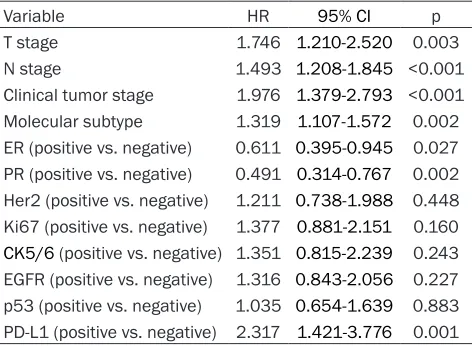
Variable HR 95% CI p
T stage 1.746 1.210-2.520 0.003
N stage 1.493 1.208-1.845 <0.001 Clinical tumor stage 1.976 1.379-2.793 <0.001 Molecular subtype 1.319 1.107-1.572 0.002 ER (positive vs. negative) 0.611 0.395-0.945 0.027 PR (positive vs. negative) 0.491 0.314-0.767 0.002 Her2 (positive vs. negative) 1.211 0.738-1.988 0.448 Ki67 (positive vs. negative) 1.377 0.881-2.151 0.160
[image:6.612.91.327.335.445.2]CK5/6 (positive vs. negative) 1.351 0.815-2.239 0.243 EGFR (positive vs. negative) 1.316 0.843-2.056 0.227 p53 (positive vs. negative) 1.035 0.654-1.639 0.883 PD-L1 (positive vs. negative) 2.317 1.421-3.776 0.001
Table 3. Multivariate COX regression analysis of clinical pathological factors and prognosis of patients with invasive breast cancer
Variable HR 95% CI p
T stage 1.438 0.878-2.356 0.149
N stage 1.160 0.769-1.750 0.480
Clinical tumor stage 1.598 0.727-3.514 0.244
Molecular subtype 1.415 0.970-2.065 0.071
ER (positive vs. negative) 2.559 0.976-6.708 0.056
PR (positive vs. negative) 0.518 0.287-0.936 0.029
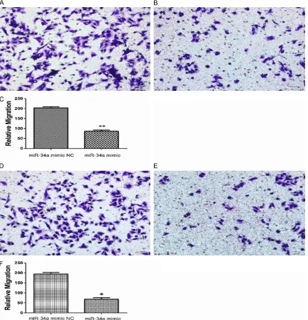
Transwell insert experiments were used to de- tect the effect of miR-34a overexpression on migration and invasion of MDA-MB-231 cells. The results that are illustrated in Figure 3 show that MDA-MB-231 cells transfected with a
miR-34a mimic have a significantly weakened
mi-gration ability compared with those of the miR-34a mimic NC group (p<0.05). A similar result was observed in the invasive ability experiment, where the number of cells transfected with a
miR-34a mimic was significantly less than that
of the NC group (p<0.05). Therefore, high ex- pression of miR-34a was able to weaken the migration and invasive ability of MDA-MB-231 cells.
In vivo experiment
After tumor challenge, drug injection, and ob- servation, the volume and growth rate of the nude mice injected with the miR-34a agomir
were analyzed and found to be significantly
lower than those injected with the NC and the
mock. RT-PCR results confirmed that miR-34a expression is significantly higher in tumors of
the miR-34a agomir group, than that of the NC group and Mock group. As for the protein, Western blots shows that the expression level of PD-L1 in the miR-34a agomir group was
sig-nificantly lower than that of the other two gr-oups and are statistically significant (p<0.05),
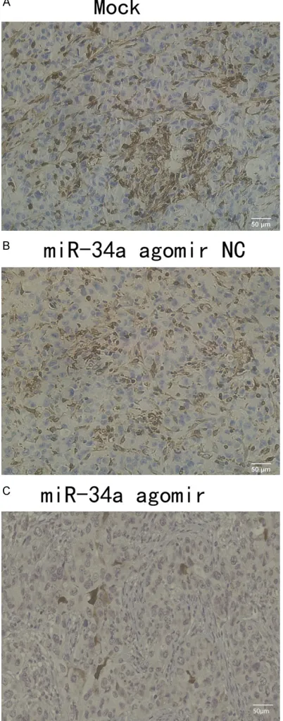
as shown in Figure 4. Additionally, expression of PD-L1 in the miR-34a agomir group was
sig-nificantly lower than that of the NC and the
Mock groups, while the IHC results of the peeled tumors in three groups are shown in Figure 5. In brief, negative regulation of miR-34 and PD-L1 expression were found using a xeno-graft mouse model.
Discussion
PD-L1 mRNA has been found in a variety of nor-mal tissues in the body, including thymus, bone
marrow, lymph nodes, heart, lungs, kidneys, li- ver, muscles, and nerve tissues. However, prev-alence of its mRNA expression differs from th- at of its protein expression [25]. The PD-L1 pro-tein is mainly expressed in antigen presenting cells (APCs), T cells, B cells, epithelial cells, myocytes, endothelial cells, as well as a variety
of tumor cells and tumor infiltrating lympho -cytes, and may participate in multiple tumor cell-associated immune responses [26]. Re- cent studies have shown that overexpression of PD-L1 may lead to immune escape in some tumors [27]. The binding of PD-L1 on tumor cells or APCs to the PD-1 receptor on T cells induces apoptosis, inhibits proliferation, and
suppresses T cell IFN-γ, IL-4, and IL-2 cytokine
release, impairing the function of T cells, while promoting the differentiation of CD4+/CD25-/
Foxp3- T cells into Foxp3+ regulatory T cells
(Treg) and inducing apoptosis of tumor-specific
T cells. Regulatory T cells have the ability to limit the growth of tumor cells, whereas the PD-1/PD-L1 signaling pathway in tumor cells can produce an immune escape by inhibiting
regulatory T cells [28]. The expression profile of
PD-L1 is slightly broader than that of PD-1 and is more involved in various links mediated by tumor cells. Therefore, immunohistochemistry was used to detect the expression level of PD-L1 protein in tissue microarrays and com-bine that with survival data of patients, in order to analyze the effect of PD-L1 on prognosis. Numerous studies indicate that miRNAs are closely related with the occurrence and pro-gression of various diseases in humans [29]. miRNAs can suppress cancer by downregulat-ing oncogene mRNA levels, as well as promote carcinogenesis through downregulation of anti-oncogene mRNA levels [30]. This phenomenon suggests that miRNAs may be deeply involved in the process of tumor development. Therefore,
it is of great significance to explore the biologi -cal functions of miRNAs in various tumors. The Figure 2. A. miR-34a expression was significantly increased in miR-34a mimic group and significantly reduced increased in miR-34a inhibitor group, p<0.05. B. PD-L1 mRNA expression was significantly decreased in miR-34a mimic group and significantly increased in miR-34a inhibitor group, p<0.05. C. PD-L1 protein expression was re
-duced with statistical significance (p<0.05) in the cells transfected with miR-34a mimic. D. PD-L1 protein expression was increased significantly (p<0.05) in the miR-34a inhibitor group. E. Sequence of 3’UTR of PD-L1 wild type (WT)
miR-34 family contains three members, namely miR-34a, miR-34b and miR-34c. miR-34a is lo- cated on chromosome 1p36.23, while miR-34b and miR-34c are located on chromosome 11- q23. Both gene loci are susceptible to change and are associated with fragile sites of the tumor genome [31]. In normal tissues, miR-34a is widely found in most tissues and organs, while miR-34b and miR-34c are found only in
[image:9.612.92.526.72.523.2]the lung, trachea, ovary and prostate. miR-34a may be involved in targeting a variety of onco-genic genes that are associated with prolifera-tion, apoptosis, and invasion [19, 32]. Nume- rous studies have demonstrated that miR-34a plays an important role in chemotherapy and chemotherapeutic resistance of tumors, as we- ll as tumor immunotherapy and immune resis-tance [33, 34].
Figure 3. A. The example of cells with miR-34a mimic NC migrated to the lower chamber. B. The example of cells with miR-34a mimic migrated to the lower chamber. C. The number of cells with miR-34a mimic migrated to the
lower chamber was significantly lower than the number of NC group, the p value <0.05. D. The example of cells with miR-34a mimic NC invaded to the lower chamber. E. The example of cells with miR-34a mimic invaded to the lower
chamber. F. The number of cells with miR-34a mimic invaded to the lower chamber was significantly lower than the
As for the regulatory association between miR-34a and PD-L1, there are studies conducted on acute myeloid leukemia that demonstrating that miR-34a can target PD-L1 mRNA to regu-late PD-L1 expression [35]. Interaction between PD-1 and its ligands, PD-L1 and PD-L2, can control the magnitude and duration of T cell re- sponses, inhibit T cell proliferation, activation, and cytokine secretion. Therefore, it can medi-ate tumor immune escape [36]. However, the
role and regulatory mechanism of miR34a and PD-L1 in invasive breast cancer are still unclear. In this study, high expression of PD-L1 was ob- served in 57.69% of invasive breast carcinoma
patients. There was significant correlation
be-tween PD-L1 expression and ER, PR, Her-2,
Ki-67 and molecular classification, but no sig
-nificant relation between age at diagnosis,
[image:10.612.91.518.70.489.2]tu-mor location, tutu-mor stage, histological grade, Figure 4. Tumor growth was monitored every 3 days after breast cancer cells subcutaneously injection and mice
were sacrificed after 54 days, those had not formed tumor were excluded. A. The final mice of each group. B. Peeled
off tumors of three groups. C. Growth curve of mean tumor volume of each group. D. RT-PCR result of miR-34a expression level in tumors from miR-34a agomir, miR-34a agomir NC and Mock group. E. RT-PCR result of PD-L1 expression level in tumors from miR-34a agomir, miR-34a agomir NC and Mock group. F. Western blot result of PD-L1 protein expression in the tumor samples of miR-34a agomir, miR-34a agomir NC and mock group. G. Expression
tumor size, lymph node metastasis, CK5/6, p53, EGFR or other indicators. Subsequent Ka- plan-Meier survival analysis demonstrated th- at high PD-L1 expression patients had a lower overall survival rate, compared with low sion level patients. Thus, high PD-L1
expres-sion was found to be significantly associated
with a poor prognosis of invasive breast carci-noma patients. Additionally, PD-L1 expression was found to be statistically associated with patient prognosis both in the single-factor COX regression and multi-factor COX regression analysis, which included T stage, N stage, tu-
mor clinical stage, molecular classification, ER,
PR, PD-L1 and 7 other indicators. Therefore,
high PD-L1 expression may be a specific pre -dictor of poor prognosis for patients with inva-sive breast cancer. To further analyze the rela-tionship between PD-L1 and other common clinicopathological features, a subgroup
analy-sis was performed to explore the influence of
PD-L1 expression on prognosis under different
conditions. A significant association between
high PD-L1 expression and a poor survival rate in groups of patients with ER+, PR+, Ki67>14%,
Ki67≤14%, and Her-2+ was found. Therefore,
high PD-L1 expression in hormone receptor-positive and Her-2 receptor-positive patients may imply poor prognosis, which means that PD-L1 tar-geted therapy may be a new treatment choice for patients with high PD-L1 expression in order to overcome endocrine treatment resistance and inability of performing Herceptin therapy. The human breast cancer cell line MDA-MB-231 was chosen for basic experimental research because of its relatively high expression of PD-L1. After transfection of miR-34a mimic, 34a mimic NC, 34a inhibitor and miR-34a inhibitor NC to MDA-MB-231 cells, PD-L1 mRNA was found to be downregulated when miR-34a is upregulated, while when PD-L1 pro-tein was found to be downregulated when miR-34a is upregulated, and vice versa. Detection of mRNA and protein changes implies that th- ere is an inverse relationship between them. To further demonstrate whether miR-34a directly regulates PD-L1, the 3’UTR ends of mutant and wild type PD-L1 were cloned into psiCHECK-2 and simultaneously transfected MDA-MB-231 cells with miR-34a mimics. Luciferase activity of cells transfected with PD-L1 WT plasmids
decreased significantly, which indicates that
[image:11.612.89.290.74.585.2]PD-L1 is a direct target gene of miR-34a. Ad- ditionally, transfection of a miR-34a mimic into MDA-MB-231 cells attenuated the prolifera-tion, migration and invasive abilities of the cell, while transfection of a miR-34a inhibitor en- hanced the same. The regulatory relationship between miR-34a and PD-L1 is in accord with Figure 5. Immunohistochemistry graph presenting
our hypothesis that mRNA-34a can target PD- L1 and that these changes can affect impor-tant biological processes of the breast carci-noma cell line, which may be an important mechanism behind breast tumor cell immune evasion.
Based on the above findings using a tumor bur -den model of nude mice there can be several conclusions. First, the tumor volume and gr- owth rate of the nude mice injected with a
miR-34a agomir were significantly lower than that of
those injected with a miR-34a agomir NC or
Mock. It was confirmed that miR-34a expres
-sion in the agomir group was found to be signifi -cantly higher than that of the agomir NC and Mock groups, while the PD-L1 mRNA
expres-sion level was found to be significantly lower
than that of the miR-34a agomir NC group and Mock group. Second, the agomir group showed lower expression of PD-L1 protein compared with that of the agomir NC and Mock groups. This indicates that miR-34a can downregulate mRNA and protein expression of PD-L1 in a xenograft model, as well as inhibit the growth of tumors in vivo.
Although the analysis of our samples found that high expression of PD-L1 is associated with a poor prognosis for invasive breast carci-noma, there have been several references in recent literature that claim that high expressi- on of PD-L1 is associated with a good prog- nosis for breast cancer patients [23, 37-39]. However, several other reports have indicated that high PD-L1 expression is associated with a poor prognosis for breast cancer, since it is also
combined with the downregulation of infiltra -tion of lymphocytes [40] and upregula-tion of Foxp3+ regulatory T cells [41]. The difference
between conclusions may be related to a vari-ety of factors, including tumor heterogeneity and patient inclusion criteria. This study includ-ed patients with various molecular types, while most of these other studies only included either triple-negative breast cancer patients or Her2 overexpression patients. The choice of experi-mental subjects and reagents may lead to dif-ferent results. For example, the choice of anti-bodies from different companies and the crite-ria of the PD-L1 high expression may lead to different conclusions [42]. Race and ethnic dif-ferences can also bring about difdif-ferences in results. More large-scale, multi-center research
and clinical trials related to the treatment of PD-L1 inhibitors are needed to determine not only gold standard antibodies and the most appropriate cut-off value, but also to further clarify the relationship between PD-L1 and the prognosis of breast cancer. On the other hand, the regulatory relationship between miR-34a and PD-L1 has now been demonstrated, while
the specific mechanisms of downstream genes
of PD-L1 and signaling pathways still need to be explored. Furthermore, the precise mecha-nism of miR-34a in inhibiting breast tumor cell proliferation, migration and invasion has not been studied in detail. Last but not the least, the in vivo experiment was only conducted on the upregulated expression of miR-34a, and
the specificity of downregulation of miR-34a
expression was not investigated. Therefore, in subsequent research plans, future studies will address these inadequacies and deepen com-prehension of the regulatory mechanism of miR-34a on PD-L1, so as to further explore how to apply tumor immunotherapy targeting of PD-L1 and miR-34a for the treatment of inva-sive breast cancer patients.
In summary, this study found that PD-L1 over-expression is associated with a poor prognosis for invasive breast cancer patients and can be used as an independent hazard predictor of prognosis. It shows that there is a reverse cor-relation between the expression of PD-L1 and miR-34a, while upregulation of miR-34a can inhibit the proliferation and invasive ability of the MDA-MB-231 cell line. Considering these results, miR-34a may serve as a promising treatment target for invasive triple-negative bre- ast cancer patients with overexpressed PD- L1, but a large number of experiments are still
required still require to verify its specific mech
-anism of action and efficacy.
Disclosure of conflict of interest
None.
References
[1] Torre LA, Bray F, Siegel RL, Ferlay J, Lortet-Tieulent J and Jemal A. Global cancer statis-tics, 2012. CA Cancer J Clin 2015; 65: 87-108. [2] Chen WQ, Zheng RS, Baade PD, Zhang SW,
Zeng HM, Bray F, Jemal A, Yu XQ and He J. Cancer statistics in China, 2015. CA Cancer J Clin 2016; 66: 115-132.
[3] Goldhirsch A, Winer EP, Coates AS, Gelber RD, Piccart-Gebhart M, Thürlimann B, Senn HJ; Panel members. Personalizing the treatment of women with early breast cancer: highlights of the St gallen international expert consensus on the primary therapy of early breast cancer 2013. Ann Oncol 2013; 24: 2206-2223. [4] Coates AS, Winer EP, Goldhirsch A, Gelber RD,
Gnant M, Piccart-Gebhart M, Thurlimann B and Senn HJ. Tailoring therapies-improving the management of early breast cancer: St gallen international expert consensus on the primary therapy of early breast cancer 2015. Ann Oncol 2015; 26: 1533-1546.
[5] Mohammed ZMA, Going JJ, Edwards J, Els- berger B, Doughty JC and McMillan DC. The re-lationship between components of tumour
in-flammatory cell infiltrate and clinicopathologi -cal factors and survival in patients with prima-ry operable invasive ductal breast cancer. Br J Cancer 2012; 107: 864-873.
[6] Schalper KA. PD-L1 expression and tumor-infil -trating lymphocytes: Revisiting the antitumor immune response potential in breast cancer. Oncoimmunology 2014; 3: e29288.
[7] Mittendorf EA, Philips AV, Mericbernstam F, Na Q, Yun W, Harrington S, Su X, Ying W, Gon- zalezangulo AM and Akcakanat A. PD-L1 ex-pression in triple negative breast cancer. Cancer Immunol Res 2014; 2: 361-370. [8] Morgensztern D, Campo MJ, Dahlberg SE,
Doebele RC, Garon E, Gerber DE, Goldberg SB, Hammerman PS, Heist RS, Hensing T, Horn L, Ramalingam SS, Rudin CM, Salgia R, Sequist LV, Shaw AT, Simon GR, Somaiah N, Spigel DR, Wrangle J, Johnson D, Herbst RS, Bunn P, Govindan R. Molecularly targeted therapies in non-small-cell lung cancer annual update 2014. J Thorac Oncol 2015; 10: 1-63.
[9] Shiao SL, Chu CY and Chung LW. Regulation of prostate cancer progression by the tumor mi-croenvironment. Cancer Lett 2016; 380: 340-348.
[10] Tarhini AA, Zahoor H, Yearley JH, Gibson C, Rahman Z, Dubner R, Rao UN, Sander C, Kirkwood JM. Tumor associated PD-L1 expres-sion pattern in microscopically tumor positive sentinel lymph nodes in patients with melano-ma. J Transl Med 2015; 13: 1-7.
[11] Bertucci F, Finetti P, Mamessier E, Pantaleo
MA, Astolfi A, Ostrowski J and Birnbaum D.
PDL1 expression is an independent prognos- tic factor in localized GIST. Oncoimmunology 2015; 4: e1002729.
[12] Bigelow E, Bever KM, Xu H, Yager A, Wu A, Taube J, Chen L, Jaffee EM, Anders RA and Zheng L. Immunohistochemical staining of
B7-H1 (PD-L1) on paraffin-embedded slides of
pancreatic adenocarcinoma tissue. J Vis Exp 2013; e4059.
[13] Muenst S, Schaerli AR, Gao F, Däster S, Trella E, Droeser RA, Muraro MG, Zajac P, Zanetti R and Gillanders WE. Expression of programmed death ligand 1 (PD-L1) is associated with poor prognosis in human breast cancer. Breast Cancer Res Treat 2014; 146: 15-24.
[14] Xu F, Xu L, Wang Q, An G, Feng G and Liu F. Clinicopathological and prognostic value of programmed death ligand-1 (PD-L1) in renal cell carcinoma: a meta-analysis. Int J Clin Exp Med 2015; 8: 14595-14603.
[15] Lau NC, Lim LP, Weinstein EG and Bartel DP. An abundant class of tiny RNAs with probable regulatory roles in caenorhabditis elegans. Science 2001; 294: 858-862.
[16] Kato M and Paranjape TR. The mir-34 microR-NA is required for the DmicroR-NA damage response in vivo in C. elegans and in vitro in human breast cancer cells. Oncogene 2009; 28: 2419-2424. [17] Maegdefessel L, Azuma J, Toh R, Merk DR,
Deng A, Chin JT, Raaz U, Schoelmerich AM, Raiesdana A and Leeper NJ. Inhibition of mi-croRNA-29b reduces murine abdominal aortic aneurysm development. J Clin Invest 2012; 122: 497-506.
[18] Zhang Y, Schiff D, Park D and Abounader R. MicroRNA-608 and microRNA-34a regulate chordoma malignancy by targeting EGFR, Bcl-xL and MET. PLoS One 2014; 9: e91546. [19] Cole KA, Attiyeh EF, Mosse YP, Laquaglia MJ,
Diskin SJ, Brodeur GM and Maris JM. A
func-tional screen identifies miR-34a as a candi -date neuroblastoma tumor suppressor gene. Mol Cancer Res 2008; 6: 735.
[20] Tazawa H, Tsuchiya N, Izumiya M and Na- kagama H. Tumor-suppressive miR-34a induc-es seninduc-escence-like growth arrinduc-est through mod-ulation of the E2F pathway in human colon cancer cells. Proc Natl Acad Sci U S A 2007; 104: 15472-15477.
[21] Shi Y, Liu C, Liu X, Tang DG and Wang J. The microRNA miR-34a inhibits non-small cell lung cancer (NSCLC) growth and the CD44hi stem-like NSCLC cells. PLoS One 2014; 9: e90022. [22] Welch C, Chen Y and Stallings RL.
MicroRNA-34a functions as a potential tumor suppressor by inducing apoptosis in neuroblastoma cells. Oncogene 2007; 26: 5017-5022.
triple-negative breast cancer is associated
with tumour-infiltrating lymphocytes and im -proved outcome. Histopathology 2015; 69: 25-34.
[24] Hammond ME, Hayes DF, Dowsett M, Allred DC, Hagerty KL, Badve S, Fitzgibbons PL, Fr- ancis G, Goldstein NS, Hayes M, Hicks DG, Lester S, Love R, Mangu PB, McShane L, Miller K, Osborne CK, Paik S, Perlmutter J, Rhodes A, Sasano H, Schwartz JN, Sweep FC, Taube S, Torlakovic EE, Valenstein P, Viale G, Visscher D, Wheeler T, Williams RB, Wittliff JL and Wolff AC. American society of clinical oncology/col-lege of American pathologists guideline recom-mendations for immunohistochemical testing of estrogen and progesterone receptors in br- east cancer (unabridged version). Arch Pathol Lab Med 2010; 134: e48-72.
[25] Saresella M, Rainone V, Al-Daghri NM, Clerici M and Trabattoni D. The PD-1/PD-L1 pathway in human pathology. Curr Mol Med 2012; 12: 259-267.
[26] Haynes D. B7-H1, a third member of the B7 family, co-stimulates T-cell proliferation and in-terleukin-10 secretion. Nat Med 1999; 5: 1365-1369.
[27] Dong H, Strome SE, Salomao DR, Tamura H, Hirano F, Flies DB, Roche PC, Lu J, Zhu G and Tamada K. Tumor-associated B7-H1 promotes T-cell apoptosis: a potential mechanism of im-mune evasion. Nat Med 2002; 8: 793. [28] Merelli B, Massi D, Cattaneo L and Mandalà M.
Targeting the PD1/PD-L1 axis in melanoma: biological rationale, clinical challenges and op-portunities. Crit Rev Oncol Hematol 2014; 89: 140.
[29] Hartmann D, Fiedler J, Sonnenschein K, Just A, Pfanne A, Zimmer K, Remke J, Foinquinos A, Butzlaff M and Schimmel K. MicroRNA-based
therapy of gata2-deficient vascular disease.
Circulation 2016; 134: 1973.
[30] Fan D, Wang Y, Qi P, Chen Y, Xu P, Yang X, Jin X and Tian X. MicroRNA-183 functions as the tu-mor suppressor via inhibiting cellular invasion and metastasis by targeting MMP-9 in cervical cancer. Gynecol Oncol 2016; 141: 166-174. [31] Calin GA, Sevignani C, Dumitru CD, Hyslop T,
Noch E, Yendamuri S, Shimizu M, Rattan S, Bullrich F and Negrini M. Human microRNA genes are frequently located at fragile sites and genomic regions involved in cancers. Proc Natl Acad Sci U S A 2004; 101: 2999-3004. [32] Dapeng C, Ying L, Yan M, Wenjia G, Jurong Y,
Quan H, Zhe F, Guangyan C, Hanyu Z and Suozhu S. miR-34a regulates mesangial cell
proliferation via the PDGFR-β/Ras-MAPK sig -naling pathway. Cell Mol Life Sci 2014; 71: 4027-4042.
[33] Bader AG. miR-34 - a microRNA replacement therapy is headed to the clinic. Front Genet 2012; 3: 120.
[34] Nosirov B, Billaud J, Vandenbon A, Diez D, Wijaya E, Ishii KJ, Teraguchi S and Standley DM. Mapping circulating serum miRNAs to their immune-related target mRNAs. Adv Appl Bioinform Chem 2017; 10: 1.
[35] Wang X, Li J, Dong K, Lin F, Long M, Ouyang Y, Wei J, Chen X, Weng Y, He T, Zhang H. Tumor suppressor miR-34a targets PD-L1 and func-tions as a potential immunotherapeutic target in acute myeloid leukemia. Cell Signal 2015; 27: 443-452.
[36] Dunn GP, Bruce AT, Ikeda H, Old LJ and Sch- reiber RD. Cancer immunoediting: from immu-nosurveillance to tumor escape. Nat Immunol 2002; 3: 991-998.
[37] Botti G, Collina F, Scognamiglio G, Rao F, Peluso V. Programmed death ligand 1 (PD-L1) tumor expression is associated with a better prognosis and diabetic disease in triple nega-tive breast cancer patients. Int J Mol Sci 2017; 18: 459.
[38] Bae SB, Cho HD, Oh MH, Lee JH, Jang SH, Hong SA, Cho J, Kim SY, Han SW and Lee JE. Expression of programmed death receptor
li-gand 1 with high tumor-infiltrating lymphocytes
is associated with better prognosis in breast cancer. J Breast Cancer 2016; 19: 242. [39] Tsang JY, Au WL, Lo KY, Ni YB, Hlaing T, Hu J,
Chan SK, Chan KF, Cheung SY and Tse GM.
PD-L1 expression and tumor infiltrating PD-1+
lymphocytes associated with outcome in HE- R2+ breast cancer patients. Breast Cancer Res Treat 2017; 162: 19-30.
[40] Mori H, Kubo M, Yamaguchi R, Nishimura R, Osako T, Arima N, Okumura Y, Okido M, Ya- mada M and Kai M. The combination of PD-L1
expression and decreased tumor-infiltrating
lymphocytes is associated with a poor progno-sis in triple-negative breast cancer. Oncotarget 2017; 8: 15584.
[41] Li Z, Dong P, Ren M, Song Y, Qian X, Yang Y, Li S, Zhang X and Liu F. PD-L1 expression is as-sociated with tumor FOXP3+regulatory T-cell
infiltration of breast cancer and poor prognosis
of patient. J Cancer 2016; 7: 784-793. [42] Sun WY, Yu KL and Koo JS. Expression of