y. Exp. Biol. (1961), 38, I3S-IS2 I 3 5 ) With 9 text-figures
Printed in Great Britain
STUDIES ON IONIC REGULATION IN
CARCINUS MAENAS (L.)
I. SODIUM BALANCE
BY J. SHAW
Department of Zoology, University of Durham, King's College, Newcastle upon Tyne
{Received 18 July i960)
INTRODUCTION
The fact that the body surface of the common shore, crab, Carcinus maenas, is permeable to inorganic ions was established long ago. Bethe (1929, 1930) showed that changes in the external concentration of Ca, Mg and K were reflected in corre-sponding changes in the blood concentration of these ions. Following the demon-stration by Schlieper (1929) that the body weight of Carcinus did not change when the animals were transferred to dilute sea water, Margaria (1931) correctly deduced that the body surface was permeable to the major blood ions (Na and Cl) from the fact that the total blood concentration decreased in a logarithmic manner when the animals were placed in dilute sea water.
The classical studies of Duval (1925) first showed that Carcinus could maintain its blood concentration well above that of the dilute sea water in which it was living. This was confirmed by Schlieper (1929) and subsequently by many other workers. For a number of years various explanations were put forward in an attempt to reconcile this fact with the conception of a freely permeable body surface. The correct explanation was suggested by Nagel (1934), who demonstrated that if animals adapted to a dilute medium were transferred to a medium of high con-centration but still below that of the blood, the blood concon-centration was neverthe-less increased. He postulated the inward secretion of salts from the medium to the blood, probably by cells of the branchial epithelium. Although, as Krogh (1939) pointed out, these experiments were not in themselves completely conclusive they provided strong evidence in favour of an active mechanism, which could transfer salts against a concentration gradient, participating in the maintenance of salt balance in these animals. This concept was finally put on a firm footing by the conclusive demonstration by Krogh (1937a, b, 1938) of the importance of such mechanisms in maintaining the relatively high blood concentrations found in many freshwater animals.
Finally, from a study of the concentration of individual ions in the blood of
Carcinus, Webb (1940) showed that concentration differences were present even in
136 J. SHAW
(Na, Cl, K and Ca) were operating even in these animals, and that they formed part of the over-all mechanism of ionic regulation, a process which underlay that of osmotic regulation.
In the process of osmotic regulation the quantitatively most important mechanisms are those responsible for the movement of sodium and chloride ions since these account for the bulk of the osmotic pressure exerted by the blood. The present study is concerned with a quantitative evaluation of those factors responsible for the maintenance of the blood sodium concentration in Carcinus.
Sodium balance in an animal results from the equality between the rates of sodium uptake and loss, each of which comprises the sum of the rates of sodium movement through a number of different channels. Thus sodium uptake is shared between a passive component due to diffusion through the permeable body surface and an active component represented by the active transport system. Sodium loss is partitioned between passive diffusion, sodium loss through the excretory organ and possibly also the active secretion of sodium. The level at which the blood sodium is set will depend on the values of these components, any of which may be variable. The fact that animals adapted to dilute sea water surpass those from normal sea water in their ability to maintain their blood sodium concentration above that of the medium may be due to the former having (a) reduced their surface permeability to sodium, (b) reduced sodium loss through the excretory organ, or (c) increased the rate of active sodium uptake. The use of the radioactive tracer technique allows these various possibilities to be investigated.
MATERIAL AND METHODS
The animals, supplied by the Dove Marine Laboratory, were collected from the Northumberland coast. They were subsequently kept in a laboratory aquarium supplied with circulating sea water. Over-all rates of sodium uptake and loss were determined by a tracer method using the sodium isotopes, MNa and 22Na, and also by chemical measurements.
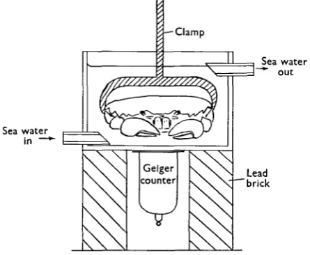
Sodium outflux was measured with ^Na. The animals were loaded with the isotope by keeping them in 11. of sea water containing approximately lOfic. ^NaCl for 1 day. The animals were then transferred to a Perspex bath through which non-radioactive sea water could flow freely and were held firmly in position by means of a screw clamp across the breadth of the carapace. The radioactivity of the whole animal was measured by means of a Geiger counter placed immediately beneath the bath and the decrease in radioactivity as the tracer was washed out was recorded by means of a recording ratemeter. The apparatus is shown in Fig. 1. During a period of washing the animal's radioactivity decreased exponentially according to the relation y = yoe~tlT, where y is the recorded activity at time t; y0 is the initial activity and T is the time constant. Hence the outflux, Moub, could
be calculated from the relation Mont = A/T, where A is the total freely exchangeable
internal sodium.
Studies on ionic regulation in Carcinus maenas (L.). / 137
previously for influx determinations on Astacus pallipes (Shaw, 1960 a). With this method it is essential that the internal sodium content should greatly exceed that in the external solution and therefore it could only be used for low external sodium concentrations. At higher concentrations the influx was calculated from chemical measurements of the net sodium loss rate using the relation: influx = outflux minus net sodium loss rate. The measured influx was made up of both active trans-port and passive diffusion components.
[image:3.451.113.336.184.367.2]Sea water in —*
Fig. 1. The apparatus for the measurement of 24Na outflux.
The rate of urine production was calculated from the rate of penetration of sulphate through the body surface and from measurements of the concentration of sulphate in the blood and urine. The calculation is described in a later section. Rate of penetration of sulphate and its relative concentration in blood and urine were measured by means of the sulphur isotope, 35S, in the form of inorganic sulphate. The radioactivity of blood and urine samples was determined on diluted samples evaporated to dryness in a standard planchet and counted by means of a very thin-walled end-window Geiger counter.
Sodium concentrations were measured by means of an EEL flame photometer after suitable dilution of the sample.
RESULTS
(a) Blood sodium concentration
This agrees with the results of Webb (1940), although the concentration difference was never quite as great as he had found. As the external concentration decreased the blood concentration first fell and was then held relatively constant at about 375 mM./l. Na as the external concentration decreased by a further 100 miw./l. At external concentrations below 250 mM./l. the blood sodium concentration decreased steadily and the rate of fall was approximately proportional to that of the outside concentration. The significance of this behaviour is discussed in detail below after the result of the outflux and influx measurements have been presented. A similar behaviour with respect to the blood chloride has been previously described (Shaw, 1955 b).
500 r
400
fi 300
1 200
100
100 200 300 400
External sodium concn. (mM/l.)
500
Fig. 2. The relation between the sodium concentration of the blood and that of the external solution.
(b) Sodium outflux
The first problem was to measure the rate of sodium loss in crabs from normal sea water. The 24Na outflux in these animals was determined by the method de-scribed on p. 136 and the results of a number of the experiments are shown in Fig. 3. Here the logarithm of the measured radioactivity of the animal is related to the time after washing with non-active sea water had commenced. This relationship was linear in all cases so that the time constant for the loss of the isotope could be deter-mined from the slope of the lines by the equation In (yly0) = —t/T after correction
for the decay of the isotope. Values of the time constant, T, are shown in Table 1. The mean value was 6-9 hr.
The outflux, Mo u t, was calculated from the relation, Moui = AjT, where A is the
total internal sodium which freely exchanges with sodium in the medium. The internal sodium is largely confined to the blood and since the blood volume in
Carcinus has been estimated at 35 % of the body weight (Webb, 1940) it is possible
Studies on ionic regulation in Carcinus maenas (L.). / 139
muscle fibres contain 54 mM. sodium/kg, water (Shaw, 1955 a) and this sodium exchanges freely with that in the blood (Shaw, 1958). If the tissue volume as a whole is similar to the blood volume and has the same sodium concentration as that found in the muscle fibres then the tissue sodium content is 35 x (460/54) = 3 % of the body weight of a solution of the same sodium concentration as the blood (460 mM./l.). This gives the total internal sodium content as a volume of solution containing 460 mM./l. Na equivalent to 38% body weight (v/w). A comparable figure of
[image:5.451.121.330.190.410.2]1 2 3 Time after washing started (hr.)
Fig. 3. The relation between the radioactivity of 24Na-loaded animals and the time after washing with non-radioactive sea water.
Table 1. The time constants for sodium outflux in animals
from normal sea water
Crab no.
2
3 4 7 9
io
II
12
13 14 IS
Mean s.D.
Time constant
(hr.) 7-S 7-S
90
8-o 6-5 6 0 7-4 5-8 6-8 4-4 8-6
140 J. SHAW
37-1 % was found in the crayfish (Shaw, 1959). For a 50 g. crab the total internal sodium would, therefore, be 50x0-38 X460 = 8740 fiM. Na. Thus the outflux for animals from normal sea water is 8740/6-9 = i267/xM./hr. This is a rapid rate of exchange and represents a loss of 14-5 % of internal sodium per hour.
It may be argued that tracer outflux measurements do not give a true measure of the rate of sodium loss because of the possible presence of an exchange diffusion component in the outflux. This would lead to an overestimation of the loss rate. The validity of the tracer method was therefore checked by chemical measurements on the rate of fall of blood sodium concentration when animals were transferred to dilute sea water. Small blood samples were withdrawn from the animals at intervals and the sodium concentration was measured. This was continued until a constant blood concentration was reached. If sodium was diffusing out through the body surface then the blood sodium concentration, C, should follow the relation:
(-1
,
s^
u
o £
o </> 8 CO
500'
400
300
200
100 j
'p
o.
-I i <
'•^
^ • - T=6-5
* ' - • --O---O
t i >
o<i T=6-5 "••-—•o-o
i i
0 10 20 30 0 10 20 30 0 10 20 30
Time in dilute sea water (hr.)
Fig. 4. The decrease in blood sodium concentration in animals transferred from normal to dilute sea water.
where Cz is the initial and CF the final blood sodium concentrations. The experi-mental results are shown in Fig. 4, together with curves fitting these results and obeying the above relation. In the three animals values of T = 6-5, 6-5 and 7-0 hr. gave a good fit and were in excellent agreement with the values of T determined by the tracer method. Therefore there is good reason to believe that the latter method gives a true measure of the rate of sodium loss. It also may be noted that Margaria (1931), who measured the rate of decline of the total blood concentration in
Carcinus when they were transferred to dilute sea water, obtained results which were
almost identical with the present ones for sodium.
Studies on ionic regulation in Carcinus maenas (L.). / 141
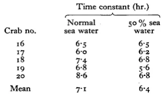
[image:7.451.144.306.209.304.2]normal sea water as before; the animals were then adapted to 50 % sea water and the measurements were repeated. The results are shown in Table 2. There was no evidence of an increase in the time constant so that a reduction in the permeability of the surface membrane cannot play a significant part in the regulatory process. In most cases the time constant was actually decreased and this can almost certainly be explained by the increased rate of urine production found under these circum-stances (see next section).
Table 2. The time constants for sodium outflux in animals from
normal and 5 0 % sea water Time constant (hr.)
:b no. 16 17 18 19
2 0
Normal sea water
6-5 6-o 7-4 6-8 8-6
50 % sea water
6-5
6 2
6-8 S-6 6-8
Mean 7-1 6-4
(c) Rate of urine production
Nagel (1934) showed that Carcinus from normal and from dilute sea water pro-duced an excretory fluid which was similar in composition to the blood both with respect to the total concentration and to the concentration of chloride. It therefore follows that if urine production plays a role in the regulatory mechanism then in dilute sea water the rate of production must be less than that in normal sea water. Previous measurements make this unlikely. Thus Nagel (1934) found the rate of flow in normal sea water was 10% body weight per day and this was increased in 50% sea water to 17% body weight per day. Bethe, von Hoist & Huf (1935) found a rate of 7*2% body weight per day for animals from 65 % sea water. Webb (1940) argues that the rate found by Nagel (1934) for normal animals was too high and, himself, obtained a much lower figure of 4-7 %. In view of these discrepancies it is not possible to be certain that the loss through the urine is not reduced in dilute sea water.
All the previous measurements of urine flow have been made by measuring the increase in body weight after the excretory pores have been blocked by dental cement. This method is open to some obvious objections particularly at high rates of flow and it was therefore decided to attempt to measure the rate by another method in order to obtain independent confirmation of the figures, together with extended measurements of the rate of flow in dilute sea water.
environ-142 J. SHAW
ment such a situation is already at hand. The concentration of sulphate in the blood is maintained below that of the external medium by the activity of the excretory organ, and it will be assumed that the excretory organ is the sole agency whereby this difference in concentration is maintained. If the permeability of the body surface to sulphate is known it is only necessary to measure the concentration of sulphate in the urine and in the blood relative to that in the medium for a calculation of urine flow to be made.
50 r
40
30
•g S20
8 O
o
© , - - ' " 0 o -'©
©
0 2 4 6 8 10 12 14 16 Time in sea water containing Na2
35
SO4 (days)
Fig. 5. The rate of penetration of inorganic sulphate from the external solution into the blood, measured with uS-labelled sulphate.
The first step was to measure the rate of penetration of inorganic sulphate. This was accomplished by keeping an animal in 400 ml. of normal sea water containing 50/x.c. of 35S in the form of inorganic sulphate. Blood samples were withdrawn at intervals and the radioactivity of the sample was compared with that of a sample of the sea water of equal volume. The results from several animals were pooled and are shown in Fig. 5. The points were rather scattered but can be approximately fitted by the relation, A = AF (1 — e~'/r), where A is the radioactivity of the blood as a percentage of the activity of the medium, AF is the final percentage activity of the blood and T is the time constant, equal to 5 days. It may be noted that the rate of penetration may vary from one animal to another, and that by using an average value for the time constant in the calculation of the urine flow the value so obtained has no significance for a given individual but only for the group mean.
con-Studies on ionic regulation in Carcinus maenas (L.). I 143
centration as a percentage of the concentration in the medium. Now sulphate influx is MVbjT.
Assuming that sulphate is confined to the blood and that the blood volume is 35% of the body weight (Webb, 1940), then the influx is 0-35 WM/T1 which in a steady state must equal the outflux. The outflux must be partitioned between diffusion and loss through the urine in the proportion of Bpjioo — Bp.
Thus sulphate outflux through the excretory organ is
100-.Bp 0-3 5 WM
~~Too T '
but the outflux must also be Vu. Up{MI 100).
H e n c e _
y *f —u
Up T
and hence the urine flow as a percentage of the body weight
= 100-^35
Up
T-Bp and Up were measured for each animal and the mean value of 5 days for T was used in all cases.
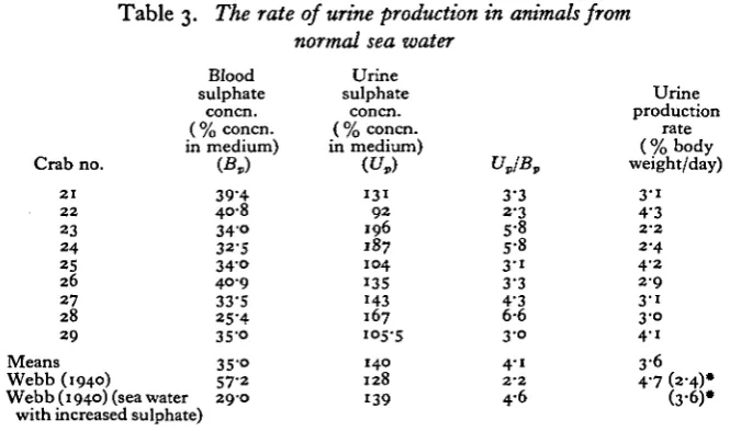
[image:9.451.55.388.417.613.2]Calculations of the rate of urine production for nine animals from normal sea water are shown in Table 3. The mean value was 3-6% body weight per day. The rate estimated in this way is clearly much lower than that found by Nagel (1934) and slightly lower than found by Webb (1940). It may be noted also that the blood sulphate concentration is well below that found by Webb, although it is similar to the mean value found by Gilbert (1959) in animals collected from the same locality as the present ones.
Table 3. The rate of urine production in animals from
normal sea water
Crab no. 2 1 2 2 23 2 4 25 26 27 28 29 Means Webb (1940) Blood sulphate concn.
( % concn. in medium)
(£*)
39-4 40-8
3 4 °
32-5
3 4 ° 4 0 9
33-S 25-4 35-o 3S-o 57-2 Webb (1940) (sea water 29-0
with increased sulphate)
Urine
sulphate concn. ( % concn. in medium)
(t/,)
1 3 1
9 2 196 187 104 135 143 167 105-5 140 128 139 VJB, 3-3 2-3 5-8 5-8
3 - i 3-3 4'3
6-6
3 0
4-i
2 - 2
4-6 Urine production rate (%body weight/day)
3 - i 4-3
2 - 2
2-4 4-2 2 9 3 1 3-o 4 1 3 6 4-7 (2-4)* (3'6)»
144 J- SHAW
Using Webb's values for the sulphate concentration of blood and urine, and the value of the time constant for sulphate penetration as here determined (5 days), the rate of urine production calculated by this method comes out at 2-4 % body weight/ day for his animals. Thus they must either have had a lower permeability to sulphate than the present ones or Webb overestimated the rate of urine production. He also measured the sulphate concentration in blood and urine of animals kept in sea water with increased sulphate content. From his figures it is clear that the rate of urine production must have been greater than in normal crabs and one may conclude that in crabs from normal sea water the rate of urine production is closely geared to the regulation of the blood sulphate concentration. If this is so then the validity of the weighing method for measuring urine flow is open to question. It seems quite clear that the water required for urine production in Carcinus in full-strength sea water is not taken up osmotically since often no osmotic gradient exists and one must suppose, therefore, that the water is absorbed by some active process to an extent which is determined by the blood sulphate level. Now the prevention of sulphate excretion by blocking the excretory pores must lead to an increase in the blood sulphate concentration and, hence, the rate of water absorption would be increased. Thus the weighing method, which measures the rate of water absorption, is likely to give a value for urine flow in excess of normal.
Table 4. The rate of urine production in animals from dilute sea water
Crab no. 3° 31 32 33 34 35 36 37 38 39 40 41 42 External medium (% normal sea water) 9 0 8 0 75 65 65 6 0 5° 5° 5 0 4 0 4 0 4 0 4 0
K
38 25 23-1 15 18-1 20-5 14-1 20-9 23-1 12 10-3u - 4
1 6 6
u,
69 46-3
46-1 i 6 -7
41-7 26-s 35 33-6 3° 17 17 17-5 27-1 UJB, i-8 1-9 2-O I - I
2-3 i-3 2-5 i-6 i-3 i-4 1-6 i"5 1 6 Urine production rate (%body weight per day) 6 0 io-8 I I - I 33-9 13-1 19-9 16-4 15-7 17-9 34-5 35-2 33-7 20-5
Studies on ionic regulation in Carcinus maenas (L.). / 145
that U/B ratio is decreased, in some cases almost to one. The relation between the external concentration and the urine production rate is shown in Fig. 6.With respect to the maintenance of sodium balance these results show that regulation in dilute sea water is not assisted by urine production; in fact, the reverse is the case. The loss of sodium through the urine is increased in dilute sea water.
5M0
- 3 0
20
10 __,©--©
, - - © " '
I
S 100 90 80 70 60 50 40 Concn. of external soln. (% normal sea water)
Fig. 6. T h e relation between the rate of urine production and the concentration of the external solution.
(d) The partition of sodium loss
Since sodium is lost through both the body surface and the urine, it is necessary to establish the relative importance of the two pathways. This can be readily estimated from the knowledge of the total sodium outflux, the blood sodium con-centration and the rate of urine production at a known external concon-centration. Calculation of the sodium loss in the urine as a percentage of the total sodium loss is shown in Table 5 for animals adapted to normal, 50 and 40 % sea water. The mean urine production rate was taken from Fig. 6.
In animals from normal sea water the loss through the urine is negligible com-pared with the total loss rate. In dilute sea water it is more important but the body surface is still the major route for sodium loss. This is confirmed by the previous measurements of tracer outflux (Table 2). The small decrease in the time constant for sodium loss in dilute sea water can be explained by the additional loss through the excretory organ.
Table 5. The partition of sodium loss
External medium (% normal sea water)
1 0 0
5°
4 0
Mean blood sodium
concn. (mM./l.)
460
34° 3 0 0
Total outflux for 50 g.
animal
(jiM./hr.) 1267 1009
891
Mean urine production
rate (% body wt./day)
3-6
23
3°
Sodium loss through
urine for 50 g.
animal
(jiM./hr.)
34-5
163 188
Loss through urine as % total outflux
2-7
1 6 1
2I-I
[image:11.451.126.323.162.304.2]146 J. SHAW
(e) Sodium influx
The fact that the permeability of the body surface to sodium is not significantly decreased during adaptation to dilute sea water and that urine production does not assist in the regulatory process means that regulation must be achieved by variations in the rate of active sodium uptake. Before this can be expressed in quantitative terms, however, it is necessary to know more about the properties of the active uptake mechanism and particularly the relation between the rate of uptake and the external sodium concentration.
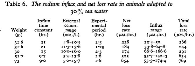
For the influx measurements the animals were first adapted to 30 % sea water so that the uptake mechanism was fully activated. For low external sodium concen-tration the influx was measured with 22Na. Each crab was placed in the animal chamber of the influx apparatus (Shaw, 1960a), together with 100 ml. of water which was circulated through the flow counter. At the start of each experiment the water contained about 5 mM./l. NaCl with added tracer and the decrease of radio-activity of the external solution was recorded over a period of about 2 hr. During this time the animal lost sodium and the external concentration had increased to about 15 mM./l. by the end of the experiment. The net loss rate of sodium during the experiment was calculated from the rate of increase of the external concentration and this value, together with the measured influx, allowed the total loss rate to be calculated. The influx was determined from the rate of decrease of radioactivity in the external solution for the relation: ln(j/_y0) = —t/T, where T was the time
[image:12.451.71.408.394.509.2]con-stant, and hence the influx M^ = A/T, where A was the total external sodium content (Shaw, 1959). The results are shown in Table 6. At the range of external
Table 6. The sodium influx and net loss rate in animals adapted to 30% sea water
Crab no. 43 43 44 45 46 Weight (g-) 3 i 6 3 1 6
3° 51-7 73 Influx time constant (hr.) 2 1 2 1 15 9 7 9-0 External concn. range (mM./l.) 4-8-10-5 11-3-13-6 10-0-16-0 5-4-13-8 5-0-15-7 Experi-mental period (hr.) 2-5 1 2 5 2-3 I "O I *O Net loss rate (/tM./hr.)
2 2 8 184 174 525 654 Influx range (ftM./hr.) 22-4-50 53-8-64-8 66-6-166-6 55-7-142-3 55-5-I74-4 Total loss rate (/tM./hi
2 6 6 2 4 4 291 6 2 4
769
Studies on ionic regulation in Carcinus maenas (L.). / 147
the net loss rate. In this way the relation between the sodium influx and the external sodium concentration was determined.
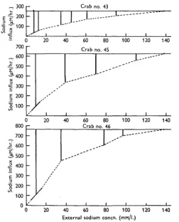
The results for three different animals are shown in Fig. 7. It can be seen that the influx is not proportional to the external concentration and the rate of increase of influx declines as the external concentration is increased.
IS-300
200
100
-- I I
-
M---Crab
J-.-•^ \ i i no. 43
i i I i
-. -. i f ( 700 600 500
^ • 4 0 0
X infl u £ odi i 300 200 100
) 20 40
— — _ -_ / I 1 60 Crab 80 no. 45
1
J--i 100 i 120 140_. " "
I i l/hr . ( 800 700 600 3-500 I 400 c Sodiu m i i 300 200 100 n ) -20 / 40 i 60 Crab j 80 no. 46
1
I 100 ,..L 120 140 i20 40 60 80 100 External sodium concn. (mM/l.)
[image:13.451.104.357.159.472.2]120 140
Fig. 7. The relation between the sodium influx and the external sodium concentration in three animals. The dotted line represents the calculated influx. The influx at low external concentra-tions was measured directly with !2Na. The vertical lines represent the net sodium loss rate.
influx at all external concentrations and, hence, to derive the relation between the active component of the influx and the external sodium concentration.
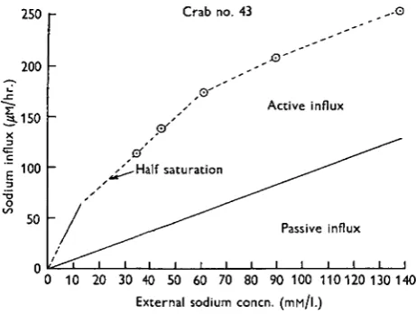
[image:14.451.107.338.221.395.2]This has been done in the case of crab no. 43 using the total influx curve from Fig. 7 and the results are shown in Fig. 8. The upper curve is that of the total influx and the lower that of the passive component of the influx. The vertical distance between the curves represents the active uptake component. It can be seen that the active influx increases rapidly at low external concentrations and then levels off reaching a maximum value at an external concentration of about 70 mM./l. The same result is obtained if the same procedure is adopted for the other animals shown in Fig. 7.
250
200
.C ^-» 1-150
| 100
•oo • J
50
n
Crab no.
, © ' '
~ ^ ^ - H a l f saturation
*
^-^i i i i i i i
43
i
, - - ' "G
Active influx
Passive influx
i i i I i
0 10 20 30 40 50 60 70 80 90 100 110120 130 140
External sodium concn. (IHM/I.)
Fig. 8. The relation between the sodium influx and the external sodium concentration in crab no. 43. The influx is divided into active and passive components.
The conclusion is therefore reached that one of the characteristic features of the active uptake mechanism is that it is fully saturated at an external concentration of about 70 mM./l. Now it may be argued that this conclusion is reached by calcula-tions which are admittedly indirect, although no assumpcalcula-tions of doubtful validity have been made. However, the conclusion is fortified by the fact that direct measure-ments of the active influx in a number of other crustaceans have already led to the same result. Thus the saturable nature of the active uptake mechanisms has been demonstrated in Astacus pallipes (Shaw, 1959), Gammarus pulex and G. duebeni (Shaw & Sutcliffe, i960) and in Eriocheir sinensis (Shaw, 19605). The special feature of the mechanism in Cardnus is the relatively high external concentration required for saturation. The external concentration for half-saturation (Km) is about 20 mM./l.
compared with concentrations of between 0-15 and 1-5 mM./l. for the other Crustacea.
Studies on ionic regulation in Carcinus maenas (L.). / 149
in regulating the blood sodium concentration it is not necessary to consider further the direct effect of the external sodium concentration on the rate of sodium uptake.(/) The regulatory nature of the active uptake mechanism
The arguments presented in the previous sections show that the regulation of the blood sodium concentration in Carcinus is brought about largely by variations in the rate of active uptake of sodium. Since the uptake mechanism is saturated at all physiological external concentrations then it follows that these variations must result from changes in the maximum rate of transport. This must be the result of different states of activation of the uptake mechanism. The relation between the rate of active uptake and the blood sodium concentration can now be put in quanti-tative terms.
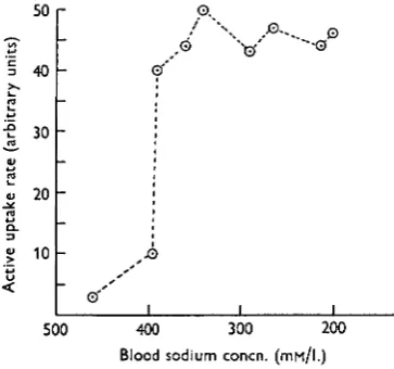
Referring back to Fig. 2 it is now clear that at any external concentration within the physiological range the rate of active uptake must be proportional to the dif-ference between the concentrations of sodium in the blood and in the medium. This may be expressed as a function of the blood sodium concentration. This has been done in Fig. 9, where the difference between the mean blood sodium con-centration and that of the external solution is related to the blood sodium level.
50
40
30
20
10
O.
A-.. 0
v •"s
0 ' '
500 400 300 200
[image:15.451.135.317.327.497.2]Blood sodium concn. (mf-i/l.)
Fig. 9. The relation between the rate of active uptake of sodium and the blood sodium concentration.
150 J. SHAW
factor of about fifteen. A further decrease in blood concentration cannot increase the rate of transport beyond this and, hence, from there on the blood concentration must fall in proportion to the external concentration.
DISCUSSION
The results presented in this paper show that the regulation of the blood sodium concentration in Carcinus is brought about largely by variations in the rate of active uptake of sodium. No evidence was found of control over the permeability of the body surface and, although the increased rate of urine production in dilute sea water tends to impede regulation, the amount of sodium lost through the urine was never more than about 20 % of that lost through the body surface. The properties of the regulating system may therefore be denned by the behaviour of the active uptake mechanism. Two features are particularly worthy of note. The first is that the active uptake rate is a function of the level of sodium in the blood. The precise nature of this relationship determines, in its turn, the level at which the blood sodium is set. Thus a fall in blood sodium concentration of 60 mM./l. from its normal value only induces a small increase in the active uptake rate, but a further small decrease below this fully activates the uptake mechanism. It thus follows that the regulatory mechanism is not so much concerned with the maintenance of the blood sodium concentration at its normal level as with the prevention of a fall in concentration below a certain limit. In terms of the function of the regulatory system as a whole this is of some importance. If a small drop in blood concentration below the normal level led immediately to a full activation of the uptake mechanism then the mechanism would have to be continually turned on and off. Because of the inevitable ' inertia' of the system as a whole, this would lead to the type of instability experienced in a simple on/off thermoregulator when the regulated temperature is only slightly above that of the outside. This kind of response is prevented in Carcinus by the fact that the uptake mechanism is only fully operative when the blood concentration is considerably greater than that of the external solution.
The other feature of the uptake mechanism of importance to the regulatory system as a whole is the fact that when fully activated it is limited to a maximum rate of transport. This means that precise regulation of the blood concentration can only be maintained over a relatively small range of external concentrations. When the external concentration falls below a certain value the uptake rate is insufficient to main-tain the blood concentration and, thereafter, the blood concentration must fall in pro-portion to the decrease in external concentration, eventually reaching the lethal level. Clearly then, for a given surface permeability, the extent to which Carcinus can survive in dilute sea water depends on the maximum rate attained by the uptake mechanism when this is fully activated.
Studies on ionic regulation in Carcinus maenas (L.). I 151
by changes in the active uptake rate, and (6) that at a critical blood concentration a small decrease leads to a great increase in the uptake rate. The main difference between the two systems lies in the fact that whereas in Carcinus the active uptake mechanism is always fully saturated under normal physiological conditions, this is not the case in Astacus, although saturation is reached at a much lower external concentration. The significance of this difference seems clear. In Carcinus balance is maintained at a high external concentration and, hence, diffusion is a major component of the influx. Thus in animals adapted to dilute sea water an increase in the external concentration leads to an increase in the influx due to the diffusion component and in spite of the fact that the uptake mechanism is saturated. Hence the normal blood concentration is soon restored. On the other hand, in Astacus balance is maintained at very low external concentrations and hence diffusion is only a very small component of the total influx. An increase in external concentration within the normal range of concentrations in its environment gives rise to only a very small increase in influx due to this cause. It follows, therefore, that if the uptake mechanism was also saturated at these concentrations then the influx would be largely independent of the external concentration. In fact, at very low external con-centrations, the balance point is on the steep part of the influx curve (about one-fifth of the saturation value) so that a small increase in external concentration leads to a substantial increase in the influx and, hence, a rapid restoration of the normal blood concentration.
These considerations emphasize again that in Astacus, as well as in Carcinus, the regulation of blood sodium, brought about by the activation of the uptake mechanism, is not concerned with the maintenance of the normal blood sodium level as such, but is a protective device which prevents the blood concentration falling below a certain limiting value, which is itself below the normal blood concentration. The mechanism in both cases is adapted to restoring the normal blood concentration under favourable conditions, viz. when the external concentration rises again.
SUMMARY
1. The mechanism of sodium balance in Carcinus maenas has been investigated. 2. Measurements of sodium outflux showed no evidence of a decrease in surface permeability to sodium in dilute sea water.
3. The rate of urine production in normal sea water was 3*6% body weight per day and the sodium loss through the urine was insignificant compared with the total sodium loss. In 40% sea water the urine rate was increased to 30% body weight per day and the loss in the urine accounted for 20 % of the total loss.
4. Measurements of sodium influx and calculation of the active component showed that the active uptake mechanism was fully saturated at all external con-centrations in which the animals could survive.
Part of the expenses of this work was defrayed by a grant from the Royal Society.
REFERENCES
BETHE, A. (1929). Ionendurchlassigkeit der Korperoberflache von wirbellosen Tiere des Meeres als Ursache der Giftigkeit von Seewasser abnormer Zusammensetzung. Pfliig. Arch. ges. Physiol.
221, 344-62.
BETHE, A. (1930). The permeability of the surface of marine animals. J. Gen. Physiol. 13, 437—44. BETHE, A., HOLST, E. & HUF, E. (1935). Die Bedeutung des mechanischen Innendruck fur die Anpassung gepanzerter Seetiere an Anderungen des osmotischen Aussendrucks. Pfliig. Arch. ges. Physiol. 235, 330-44.
DUVAL, M. (1925). Recherches physico-chimiques et physiologiques sur le milieu inte>ieur des animaux aquatiques. Modifications sous l'influence du milieu ext£rieur. Ann. Inst. oceanogr.
2, 232-407.
GILBERT, A. B. (1959). The composition of the blood of the shore crab, Carcinus moenas Pennant, in relation to sex and body size. II. Blood chloride and sulphate. J. Exp. Biol. 36, 356-62. KROGH, A. (1937a). Osmotic regulation in the frog (R. esculenta) by active absorption of chloride
ions. Skand. Arch. Physiol. 76, 60-73.
KROGH, A. (19374). Osmotic regulation in fresh water fishes by active absorption of chloride ions. Z. vergl. Physiol. 24, 656-66.
KROGH, A. (1938). The active absorption of ions in some freshwater animals. Z. vergl. Physiol. 25,.
33S-5O-KROGH, A. (1939). Osmotic Regulation in Aquatic Animals. Cambridge University Press.
MARGARIA, R. (1931). The osmotic changes in some marine animals. Proc. Roy. Soc. B, 107,.
60—24.
NAGEL, H. (1934). Die Aufgaben der Exkretionsorgane und der Keimen bei der Osmoregulation von Carcinus maenas. Z. vergl. Physiol. 21, 468-91.
SCHLIEPER, C. (1929). Uber die Einwirkung miederer Salzkonzentrationen auf marine
Organismen-Z. vergl. Physiol. 9, 478—514.
SHAW, J. (1955 a). Ionic regulation in the muscle fibres of Carcinus maenas. I. The electrolyte composition of single fibres. J. Exp. Biol. 32, 383—96.
SHAW, J. (19556). Ionic regulation in the muscle fibres of Carcinus maenas. II. The effect of reduced blood concentration. J. Exp. Biol. 32, 664-80.
SHAW, J. (1958). Further studies on ionic regulation in the muscle fibres of Carcinus maenas. J. Exp. Biol. 35, 902-19.
SHAW, J. (1959). The absorption of sodium ions by the crayfish, Astacus pallipes Lereboullet. I. The effect of external and internal sodium concentrations. J. Exp. Biol. 36, 126-44. SHAW, J. (1960a). The absorption of sodium ions by the crayfish, Astacus pallipes Lereboullet.
II. The effect of the external anion. J. Exp. Biol. 37, 534-47.
SHAW, J. (1960A). Sodium balance in Eriocheir sinensis (M. Edw.). The adaptation of the Crustacea to fresh water. J. Exp. Biol. 38, 153-62.
SHAW, J. & SUTCLIFFE, D. W. (1961). Studies on sodium balance in Gammarus duebeni Lilijeborg; and Gammarus pulex pulex (L.). J. Exp. Biol. 38, 1—15.