Original Article
Factors associated with improved Karnofsky
performance status after surgery for petroclival
meningiomas: a 10-year follow-up retrospective study
Li Qiao1,2, Chunjiang Yu1, Hongwei Zhang1, Guang Han2, Tian Lan1, Yubo He1
1Department of Neurosurgery, Sanbo Brain Hospital, Capital Medical University, Beijing, China; 2Department of
Neurosurgery, Gansu Provincial Hospital, Lanzhou, China
Received December 25, 2018; Accepted April 9, 2019; Epub July 15, 2019; Published July 30, 2019
Abstract: Objectives: Petroclival meningiomas, generally, have a good prognosis. However, neurological dysfunction, complications, and recurrence/progression after surgery seriously affect the long-term quality of life of patients. The aim of the current study was to determine factors involved in improvement of Karnofsky performance scores (KPS) after surgery for petroclival meningiomas. Methods: A retrospective study of 163 patients with petroclival menin-giomas was conducted. Patients underwent surgery between May 2006 and October 2015 at Sanbo Brain Hospital (China). According to changes in KPS during long-term follow-ups, the patients were divided into improvement and no improvement groups. Prognostic factors associated with improvements in KPS were identified. Results: Com -pared with the no improvement group, the KPS improvement group had lower preoperative KPS scores (P < 0.001), higher postoperative KPS scores (P = 0.021), and higher frequencies of cranial nerve 1 involvement (P = 0.029). Compared with the no improvement group, the improvement group showed higher postoperative KPS scores (P < 0.001), as well as higher rates of gross total resection, subtotal resection + radiotherapy, and subtotal resection (all P < 0.001). Multivariable analysis revealed that duration of symptoms (OR = 0.985, 95% CI: 0.972-0.998, P = 0.021), preoperative KPS (OR = 0.798, 95% CI: 0.710-0.860, P < 0.001), and postoperative KPS (OR = 1.153, 95% CI: 1.092-1.218, P < 0.001) were independently associated with improvements in long-term KPS after treatment. Cranial nerve involvement, surgical approach, and extent of resection were not associated. Conclusion: Present results suggest that duration of symptoms, preoperative KPS, and postoperative KPS are associated with improved long-term KPS after surgery for petroclival meningiomas.
Keywords: Petroclival meningioma, recurrence/progression, surgical treatment, prognostic factors, quality of life, Karnofsky performance score
Introduction
Petroclival meningiomas account for approxi-mately 3-10% of posterior fossa meningiomas [1]. These tumors have a total surgical resec-tion rate of 32-61%, mortality rate of 0-1.2%, complication rate of 31-65.9%, and neurologi-cal dysfunction rate of 22-37.8% [1-7]. Indeed, surgical resections of petroclival meningiomas are extremely difficult and challenging due to tumor characteristics, the deep anatomical location in the skull base, and proximity to com-plex and important nerves and blood vessels. Petroclival meningiomas, generally, have a good prognosis. However, long-term neurologi-cal dysfunction and severe complications after surgery seriously affect the long-term quality of life of patients.
observa-tion factors [3, 5, 7-9]. These studies also lacked detailed comparisons of neurological dysfunction and analysis of complications before and after surgery.
Therefore, the present retrospective study aimed to analyze general conditions, tumor characteristics, treatment modality, and short- and long-term prognostic indicators of pati- ents with petroclival meningiomas, determin- ing factors involved in improvement of long-term Karnofsky performance scores (KPS) [10]. Current results should help clinicians in em- ploying optimal individualized treatment strate-gies, aiming to achieve maximum tumor resec-tions while preserving important neurovascular structures, reducing postoperative complica-tions and neurological dysfunction, and improv-ing the long-term quality of life of patients. Materials and methods
Study design
The current retrospective study was conducted for patients undergoing surgery for petroclival meningiomas, between May 2006 and Octo- ber 2015, at Sanbo Brain Hospital (China). The study design complied with ethical principles of the Declaration of Helsinki and was approved by the Sanbo Brain Hospital Medical Ethics Committee of Capital Medical University. Patients
Inclusion criteria: 1) age ≥ 18 years old; 2) con -firmed diagnosis of petroclival meningiomas; and 3) underwent scheduled surgery. Patients with incomplete data were excluded.
Grouping
According to changes in KPS during follow-ups, the patients were divided into improvement and no improvement groups.
Neuroradiological imaging evaluation
All patients underwent computed tomography (CT) and magnetic resonance imaging (MRI) scans before surgery. Based on the tumor eq- uivalent diameter (TED) formula [(D1×D2× D3)1/3], the maximum diameter of the tumor on the sagittal, coronal, and axis planes was defined as D1, D2, and D3, according to the enhanced MRI. Petroclival meningiomas were
divided into four categories, including small (< 1.0 cm), medium (1.0-2.4 cm), large (2.5-4.4 cm), and giant (≥ 4.5 cm) [1]. Brain CT with a bone window can be used to determine osteop-roliferation or tumor invasion at the skull base, as well as the severity of cerebellar tonsil her-niation. Contrast-enhanced brain MRI scans can identify the origin of the tumor base. This can be used to divide petroclival meningiomas into the three regions, including clival, petro-clival, and sphenopetroclival portions [1, 8]. T1-weighted MRI images can determine tumor blood supply and consistency. T2-weighted im- ages can determine the presence of peritumor-al brain edemas and the tumor border, particu-larly the arachnoid border between the brain-stem and the tumor. Postoperative enhanced MRI images were obtained for extent of resec-tion (EOR) determinaresec-tion in each patient within 1 month of surgery. EOR was divided into gross total resection (GTR), subtotal resection (STR), and partial resection (PTR).
Treatment strategies
Before admission, treatment strategies includ-ed surgery, surgery plus radiotherapy, surgery plus gamma-knife treatment, and careful ob- servation. Most patients received conservative careful observation. All patients underwent sur-gical treatment after admission. A small num-ber of patients underwent a second surgery. Adjuvant radiotherapy or gamma-knife treat-ment was given due to postoperative residual tumor tissues within 3 months of surgery. Pa- tients undergoing STR or PTR (Simpson Grade III-IV) and GTR (Simpson Grade I/II) with a path-ological grade II-III meningiomas, according to the 2016 World Health Organization (WHO) classification of tumors of the central nervous system [11], received adjuvant radiotherapy or gamma-knife treatment within 3 months of sur-gery. No adjuvant radiotherapy or gamma-knife treatments were given to patients that under-went GTR (Simpson Grade I/II) with a pathologi-cal grade I meningioma.
Surgical treatment
pre-operative imaging evaluations (CT/MRI). Indi- cations for selection of the surgical approach were as follows. A temporooccipital craniotomy sub-temporal transtentorial petrosal apex ap- proach was used for the removal of petroclival meningiomas originating from the upper third of the clivus or the lower edge of the petroclival fissure base, not lower than the level of the plane of the internal auditory canal and extend-ing toward the interpeduncular fossa, side of the brain stem, the anterior or posterior petrous apex, the superior or inferior tentorial notch, and the posterolateral wall of the cavernous sinus. A frontotemporal craniotomy pterional or orbitozygomatic approach was used for the removal of tumors extending toward the supra-sellar cistern, optic chiasm, superior and inferi-or inferi-orbital pinferi-ortion, lateral wall of the inferi-orbit, dinferi-or- dor-sum sellae, parasellar, lateral wall of the ca- vernous sinus, or the base of the middle cranial fossa. A suboccipital retrosigmoid approach or superior and inferior tentorium transpetrosal presigmoid approach was used for the removal of tumors originating from the upper two-thirds of the clivus or the lower edge of the petroclival fissure base, not lower than the level of the jugular foramen and extending toward the ven-tral pons, cerebellopontine angle and prepon-tine cistern, dorsum of the petrous bone, and inferior tentorial notch. A far-lateral transcondy-lar approach was used for the removal of tumors that extended below the level of the jugular foramen to the inferior clivus or the fo- ramen magnum. A combined subtemporal tr- anstentorial and suboccipital retrosigmoid ap- proach or combined suboccipital retrosigmoid and far-lateral transcondylar approach was us- ed for the removal of giant or large petroclival meningiomas that had a wide base and involved two or more cranial fossa.
Operative time was defined as the time from incision of the scalp to closure of the scalp. Fluids were aspirated into a reservoir bottle for blood recycling. The amount of intraopera-tive blood loss was determined as the fluid amount in the reservoir bottle subtracted from the amount of rinsing fluid. Adhesive drapes were used to cover the operative field and surgical sheets were used to avoid the out- flow of blood or blood absorbance by surgical sheets. The bag below the operative field completely collected the blood and rinsing fl-uid.
Follow-ups
Thirteen patients (7.4%) were lost to follow-up. Their mailing addresses or phone numbers had changed. One hundred sixty-three out of the 176 patients (92.6%) completed follow-ups in July 2016. The median follow-up was 38.5 months (interquartile range: 44.8 mo- nths). Follow-ups were performed via outpa-tient visits, phone calls, and mailed question-naires. Follow-up visits mainly included an as- sessment of enhanced brain MRI scans, clini-cal symptoms, and signs. The function of the facial nerves was evaluated using the House and Brackmann (HB) score grading system. Quality of life was assessed using KPS, preop-erative KPS, postoppreop-erative KPS, and final KPS (the last follow-up). KPS improvement indicates that the final KPS was higher than the preoper -ative KPS. No KPS improvement indicates that the final KPS was lower than or equal to the preoperative KPS.
Tumor recurrence is defined as any newly iden -tified enhancement after GTR (Simpson Grade I/II). Tumor progression is defined as any am-ount of increase of enhancing tumor volume after STR (Simpson Grade III/IV) or PTR (Si- mpson Grade IV) [12].
Statistical analysis
Results
Patient characteristics
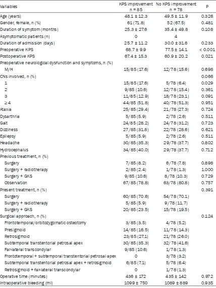
Clinical data of 227 patients that underwent surgical treatment for petroclival meningiomas, between May 2006 and October 2015, was retrieved. Fifty-one patients were excluded ac- cording to the diagnostic criteria of occurrence and origin of petroclival meningiomas. Thirteen patients were excluded because of loss to fol-low-up. Therefore, 163 patients were included in this study. Based on KPS, the patients were classified as KPS improvement (n = 85) or no KPS improvement (n = 78) (Table 1). There were no differences between the two groups regarding age, gender, duration of symptoms, hospital stay, preoperative neurological dys-function, symptoms, previous treatments, pres-ent treatmpres-ents, extpres-ent of resection, operative time, and intraoperative bleeding (all P > 0.05). Compared with the no improvement group, the KPS improvement group had lower preopera-tive KPS scores (P < 0.001), higher postopera-tive KPS scores (P = 0.021), and higher fre-quencies of cranial nerve 1 involvement (P = 0.029).
Tumor characteristics
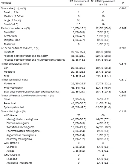
Table 2 shows tumor characteristics. There were no differences between the two groups regarding tumor size, peritumoral edema, AB between tumor and NVS, tumor consistency, tumor vascularity, skull base bone changes, tumor differentiation, and tumor histology (all P > 0.05).
Postoperative clinical characteristics
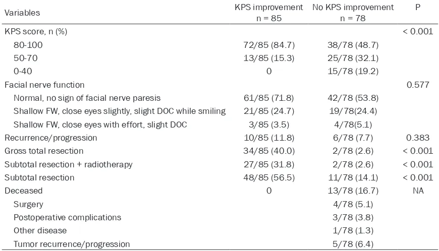
Table 3 shows postoperative characteristics of the patients. Compared with the no improve-ment group, the improveimprove-ment group showed higher postoperative KPS scores (P < 0.001), as well as higher rates of gross total resection, subtotal resection + radiotherapy, and subtotal resection (all P < 0.001). There were no differ-ences between the two groups regarding re- currence/progression (P = 0.383) and facial nerve function (P = 0.577).
Multivariable analysis
Factors associated with KPS improvement in univariable analyses were included in multivari-able analysis. Duration of symptoms was in-
cluded to adjust results. It may affect outcomes since it is associated with the aggressiveness of the disease [13]. Multivariable analysis re- vealed that duration of symptoms (OR = 0.985, 95% CI: 0.972-0.998, P = 0.021), preoperative KPS (OR = 0.798, 95% CI: 0.710-0.860, P < 0.001), and postoperative KPS (OR = 1.153, 95% CI: 1.092-1.218, P < 0.001) were inde- pendently associated with improvements in KPS after treatment. Cranial nerve involve-ment, surgical approach, and extent of resec-tion were not associated (Table 4).
Discussion
long-term quality of life. On the other hand, age, intraoperative bleeding, symptomatic time, ho- spitalization time, preoperative neurological dy- sfunction and symptoms, pre-admission
[image:5.612.93.521.84.653.2]treat-ment, and surgical approach were not signifi -cantly different between the two groups. Size of the tumors, peritumoral edema, arachnoid in- terface between tumors and important cranial
Table 1. Baseline characteristics
Variables KPS improvementn = 85 No KPS improvementn = 78 P
Age (years) 48.1 ± 12.3 49.5 ± 11.9 0.326
Gender, female, n (%) 61 (71.8) 52 (67.5) 0.481
Duration of symptom (months) 25.3 ± 27.6 35.4 ± 49.8 0.108
Asymptomatic patients (n) 0 4
Duration of admission (days) 25.7 ± 11.2 30.0 ± 31.6 0.233
Preoperative KPS 68.7 ± 9.9 77.5 ± 14.1 < 0.001
Postoperative KPS 67.4 ± 15.3 60.9 ± 20.2 0.021
Preoperative neurological dysfunction and symptoms, n (%)
M/H 15/85 (17.6) 12/78 (15.6) 0.698
CNs involved, n (%) 0.066
1 15/85 (17.6) 5/78 (6.4) 0.029
2 9/85 (10.6) 12/78 (15.4) 0.361
3 11/85 (12.9) 18/78 (23.1) 0.091
≥ 4 44/85 (51.8) 40/78 (51.3) 0.951
Ataxia 25/85 (29.4) 21/78 (27.3) 0.724
Dysarthria 5/85 (5.9) 2/78 (2.6) 0.511
Gait 24/85 (28.2) 24/78 (31.2) 0.723
Dizziness 27/85 (31.8) 22/78 (28.6) 0.621
Epilepsy 5/85 (5.9) 2/78 (2.6) 0.511
Headache 30/85 (35.3) 29/78 (37.7) 0.802
Hydrocephalus 34/85 (40.0) 29/78 (37.7) 0.712
Previous treatment, n (%)
Surgery 7/85 (8.2) 6/78 (7.8) 0.898
Surgery + radiotherapy 2/85 (2.4) 1/78 (1.3) 1.000
Surgery + GKS 9/85 (10.6) 8/78 (10.3) 0.729
Observation 67/85 (78.8) 63/78 (80.8) 0.757
Present treatment, n (%) 0.391
Surgery 60/85 (70.6) 54/78 (70.1)
Surgery + radiotherapy 5/85 (5.9) 9/78 (11.7)
Surgery + GKS 20/85 (23.5) 15/78 (19.5)
Surgical approach, n (%) 0.124
Frontotemporal/orbitozygomatic osteotomy 3/85 (3.5) 4/78 (5.2)
Presigmoid 14/85 (16.5) 11/78 (14.3)
Retrosigmoid 23/85 (27.1) 21/78 (26.0)
Subtemporal transtentorial petrosal apex 30/85 (35.3) 32/78 (41.6)
Far-lateral transcondylar 9/85 (10.6) 1/78 (1.3)
Frontotemporal + subtemporal transtentorial petrosal apex 0 3/78 (3.2) Subtemporal transtentorial petrosal apex + retrosigmoid 6/85 (7.1) 5/78 (6.4)
Retrosigmoid + far-lateral transcondylar 0 1/78 (1.3)
Operative time (minutes) 436 ± 172 435 ± 142 0.972
Intraoperative bleeding (ml) 1099 ± 750 1089 ± 889 0.935
nerves and blood vessels and brainstem, tu- mor consistency, tumor blood supply, tumor erosion of skull base, and tumor location were not significantly different between the two gr-oups. Two cases of malignant petroclival men- ingiomas (WHO Grade III) were included in the
KPS no-improvement group, suggesting that the long-term quality of life of malignant petro-clival meningioma patients is not good.
Patients with good long-term quality of life (KPS ≥ 80) accounted for 84.7% in the KPS improve
-Table 2. Tumor characteristics
Variables KPS improvement n = 85 No KPS improvementn = 78 P
Tumor size (cm), n (%) 0.466
Small (< 1.0) 1 0
Medium (1.0-2.4) 8 10
Large (2.5-4.4) 54 44
Giant (≥ 4.5) 15 19
Peritumoral edema, n (%) 13/85 (15.3) 12/78 (15.6) 0.987
Brainstem 5/85 (5.9) 7/78 (9.1)
Cerebellum 4/85 (4.7) 1/78 (1.3)
Temporal lobe 4/85 (4.7) 3/78 (3.9)
Thalamus 0 1/78 (1.3)
AB between tumor and NVS, n (%) 0.296
Presence 23/85 (27.1) 13/78 (16.9)
Absence between tumor and brainstem 21/85 (24.7) 22/78 (28.2) Absence between tumor and neurovascular structures 41/85 (48.3) 43/78 (55.1)
Tumor consistency, n (%) 0.574
Soft 22/85 (25.9) 18/78 (23.4)
Moderate 20/85 (23.5) 15/78 (19.2)
Firm 43/85 (50.6) 45/78 (57.7)
Tumor vascularity, n (%) 0.572
Moderate 22/85 (25.9) 17/78 (22.1)
Hypervascularity 63/85 (74.1) 61/78 (76.9)
Skull base bone erosion/osteoproliferation, n (%) 21/85 (24.7) 16/78 (20.8) 0.523
Tumor differentiation of regions involved, n (%) 0.847
Clival 5/85 (5.9) 5/78 (6.5)
Petroclival 48/85 (56.5) 41/78 (51.9)
Sphenopetroclival 32/85 (37.6) 32/78 (41.0)
Tumor histology, n (%) 0.327
WHO Grade I 76 68
Meningothelial meningioma 48/85 (56.5) 44/78 (57.1)
Fibrous meningioma 5/85 (5.9) 4/78 (5.2)
Transitional meningioma 18/85 (21.2) 14/78 (18.2)
Psammomatous meningioma 2/85 (2.4) 2/78 (2.6)
Angiomatous meningioma 2/85 (2.4) 1/78 (1.3)
Secretory meningioma 1/85 (1.2) 3/78 (3.9)
WHO Grade II 9 8
Chordoid 2/85 (2.4) 1/78 (1.3)
Atypical 7/85 (8.2) 7/78 (9.1)
WHO Grade III 0 2
Rhabdoid 0 1/78 (1.3)
Anaplastic (malignant) 0 1/78 (1.3)
[image:6.612.89.521.88.634.2]ment group and 48.7% in the KPS no-improve-ment group. Additionally, patients with lower long-term quality of life (KPS 50-70) accounted for 15.3% and 32.1%, respectively. There was a significant difference between the two groups (P < 0.001). Patients with poor long-term quali-ty of life (KPS ≤ 40) accounted for 19.2% in the KPS no-improvement group. Results showed that most patients in the KPS improvement group had good long-term quality of life, while most patients in the KPS no-improvement gr- oup had lower long-term quality of life. Com- paring preoperative KPS scores, results indi-cated that most patients with improved KPS scores had good long-term quality of life and prognosis. There were no significant differenc -es in long-term neurological function and tumor recurrence and progression between the two
vement group had higher rates of total resec-tion and subtotal resecresec-tion. In addiresec-tion, most patients with total or subtotal resections had good long-term quality of life and prognosis. Dead patients represented 16.7% in the KPS no-improvement group: Four cases of opera-tion-related deaths (5.1%), 3 cases of postop-erative complications deaths (3.8%), 5 cases of recurrence/progression-related deaths, and 1 case of unknown death (1.3%). When the KPS score decreased after the operation, the pa- tients experienced poor long-term quality of life and prognosis, even death.
[image:7.612.88.523.85.334.2]Multivariable analysis results suggest that du- ration of symptoms, preoperative KPS, and postoperative KPS were associated with long-term improved KPS after surgical treatment for
Table 3. Final status and outcomes of the patients (n = 163)
Variables KPS improvementn = 85 No KPS improvementn = 78 P
KPS score, n (%) < 0.001
80-100 72/85 (84.7) 38/78 (48.7)
50-70 13/85 (15.3) 25/78 (32.1)
0-40 0 15/78 (19.2)
Facial nerve function 0.577
Normal, no sign of facial nerve paresis 61/85 (71.8) 42/78 (53.8) Shallow FW, close eyes slightly, slight DOC while smiling 21/85 (24.7) 19/78(24.4) Shallow FW, close eyes with effort, slight DOC 3/85 (3.5) 4/78(5.1)
Recurrence/progression 10/85 (11.8) 6/78 (7.7) 0.383
Gross total resection 34/85 (40.0) 2/78 (2.6) < 0.001
Subtotal resection + radiotherapy 27/85 (31.8) 2/78 (2.6) < 0.001
Subtotal resection 48/85 (56.5) 11/78 (14.1) < 0.001
Deceased 0 13/78 (16.7) NA
Surgery 4/78 (5.1)
Postoperative complications 3/78 (3.8)
Other disease 1/78 (1.3)
Tumor recurrence/progression 5/78 (6.4)
KPS: Karnofsky performance status; FW: forehead wrinkles; DOC: distortion of commissure.
Table 4. Long-term prognostic factors for improved KPS
Factors OR 95% CI P
Duration of symptom (months) 0.985 0.972-0.998 0.021 Preoperative KPS 0.798 0.740-0.860 < 0.001 Postoperative KPS 1.153 1.092-1.218 < 0.001 CN (two CNs involved) 0.229 0.027-1.941 0.176
Surgical approach 12.938 0.523-3.283 0.118
Extent of resection (partial resection) 0.263 0.038-1.823 0.176
OR: odds ratio; CI: confidence interval; KPS: Karnofsky performance status;
CN: cranial nerve.
[image:7.612.92.362.378.472.2]petroclival meningiomas. Taken together, resu- lts suggest that patients with low KPS before surgery had a higher probability of significant improvement after surgery. On the other hand, the association between postoperative KPS and improvement is by definition. Multivaria-ble analysis showed no significant effects of in-volvement of two or more cranial nerves, the extent of resection, and the surgical approach on KPS improvement. However, it indicated that predictive factors included shorter symp-tomatic time, lower KPS scores before the oper-ation, and higher KPS scores after the opera-tion. As a result, patients with these factors had improved KPS scores and lower neurologi-cal dysfunction and complications, leading to good long-term quality of life and prognosis. In recent years, individualized surgical appr- oaches and intraoperative neurophysiological monitoring have helped to reduce postopera-tive complications and various neurological disorders after surgery. The treatment strategy for petroclival meningiomas has, therefore, ch- anged from performing a total resection or maximum extent of resection to protecting and improving the long-term quality of life and mi- nimizing postoperative complications and ne- urosurgical dysfunction [2-5, 14-22]. In addi-tion to total resecaddi-tion of tumors, subtotal or partial resections with adjuvant radiotherapy or stereotactic radiosurgery (gamma-knife treat-ment) have become more common. These methods avoid damage to blood vessels or nerves and control or delay tumor progression and recurrence [23-27].
The long-term quality of life of patients with pet-roclival meningiomas is closely related to post-operative complications and recovery of neuro-logical dysfunction. In the current series, the complications were similar to those previously observed [3, 4, 28-31]. Sekhar et al. [32] inves-tigated the impact of preoperative and intraop-erative factors on neurological function recov-ery in 75 patients with clival meningiomas that underwent surgery. Results showed that the following preoperative factors had a significant negative impact on postoperative neurological function: male gender, lower KPS scores, tumor size ≥ 2.5 cm, compression and edema of the brain stem, unclear arachnoid border, vascular encasement, and basilar artery blood supply. Difficult resections, absence of an arachnoid border, and partial resections had a negative
impact on postoperative neurological function [32]. In addition, long-term follow-ups revealed that male gender, difficult resections, partial resections, basilar artery blood supply, and early postoperative neurological dysfunction were adverse prognostic factors for long-term neurological rehabilitation [32]. In the present series, the KPS improvement group showed that lower preoperative KPS scores were asso-ciated with KPS improvement, as in the study by Sekhar et al. [32]. However, there were no significant effects from gender, arachnoid bor -der of tumor-to-brainstem, brainstem edema, and tumor size on KPS improvement. All pa- tients in Sekhar’s study had clivus meningio-mas. The structural relationship between tu- mors and the ventral brainstem was close. Therefore, preoperative factors, such as the interface between tumors and brainstem, ed- ema, and the size of tumors, directly affected neurological function after surgery. The present study showed that KPS improvement after sur-gery was significantly associated with lower KPS scores before surgery. Since most patients in the present series had mild symptoms be- fore admission and conservative observations were performed, surgical treatment was not considered until the symptoms were severe. As a result, KPS scores were significantly lower than those before admission.
improvement and no-improvement groups. Re- sults indicated that these intraoperative fac-tors had no significant effects on KPS impro-vement in patients after surgery. Obviously, cur-rent results are not consistent with the study by Little et al. [4]. Grouping might explain, at least in part, the discrepancies. Little et al. [4] analyzed the extent of tumor resection and neurological dysfunction after the operation with the criteria of whether or not to consider the characteristics of intraoperative tumors (fibrous texture of tumors and adhesion of tu-mors to nerves and vessels). This resulted in imbalanced groups (n = 31 vs. n = 106). For hard tumors with tight adhesion and unclear interface to the brain stem or important cranial nerve and vessels, present results are consis-tent with those of Little et al. [4]. To reduce neu-rological dysfunction after the operation, subto-tal resections of the tumors is an appropriate choice. Therefore, the present study shows th- at the proportion of subtotal resections in the KPS improvement group was significantly lar-ger than that in the KPS no-improvement group. In the present study, multivariable analysis sh- owed that duration of symptoms, preoperative KPS, and postoperative KPS were indepen-dently associated with improved KPS after tre- atment for petroclival meningiomas. Cranial nerve involvement, surgical approach, and extent of resection were not associated with improved KPS after treatment for petroclival meningiomas. Discrepancies among studies could be due to many factors, including surgical experience and skill, available techniques and technologies, radiotherapy planning, and post-operative management. Meningiomas are rela-tively indolent and slow-growing tumors [33]. Therefore, duration of symptoms is an indirect indication of tumor course, with longer dura-tions likely to be associated with more advanced disease. However, this is not always the case since some tumors may display aggressive fea-tures [13]. Since the KPS indicates the extent of symptoms due to the compression of ner-vous and vascular structures by the tumor, patients with better preoperative KPS scores are more likely to have a long-term improved KPS and quality of life, compared with patients with worse KPS. Immediate postoperative KPS scores are also probably indicative of treatment success. They may be associated with better long-term KPS.
The present study had several limitations. It was a retrospective, non-randomized, and ob- servational study. Therefore, present research results need to be improved. A prospective ran-domized cohort study is needed to investigate the trends of development and prognosis of petroclival meningiomas using multivariable analysis.
Duration of symptoms, preoperative KPS, and postoperative KPS were associated with im- proved KPS after treatment for petroclival meningiomas. Surgical treatment strategies for petroclival meningiomas must involve a tra- de-off between the extent of the resection, postoperative complications, and neurological dysfunction with long-time quality of life. Disclosure of conflict of interest
None.
Address correspondence to: Chunjiang Yu, Depart- ment of Neurosurgery, Sanbo Brain Hospital, Capi- tal Medical University, Xiangshan Yikesong 50, Haidian District, Beijing 100093, China. Tel: +86-13901316186; Fax: +86-010-62856719; E-mail:
yuchunjiang2017@163.com
References
[1] Mehdorn HM. Intracranial meningiomas: a 30-year experience and literature review. Adv Tech Stand Neurosurg 2016; 139-84.
[2] Bambakidis NC, Kakarla UK, Kim LJ, Nakaji P, Porter RW, Daspit CP, Spetzler RF. Evolution of surgical approaches in the treatment of petro-clival meningiomas: a retrospective review. Neurosurgery 2007; 61: 202-209; discussion 209-11.
[3] Li D, Hao SY, Wang L, Tang J, Xiao XR, Zhou H, Jia GJ, Wu Z, Zhang LW and Zhang JT. Surgical management and outcomes of petroclival me-ningiomas: a single-center case series of 259 patients. Acta Neurochir (Wien) 2013; 155: 1367-1383.
[4] Little KM, Friedman AH, Sampson JH, Wanibu-chi M and Fukushima T. Surgical management of petroclival meningiomas: defining resection goals based on risk of neurological morbidity and tumor recurrence rates in 137 patients. Neurosurgery 2005; 56: 546-559; discussion 546-559.
[6] Seifert V. Clinical management of petroclival meningiomas and the eternal quest for preser-vation of quality of life: personal experiences over a period of 20 years. Acta Neurochir (Wien) 2010; 152: 1099-1116.
[7] Yamakami I, Higuchi Y, Horiguchi K and Saeki N. Treatment policy for petroclival meningioma based on tumor size: aiming radical removal in small tumors for obtaining cure without mor-bidity. Neurosurg Rev 2011; 34: 327-334; dis-cussion 334-325.
[8] Al-Mefty O, Abdulrauf SI and Haddad GF. Me-ningiomas. Winn HR (eds) Youmans neuro-log-ical surgery (Sixth edition), Elsevier, Philadel-phia 2011; 1442- 1444.
[9] Behari S, Tyagi I, Banerji D, Kumar V, Jaiswal AK, Phadke RV and Jain VK. Postauricular, transpetrous, presigmoid approach for exten-sive skull base tumors in the petroclival region: the successes and the travails. Acta Neurochir (Wien) 2010; 152: 1633-1645.
[10] Peus D, Newcomb N and Hofer S. Appraisal of the Karnofsky performance status and propos-al of a simple propos-algorithmic system for its evpropos-alua- evalua-tion. BMC Med Inform Decis Mak 2013; 13: 72.
[11] Louis DN, Perry A, Reifenberger G, von Deim-ling A, Figarella-Branger D, Cavenee WK, Oh-gaki H, Wiestler OD, Kleihues P and Ellison DW. The 2016 world Health organization clas-sification of tumors of the central nervous sys -tem: a summary. Acta Neuropathol 2016; 131: 803-820.
[12] Nanda A, Bir SC, Maiti TK, Konar SK, Missios S and Guthikonda B. Relevance of Simpson grading system and recurrence-free survival after surgery for world health organization grade I meningioma. J Neurosurg 2017; 126: 201-211.
[13] Vranic A and Gilbert F. Prognostic implication of preoperative behavior changes in patients with primary high-grade meningiomas. Scienti-ficWorldJournal 2014; 2014: 398295.
[14] Ichimura S, Kawase T, Onozuka S, Yoshida K and Ohira T. Four subtypes of petroclival me-ningiomas: differences in symptoms and oper-ative findings using the anterior transpetrosal approach. Acta Neurochir (Wien) 2008; 150: 637-645.
[15] Mathiesen T, Gerlich A, Kihlstrom L, Svensson M and Bagger-Sjoback D. Effects of using com-bined transpetrosal surgical approaches to treat petroclival meningiomas. Neurosurgery 2007; 60: 982-991; discussion 991-982. [16] Misra BK. The paradigm of skull base
menin-giomas: what is optimal? World Neurosurg 2012; 78: 220-1.
[17] Park CK, Jung HW, Kim JE, Paek SH and Kim DG. The selection of the optimal therapeutic
strategy for petroclival meningiomas. Surg Neurol 2006; 66: 160-165; discussion 165-166.
[18] Pichierri A, D’Avella E, Ruggeri A, Tschabits- cher M and Delfini R. Endoscopic assistance in the epidural subtemporal approach and Ka-wase approach: anatomic study. Neurosurgery 2010; 67 Suppl Operative: ons29-37; discus-sion ons37.
[19] Roche PH, Fournier HD, Sameshima T and Fu-kushima T. [The combined petrosal approach. Anatomical principles, surgical technique and indications]. Neurochirurgie 2008; 54: 1-10. [20] Samii M, Gerganov V, Giordano M and Samii A.
Two step approach for surgical removal of pet-roclival meningiomas with large supratentorial extension. Neurosurg Rev 2010; 34: 173-179. [21] Sekhar LN, Juric-Sekhar G, Brito da Silva H and
Pridgeon JS. Skull base meningiomas: aggres-sive resection. Neurosurgery 2015; 62: 30-49. [22] Tahara A, de Santana PA Jr, Calfat Maldaun
MV, Panagopoulos AT, da Silva AN, Zicarelli CA, Pires de Aguiar PH. Calfat Maldaun MV, Pana-gopoulos AT, da Silva AN, Zicarelli CA and Pires de Aguiar PH. Petroclival meningiomas: surgi-cal management and common complications. J Clin Neurosci 2009; 16: 655-9.
[23] Havenbergh TV, Carvalho G and Samii M. Natu-ral history of petroclival meningiomas. Neuro- surgery 2003; 52: 55-64.
[24] Kobayashi T, Kida Y and Mori Y. Long-term re-sults of stereotactic gamma radiosurgery of meningiomas. Surg Neurol 2001; 55: 325-331.
[25] Lee JY, Niranjan A, McInerney J, Kondziolka D, Flickinger JC and Lunsford LD. Stereotactic ra-diosurgery providing long-term tumor control of cavernous sinus meningiomas. J Neurosurg 2002; 97: 65-72.
[26] Metellus P, Regis J, Muracciole X, Fuentes S, Dufour H, Nanni I, Chinot O, Martin PM and Grisoli F. Evaluation of fractionated radiothera-py and gamma knife radiosurgery in cavernous sinus meningiomas: treatment strategy. Neu-rosurgery 2005; 57: 886; discussion 873-886.
[27] Nicolato A, Foroni R, Alessandrini F, Maluta S, Bricolo A and Gerosa M. The role of Gamma Knife radiosurgery in the management of cav-ernous sinus meningiomas. Int J Radiat Oncol Biol Phys 2002; 53: 992-1000.
[28] Jung HW, Yoo H, Paek SH and Choi KS. Long-term outcome and growth rate of subtotally resected petroclival meningiomas: experience with 38 cases. Neurosurgery 2000; 46: 567-574; discussion 574-565.
out-come of patients with skull base meningioma. J Clin Neurosci 2011; 18: 895-898.
[30] Yang J, Liu YH, Ma SC, Wei L, Lin RS, Qi JF, Hu YS and Yu CJ. Subtemporal transtentorial pe-trosalapex approach for giant petroclival me-ningiomas: analyzation and evaluation of the clinical application. J Neurol Surg B Skull Base 2012; 73: 54-63.
[31] Yang J, Yu CJ and Qi Z. Microsurgical manage-ment of giant petroclival meningiomas. Chin J Neurosurg 2008; 24: 190-192.
[32] Sekhar LN, Swamy NK, Jaiswal V, Rubinstein E, Hirsch WE Jr, Wright DC. Surgical excision of meningiomas involving the clivus: preopera-tive and intraoperapreopera-tive features as predictors of postoperative functional deterioration. J Neurosurg 1994; 81: 860-8.


