Original Article
The expression levels of procalcitonin-20 and E-cadherin
in lung cancer and their correlations with prognosis
Guang Zheng1, Tingting Han2, Jue Wang3, Yin Xiao1
1Department of Medical Oncology, The Third Affiliated Hospital of Qiqihar Medical College, Qiqihar, Heilongjiang,
China; 2Department of Obstetrics and Gynecology, Qiqihar Second Hospital, Qiqihar, Heilongjiang, China; 3Department of Oncology, The Third Affiliated Hospital of Qiqihar Medical College, Qiqihar, Heilongjiang, China
Received January 9, 2019; Accepted April 11, 2019; Epub July 15, 2019; Published July 30, 2019
Abstract: Lung cancer metastasis is correlated with prognosis. Epithelial E-cadherin (EC) inhibits tumor invasion,
and EC deficiency facilitates tumor metastasis. Protocadherin-20 (PCDH20) plays important roles in intracellular signal transduction and inhibits the growth and clonal formation potency of non-small cell lung cancer (NSCLC). This study measured PCDH20 and EC expressions in lung cancer tissues to investigate their relationship with prognosis. A total of 68 NSCLC patients were recruited during the period January 2013 to June 2015. Immunohistochemical (IHC) staining was used to measure PCDH20 and EC protein expressions to analyze their correlations with the cohort's clinical/pathological features. The Kaplan-Meier approach plotted survival curves, and the Cox ratio risk
model was employed to conduct a multi-variate survival analysis. NSCLC tissues had 20.59% and 44.12% positive
rates of PCDH20 and EC expression respectively, which were significantly lower than in adjacent tissues (58.82%
and 61.76%, p<0.05). Protein expressions were correlated with TNM stage and lymph node metastasis (p<0.05) but
not with the pathology subtype or tumor size. A Kaplan-Meier curve revealed 12.0 months of median survival time in the PCDH20-negative groups, as compared to the PCDH20-positive rate (20.0 months). The EC-negative patients
also showed a shorter median survival span than the EC-positive groups (14.0 months vs. 23.0 months, p<0.05).
The Cox regression analysis identified the TNM stage, differentiation grade, PCDH20/EC protein expression, and lymph node metastasis as independent prognostic factors for NSCLC (p<0.05). PCDH20 and EC may play important roles in NSCLC progression and might work as reference points for prognostic evaluation.
Keywords: Lung cancer, E-cadherin, protocadherin-20, prognosis
Introduction
Primary lung cancer is a common malignant tumor in clinics, with a relatively high incidence
and mortality. Based on its differential histo
-pathological features, it can be subdivided into
small cell lung cancer and non-small cell lung
cancer (NSCLC), the latter of which occupies 80-85% of total lung cancer cases. The treat
-ment strategies and prognostic evaluation of
lung cancer are correlated with early diagnosis and histology subtype [1, 2]. Lung cancer me- tastasis is correlated with prognosis, involving
multiple genes and factors, such as the vas-cular angiogenesis factor, extracellular matrix
metalloproteinase (MMP) and adhesion
mole-cules. During the invasion and metastasis of
malignant tumors, both the epithelial-mesen-chymal transition (EMT) and depressed cell
adhesion play important roles [3, 4]. EMT can potentiate cell invasion or metastasis potency, and epithelial EC can inhibit tumor metastasis
by regulating cell adhesion. EC knockdown or deficiency may facilitate tumor cell infiltration and metastasis [5, 6]. Protocadherin-20 (PCD-H20) plays important roles in the formation of intracellular signal transduction. The PCDH20
gene is located on chromosome 13q-21, and promoter methylation or cell mutation may lead
to the inactivation of gene expression. In the NSCLC cell line, PCDH20 is downregulated. The down-regulation of PCDH-PC in prostate cancer
tissues is correlated with the methylation level
in the promoter region, and PCDH20 overex -pression can inhibit prostate cancer cell grow- th [7, 8]. Previous studies showed that the
PCDH20 promoter methylation level is correlat
growth or clonal formation of NSCLC cell line
[image:2.612.89.373.71.250.2]A459, thus exerting an antitumor role in NSCLC [9, 10].
Previous studies also showed that the adhe-sion molecule E-cadherin could induce tumor angiogenesis and plays important roles in
tumor vascular formation and distal metasta -sis. In various tumors such as pancreatic can-cer and colorectal carcinoma, E-cadherin ex- pression is downregulated and is closely corre-lated with lymph node metastasis. As an endogenous negative regulator, E-cadherin at its normal expression level can inhibit cancer cell metastasis. In malignant tumors, the
deac-tivation of the E-cadherin gene is correlated
with promoter methylation [11, 12]. However, the exact role and mechanism by which E-
cadherin and PCDH20 are involved in the occur
-rence/progression of lung cancer remains poor-ly understood. This study measured PCDH20
and EC protein expression levels in lung cancer tissues to evaluate their correlation with
prog-nosis, thus providing evidence for the diagno
-sis, treatment and prognostic evaluation of
lung cancer in clinics.
Materials and methods
General information
A total of 68 NSCLC patients who were admit
-ted from January 2015 to June 2017 and diag
-nosed by pathology or histology in the Affiliated Cancer Hospital of Zhengzhou University were recruited in this study. None of the patients had
University, and all the patients gave their in-formed consent before the study began.
Inclusive criteria
All the patients received a confirmed diagnosis
by histology or pathology, including
percutane-ous puncture, lymph node biopsy, fiber bron
-choscopy biopsy, thoracic cavity fluid cytology
assay, or sputum cytology. All recruited patients
had their complete clinical information record -ed and clear stage determin-ed, and they re- ceived systemic post-op treatment (including
intravenous chemo-therapy and focal
treat-ment).
Exclusive criteria
Those patients complicated with other primary
malignant tumors or who were lost during fol -low-up were excluded.
Reagent and equipment
PCDH20, EC antibody and SP immunohisto
-chemistry (IHC) staining kits were provided by
Boster Bio (China). The secondary antibody was provided by Zhongshan Jinqiao Biotech (China).
The inverted microscope was from Olympus
(Japan). The tissue embedding equipment was
from SAKURA (Japan). The Microtome was fr-om Leica (Germany). The oscillator was frfr-om
Jinghong Equipment (China). The heat-resis-
tant glass slide rack was from Maixin Biotech,
(China). The computer-assisted image analysis
system was from HP (United States).
Figure 1. PCDH20 and EC protein expression in lung cancer tissues (×100).
received any chemo- or
radio-therapy before surgery.
Can-cer tissues samples were
col-lected from surgical resec
-tions and were confirmed by
pathology. Tumor adjacent
tis-sues with a distance of >5 cm from the cancer edge were
also collected. In the patient cohort, there were 32 males
and 36 females, aged
bet-ween 31 and 76 years (aver-age (aver-age = 60.5±6.7 years).
The study protocol was app- roved by the Research Ethics
Patient follow-ups
The patients received post-op follow-ups by
electronic charts and telephone interviews.
Progression-free survival (PFS) was evaluated from the time of confirmed diagnosis until the treatment efficacy evaluation revealed any pro
-gression of the disease (PD). Overall survival (OS) was deduced from the time of diagnosis
until mortality or January 2017, using months
as the unit. Till the endpoint of the observation
window, the survivors were treated as cut-tail
data. The correlation between PCDH20/EC
expression and clinical pathology or prognosis
was analyzed, along with survival period
follow-elds were randomly selected from each slide for recording.
Deduction of positive expression
Positive expression was deduced as brown-yel-low granules in the nucleus, cytoplasmic mem-brane or cytoplasm. The IHC staining results
were analyzed in a semi-quantitative manner.
Scores were given according to the percentage
of positive cells: 0 for no positive cells; 1 for <25% positive cells, 2 for 25%~50% positive cells, 3 for 50%~75% positive cells, and 4 for >75% positive cells. Staining intensity was gi-
ven for 0, 1, 2, and 3 for no significant
stain-Table 1. PCDH20 and EC protein expressions in lung cancer tissues Tissue type PCDH20 (%, n) EC (%, n) χ2 value P value
Tumor tissue 20.59 (14/68)* 44.12 (30/68)* 20.762 0.000
Tumor adjacent tissue 58.82 (40/68) 61.76 (42/68) 4.250 0.032
[image:3.612.89.380.83.137.2]Note: *, p<0.05 compared to control group.
Table 2. The correlation between PCDH20 and EC protein expres
-sion and clinical/pathological features
Item N positive ratePCDH20 positiveEC valueχ2 valueP Sex
Male 32 6 (18.75) 14 (43.75) 0.125 >0.05
Female 36 8 (22.22) 16 (44.44) 0.033 >0.05
Age
<60 46 10 (21.74) 18 (39.13) 0.112 >0.05
≥60 22 4 (18.18) 12 (54.54) 1.434 >0.05
Tumor size (cm)
<5 25 4 (16.00) 10 (40.00) 0.509 >0.05
≥5 43 10 (23.26) 20 (46.51) 1.809 >0.05
Pathology time
Adenoma 49 11 (22.45) 23 (46.94) 0.417 >0.05
Squamous carcinoma 19 3 (15.79) 7 (36.84) 0.824 >0.05 Differentiation grade
Low 47 13 (27.66) 25 (53.19) 4.655 <0.05 Moderate to high 21 1 (4.76) 5 (23.81) 5.083 <0.05
Infiltration depth
No reaching serosa 50 11 (22.00) 26 (52.00) 3.118 >0.05
Penetrating serosa 18 3 (16.67) 4 (22.22) 0.230 >0.05
Lymph node metastasis
Yes 16 0 (0.00) 1 (6.25) 5.425 <0.05 No 52 14 (26.92) 29 (55.77) 12.169 <0.05 Tumor TNM stage
I+II 32 10 (31.25) 24 (75.00) 23.382 <0.05 III+IV 36 4 (11.11) 6 (16.67) 4.205 <0.05
ups. The Kaplan-Meier
app-roach was used to plot the survival curve, and the Cox
ratio-risk model was em-ployed for multi-variant sur -vival analysis.
Experimental approaches
IHC staining was employed
to measure PCDH20 and EC
expression in lung cancer and adjacent tissues. In bri-
ef, the tissues were fixed in formalin, followed by being
dehydrated and then immer-
sed in paraffin for tissue embedding. Paraffin-based
tissues were cut into sec-tions, which were dried, de- hydrated, and processed in
heat antigen retrieval. After blocking, normal goat serum (NGS) was added, followed
by 1 h room temperature in-
cubation with 50 μl rabbit
human monoclonal
anti-body of PCDH20 or EC (1: 100 for both). 50 μl of sec -ondary antibody (1:100) was
then added for 10 min incu
-bation, followed by incuba
-tion with 50 μl streptavidin-peroxidase for 10 min at room temperature. After
de-velopment, quenching,
coun-ter-staining and differentia -tion, a computer-assisted im- aging system was used to
[image:3.612.91.382.183.525.2]fi-ing, light yellow, brown-yellow and dark brown,
respectively. The total score was determined
by calculating the product of staining intensity
score and the positive cell score. The product
was deduced as negative (-) for scores of 0~3, weak positive (+) for scores of 4~8, and posi
-tive (++) for scores of 9~12.
Data processing
Data were analyzed using SPSS 19.0 software.
Enumeration data were processed using a chi-square test. The measurement data were
test-ed for normality using a Kolmogorov-Smirnov
test. The biased distribution was described
using median ± quadrille (M ± Q). A rank-sum
test was used to compare the independent
samples. The measurement data that were fit -ted into a normal distribution were presen-ted
as the mean ± standard deviation (SD) and analyzed using an analysis of variance (ANOVA)
and Student’s t-test. The Kaplan-Meier
app-roach was used to plot the survival curve, and
the Cox ratio risk model was employed to do a
multi-variant survival analysis. Statistical
sig-nificance was defined as p<0.05.
Results
PCDH20 and EC protein expressions in lung cancer tissues
In NSCLC tissues, the positive rates of PCDH20
and EC protein expressions were 20.59% and
44.12%, respectively, which were significantly
lower than those in controlled tumor adjacent
tissues (58.82% and 61.76%, χ2=20.762 and
4.250, p<0.05). Positive expression of the tar
-get protein was shown as light yellow to dark brown colors. PCDH20 positive expression was
mainly shown in the nuclei and cytoplasms, but EC protein was mainly expressed in the cytoplasmic plasma and cytoplasms. T-cadhe- rin expression is mainly located on the
mem-branes, and CD34 positive expression exists
on the membranes and cytoplasms (Figure 1, Table 1).
Correlation analysis between PCDH20/EC pro
-tein expressions in NSCLC and clinical/patho
-logical features
PCDH20 and EC protein positive expressions were found to be correlated with the tumor dif
-ferentiation grade, TNM stage and lymph node metastasis (p<0.05), but not with tumor size,
age, sex, or pathological type. The patients wi-
th lymph node metastasis had a lower
PCD-H20/EC protein positive expression rate com-pared to those without lymph node metastasis.
In the advanced clinical stages, the PCDH20
and EC proteins showed decreased positive expressions (Table 2).
PCDH20 and EC expressions in lung cancer tissues and their relationship with prognosis
A total of 68 patients received post-up follow
ups until January 2017, with median overall
[image:4.612.92.289.71.276.2]sur-vival (OS) at 15 months (95% CI: 13.716~
[image:4.612.90.288.325.531.2]Figure 2. Correlation analysis between PCDH20 pro -tein expression in NSCLC and its prognosis.
17.088). Those patients with PCDH20-negative expression showed a significantly shorter sur
-vival span than the PCDH20-positive ones
(11.0 months vs. 17.0 months, p<0.05). The
EC-negative patients also showed shorter OS
than the EC-positive individuals (12.0 months vs. 18.0 months, p<0.05, Figures 2 and 3).
Cox regression analysis for the independent prognostic factors of NSCLC
Various factors, including age, sex, clinical st-age, pathological type, and PCDH20/EC protein expression were included in the Cox ratio risk model for multi-variate survival analysis, and we found that clinical TNM stage, tumor differ
-entiation grade, PCDH20/EC protein expres -sions and lymph node metastasis were
inde-pendent prognostic factors of NSCLC (p<0.05,
Table 3).
Discussion
PCDH is an important member of the cadhe-rin protein family and plays important roles in
intracellular signal transduction. Mainly expre-
ssed in the nervous system, PCDH has cell adhesion properties. Such extracellular fea
-tures of transmembrane PCDH20 exerts
im-portant roles in cell-to-cell adhesion [13, 14].
PCDH20 is a non-clustered PCDH, and its gene
is located on chromosome 13q21.2, with six extracellular cadherin domains. Previous
stud-ies showed decreased PCDH20 expressions
in the NSCLC cell line, and NSCLC tissues also
showed higher a PCDH20 gene methylation rate than normal lung tissues. After treatment with de-methylation drugs, PCDH20 expression is significantly up-regulated. The promoter me-thylation level of the PCDH20 gene is closely
correlated with its expression in NSCLC tissues [15-17]. In epithelial cell adhesion, the epitheli-al EC protein complex has important roles, and
lowly differentiated EC is correlated with tumor cell differentiation. Previous studies have gen
-erated inconsistent results regarding the rela-tionship between NSCLC prognosis and EC
expression [18, 19]. This study measured
PCD-H20 and EC expression in lung cancer tissues to investigate their correlation with prognosis.
The results showed lower PCDH20 and EC
expression in NSCLC tissues, which were high-er in tumor tissues compared to the adjacent tissues, indicating that they might be involved in NSCLC onset and progression.
Previous studies showed important roles of
tumor cell adhesion lost and separated during the tumor invasion and metastasis process.
The EC protein is a major mediator for epithelial
cell adhesion and intracellular connections [20,
21]. By transfecting highly invasive tumor cells
with the wild type EC gene, tumor invasion or metastasis was inhibited. EC down-expression is correlated with the lymph node metastasis
of various malignant tumors. A previous study showed a correlation between PCDH20 meth
-ylation levels and unfavorable cancer progno -ses [22]. In the present study, we did not reveal
a significant correlation between PCDH20/EC
protein expression and sex, age, or patients’
pathological subtypes. However, tumor differ -entiation grade, TNM stage, lymph node
metas-tasis, and EC expression affect tumor cell reat
-tachment and de-at-tachment. During tumor progression, EC down-regulation or deficiency is directly correlated with tumor infiltration and metastasis [23, 24]. This study further proved that PDH20/EC protein down-regulation or defi -ciency caused decreased cell adhesion
poten-cy, making tumor cells detach from primary lesions, thus facilitating tumor infiltration or metastasis. A Kaplan-Meier curve showed that the PCDH20-negative patients had a signifi
-cantly shorter survival period than the
PCD-H20-positive patients did, and the EC-negative patients had a shorter survival than the EC-
[image:5.612.86.526.85.168.2]positive ones, indicating possible roles of PC-DH20 and EC in NSCLC progression. PCPC-DH20
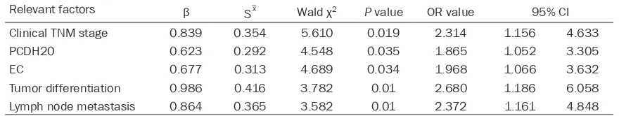
Table 3. Cox regression analysis for the adverse factors of prognosis in NSCLC patients
Relevant factors β S_x Wald χ2 P value OR value 95% CI
Clinical TNM stage 0.839 0.354 5.610 0.019 2.314 1.156 4.633
PCDH20 0.623 0.292 4.548 0.035 1.865 1.052 3.305
EC 0.677 0.313 4.689 0.034 1.968 1.066 3.632
Tumor differentiation 0.986 0.416 3.782 0.01 2.680 1.186 6.058
and EC expression thus might be used as re-
ference points for the prognostic evaluation of
NSCLC.
A Cox regression analysis showed that TNM
stage, tumor differentiation grade, PCDH20/EC
protein expression and lymph node metastasis
were independent prognostic factors for
NS-CLC, suggesting that patient prognosis could
be primarily evaluated according to PCDH20 and EC expression, which might be beneficial for the clinical evaluation of NSCLC patient prognosis. Due to the limited sample size
included in the present study, a large cohort
clinical study should be performed to demon
-strate the correlation between PCDH20/EC
expression and post-op survival time in NSCLC patients. Moreover, determining the roles and
mechanisms of E-cadherin and PCDH20 in lung cancer onset and progression is also worthy of further investigation.
Conclusion
PCDH20 and EC may play important roles in
NSCLC progression, and their expressions may
serve as reference points for evaluating the post-op prognosis of NSCLC.
Disclosure of conflict of interest
None.
Address correspondence to: Dr. Yin Xiao, Depart-ment of Oncology, The Third Affiliated Hospital of
Qiqihar Medical College, No. 27, Taishun Street, Tie-
feng District, Qiqihar, Heilongjiang, China. Tel: +86-0452-2120197; Fax: +86-+86-0452-2120197; E-mail: kqegomaw2sq@sina.com
References
[1] Birse CE, Lagier RJ, FitzHugh W, Pass HI, Rom WN, Edell ES, Bungum AO, Maldonado F, Jett JR, Mesri M, Sult E, Joseloff E, Li A, Heidbrink J, Dhariwal G, Danis C, Tomic JL, Bruce RJ, Moore
PA, He T, Lewis ME and Ruben SM.
Blood-based lung cancer biomarkers identified
th-rough proteomic discovery in cancer tissues, cell lines and conditioned medium. Clin Pro-teomics 2015; 12: 18.
[2] Korbakis D, Dimitromanolakis A, Prassas I, Da
-vis GJ, Barber E, Reckamp KL, Blasutig I and Diamandis EP. Serum LAMC2 enhances the prognostic value of a multi-parametric panel in
non-small cell lung cancer. Br J Cancer 2015; 113: 484-91.
[3] Romero-Ventosa EY, Blanco-Prieto S, Gonzalez-Pineiro AL, Rodriguez-Berrocal FJ, Gonzalez- Pineiro-Corrales G and Paez de la Cadena M. Pre-treatment levels of the serum biomarkers CEA, CYFRA 21-1, SCC and the soluble EGFR and its ligands EGF, TGF-alpha, HB-EGF in the predic
-tion of outcome in erlotinib treated
non-small-cell lung cancer patients. Springerplus 2015; 4: 171.
[4] Cingelova S, Labudova V, Berkesova D, Dien
-erova M, Dammak A, Grmanova E, Nadaska O, Vasilenkova I, Najselova E, Skarbova V, Migas
-ova M, Viktorin-ova Z, Jurga L. [Prognostic markers of advanced non-small cell lung carci
-noma - assessing the significance of onco
-markers using data-mining techiques RPA]. Klin Onkol 2014; 27: 347-52.
[5] Liu XL, Zhang XT, Meng J, Zhang HF, Zhao Y, Li C, Sun Y, Mei QB, Zhang F, Zhang T. ING5 knockdown enhances migration and invasion of lung cancer cells by inducing EMT via EGFR/ PI3K/Akt and IL-6/STAT3 signaling pathways. Oncotarget 2017; 8: 54265-54276.
[6] Enderle-Ammour K, Bader M, Ahrens TD, Franke K, Timme S, Csanadi A, Hoeppner J, Kulemann B, Maurer J, Reiss P, Schilling O, Keck T, Brabletz T, Stickeler E, Werner M, Wellner UF, Bronsert P. Form follows function:
morphological and immunohistological in-sights into epithelial-mesenchymal transition
characteristics of tumor buds. Tumour Biol
2017; 39: 1010428317705501.
[7] Feng J, Zhang X, Zhu H, Wang X, Ni S and Huang J. FoxQ1 overexpression influences
poor prognosis in non-small cell lung cancer,
associates with the phenomenon of EMT. PLoS One 2012; 7: e39937.
[8] Kinoshita Y, Takasu K, Yuri T, Yoshizawa K, Ue
-hara N, Kimura A, Miki H, Tsubura A, Shikata N. Two cases of malignant peritoneal mesothelio -ma without asbestos exposure: cytologic and
immunohistochemical features. Ann Diagn
Pathol 2013; 17: 99-103.
[9] Park J, Yang JS, Jung G, Woo HI, Park HD, Kim JW, Huh W, Ko JW, Kim H, Cho JY, Lee SY. Subunit-specific mass spectrometry method identifies haptoglobin subunit alpha as a diag
-nostic marker in non-small cell lung cancer. J
Proteomics 2013; 94: 302-10.
[10] Li M, Zheng C, Xu H, He W, Ruan Y, Ma J, Zheng
J, Ye C, Li W. Inhibition of AMPK-related kinase 5 (ARK5) enhances cisplatin cytotoxicity in
non-small cell lung cancer cells through
regu-lation of epithelial-mesenchymal transition.
Am J Transl Res 2017; 9: 1708-1719.
[11] Eisenhauer EA, Therasse P, Bogaerts J, Sch-
wartz LH, Sargent D, Ford R, Dancey J, Arbuck
S, Gwyther S, Mooney M, Rubinstein L,
Shan-kar L, Dodd L, Kaplan R, Lacombe D, Verweij J.
tu-mours: revised RECIST guideline (version 1.1). Eur J Cancer 2009; 45: 228-47.
[12] Lumachi F, Santeufemia DA, Del Conte A, Maz
-za F, Tozzoli R, Chiara GB, Basso SM.
Carboxy-terminal telopeptide (CTX) and amino-Carboxy-terminal
propeptide (PINP) of type I collagen as markers of bone metastases in patients with non-small
cell lung cancer. Anticancer Res 2013; 33: 2593-6.
[13] Shibuya M, Kogo M, Kurihara T, Shikama Y, Na
-kajima H, Yoneyama K, Kiuchi Y. [Analysis of the risk factors for severe neutropenia in ad
-vanced non-small cell lung cancer after the first course of chemotherapy with third-genera
-tion agents]. Yakugaku Zasshi 2013; 133:
703-9.
[14] Kozu Y, Maniwa T, Takahashi S, Isaka M, Ohde Y, Nakajima T. Prognostic significance of post -operative serum carcinoembryonic antigen lev-els in patients with completely resected patho-logical-stage I non-small cell lung cancer. J Cardiothorac Surg 2013; 8: 106.
[15] Tanaka K, Hata A, Kaji R, Fujita S, Otoshi T, Fujimoto D, Kawamura T, Tamai K, Takeshita J, Matsumoto T, Monden K, Nagata K, Otsuka K, Nakagawa A, Tachikawa R, Otsuka K, Tomii K and Katakami N. Cytokeratin 19 fragment pre
-dicts the efficacy of epidermal growth factor receptor-tyrosine kinase inhibitor in non-small-cell lung cancer harboring EGFR mutation. J Thorac Oncol 2013; 8: 892-8.
[16] Hur J, Lee HJ, Nam JE, Kim YJ, Hong YJ, Kim HY, Kim SK, Chang J, Kim JH, Chung KY, Lee HS and Choi BW. Additional diagnostic value of tumor markers in cytological fluid for diagnosis of non-small-cell lung cancer. BMC Cancer
2012; 12: 392.
[17] Jung M, Kim SH, Hong S, Kang YA, Kim SK, Chang J, Rha SY, Kim JH, Kim DJ and Cho BC. Prognostic and predictive value of carcinoem
-bryonic antigen and cytokeratin-19 fragments
levels in advanced non-small cell lung cancer
patients treated with gefitinib or erlotinib. Yon -sei Med J 2012; 53: 931-9.
[18] Trape J, Montesinos J, Catot S, Buxo J, Fran
-quesa J, Sala M, Domenech M, Sant F, Badal
JM and Arnau A. A prognostic score based on
clinical factors and biomarkers for advanced non-small cell lung cancer. Int J Biol Markers
2012; 27: e257-62.
[19] Li X, Asmitananda T, Gao L, Gai D, Song Z,
Zhang Y, Ren H, Yang T, Chen T and Chen M.
Biomarkers in the lung cancer diagnosis: a
clinical perspective. Neoplasma 2012; 59: 500-7.
[20] Lee DS, Kim YS, Jung SL, Lee KY, Kang JH, Park S, Kim YK, Yoo Ie R, Choi BO, Jang HS and Yoon SC. The relevance of serum carcinoem
-bryonic antigen as an indicator of brain metas -tasis detection in advanced non-small cell lung cancer. Tumour Biol 2012; 33: 1065-73. [21] Cabrera-Alarcon JL, Carrillo-Vico A, Santotoribio
JD, Leon-Justel A, Sanchez-Gil R, Gonzalez-Castro A and Guerrero JM, CYFRA 21-1 as a tool for distant metastasis detection in lung
cancer. Clin Lab 2011; 57: 1011-4.
[22] Hong YJ, Hur J, Lee HJ, Nam JE, Kim YJ, Kim HS, Kim HY, Kim SK, Chang J, Kim JH, Chung KY, Choi BW and Choe KO. Analysis of tumor markers in the cytological fluid obtained from
computed tomography-guided needle
aspira-tion biopsy for the diagnosis of non-small cell lung cancer. J Thorac Oncol 2011; 6: 1330-5.
[23] Jung M, Kim SH, Lee YJ, Hong S, Kang YA, Kim SK, Chang J, Rha SY, Kim JH, Kim DJ and Cho BC, Prognostic and predictive value of CEA and CYFRA 21-1 levels in advanced non-small cell lung cancer patients treated with gefitinib or
erlotinib. Exp Ther Med 2011; 2: 685-693. [24] Hanagiri T, Sugaya M, Takenaka M, Oka S,
Baba T, Shigematsu Y, Nagata Y, Shimokawa H, Uramoto H, Takenoyama M, Yasumoto K and Tanaka F. Preoperative CYFRA 21-1 and CEA as prognostic factors in patients with


![5 Diethylamino 2 [(E) (4 methyl 3 nitrophenyl)iminomethyl]phenol: a redetermination](data:image/gif;base64,R0lGODlhAQABAIAAAP///wAAACH5BAEAAAAALAAAAAABAAEAAAICRAEAOw==)