Case Report
Early computed tomography for detection of internal
jugular vein thrombosis after neck dissection
and/or reconstruction surgery for head
and neck cancer patients
Nobuyuki Maruyama1,2, Yusuke Shimizu3, Moriyasu Nakaema4, Kazuhide Nishihara1,5, Toshiyuki Nakasone5, Hirofumi Matsumoto6, Takeaki Kusada7, Fumikazu Nimura1, Akira Matayoshi5, Tessho Maruyama5,8, Naoki Yoshimi6,9, Akira Arasaki1,5
Departments of 1Oral and Maxillofacial Functional Rehabilitation, 3Plastic and Reconstructive Surgery, 4Thoracic
and Cardiovascular Surgery, 7Radiology, 9Pathology and Oncology, Graduate School of Medicine, University of
The Ryukyus, Nishihara, Okinawa, Japan; Departments of 5Oral and Maxillofacial Surgery, 6Pathology, University
Hospital of The Ryukyus, Nishihara, Okinawa, Japan; 2Research Fellowship for Young Scientists, Japan Society
for The Promotion of Science, Tokyo, Japan; 8Molecular Microbiology Group, Tropical Biosphere Research Center,
University of The Ryukyus, Nishihara, Okinawa, Japan
Received March 18, 2018; Accepted April 9, 2019; Epub May 15, 2019; Published May 30, 2019
Abstract: Internal jugular vein thrombosis (IJVT) is a complication that occurs after radical neck dissection (ND) and/or reconstruction surgery for patients with head and neck (H&N) cancer. As a protocol, follow-up computed tomography (CT) is performed after cancer resection to detect residual cancer, recurrence, or metastasis. In con-trast, there are no clear guidelines for the detection of IJVT after H&N cancer therapy. We report the case of a 67-year-old woman with IJVT subsequent to H&N surgery for tongue cancer. To our knowledge, this is the first case of IJVT after combined supraomohyoid ND and anterolateral thigh flap reconstruction using the internal jugular vein. Further evaluation of swelling and reddening of the right mandibular and neck area by CT scan was performed 71 h postoperatively, and IJVT was clearly detected. Routine CT for patients with H&N cancer is performed at least 4-8 weeks postoperatively, which is too late for safe detection of IJVT and worsens the prognosis of the patient because of pulmonary embolism or flap failure. Based on literature review, we suggest that CT should be performed within a week after ND and/or reconstruction surgery for H&N cancer to detect IJVT at the earliest time point.
Keywords: Head and neck cancer, tongue cancer, internal jugular vein, thrombosis, neck dissection, reconstruc-tion surgery, computed tomography, case report
Introduction
Worldwide, approximately 980,000 cases of head and neck (H&N) cancer (including thyroid) occurred per year, and approximately 40% of patients with this cancer died per year [1]. The standard method for radical treatment of this cancer is neck dissection (ND) [2], with recon-structive surgery performed to treat the defect after primary cancer resection and ND [3]. However, ND and reconstructive surgery have risks for various complications [4-6].
Internal jugular vein (IJV) thrombosis (IJVT) is one complication after radical ND and/or
We report a case of IJVT of tongue cancer after SOHND and anterolateral thigh (ALT) flap recon-struction. Contrast-enhanced CT was perfor- med owing to her clinical symptoms. IJVT was detected by CT within 3 days after the surgery. Case report
A 67-year-old female was referred to the De- partment of Oral and Maxillofacial Surgery, University Hospital of the Ryukyus (Okinawa, Japan), for treatment of right tongue lesion in August 2016. She had a 2.5 month history of tongue pain. Physical examination revealed a hard 2.3 × 1.8 cm mass with an ulcer on the right tongue extending to the oral floor. No pal-pable lymphadenopathy was found in the neck area. She underwent a resection for uterine cancer (endometrioid adenocarcinoma), i.e., total abdominal hysterectomy with bilateral salpingo-oophorectomy at our institute in 2005. No radiation or chemotherapy was per-formed for the uterine cancer. She had no his-tory of smoking and drinking. No other family history was found. The tongue mass was clini-cally suspicious for cancer. For cancer staging, contrast-enhanced CT (head to chest regions), magnetic resonance imaging (head and neck regions), upper gastrointestinal endoscopy, and 2-[18F]-fluoro-2-deoxy-D-glucose (FDG)-positr- on emission tomography (PET) were performed. Contrast-enhanced CT revealed tongue lesion; however, there were no visible radiological ch- anges in the neck lymph nodes, and the inter-nal jugular vein (IJV) was normal. Further
whole-body PET revealed increased FDG uptake in the right tongue; no neck lymph nodes had strong FDG uptake. No other malignant lesion was found, and no abnormality was found in the IJV. Additionally, no abnormal coagulation test re- sults were found before the cancer treatment. Histopathological diagnosis of the tongue mass biopsy was well-to-moderately differentiated squamous cell carcinoma. Final diagnosis was tongue to oral floor squamous cell carcinoma (T4aN0M0, Stage IVa; UICC TNM classification 7th edition, 2009). Neoadjuvant chemotherapy (TS-1 tegafur-gimeracil-oteracil potassium 100 mg/day for 2 weeks) was administered for the tongue cancer.
We performed tumor resection, right SOHND (IJV was preserved), and ALT flap reconstruc-tion to reconstruct large tongue and neck defects. On the artery side, we first planned a combination of right lateral femoral circumflex artery (LFCA) anastomosed end-to-end to the right lingual artery. However, the patient showed reduction in the blood flow to the flap, and arte-rial thrombosis was observed. Therefore, the artery anastomosis was resected, and a new anastomosis (a combination of the right LFCA anastomosed end-to-end to the right superior thyroid artery) was performed. The artery inti-ma on the flap side was partially calcified. Thrombosis immediately recurred after the anastomosis. Therefore, the anastomosis was resected; the thrombosis was removed, and the arteries were anastomosed again. After the additional treatment, a stable blood flow was observed. On the vein side, the right lateral femoral circumflex vein was anastomosed side-to-end to the IJV. Histopathological examina-tion revealed well-differentiated squamous cell carcinoma of the tongue with no metastatic lymph node at levels I-III. Central venous cath-eter was inserted in the groin area, and it remained inside even after the surgery.
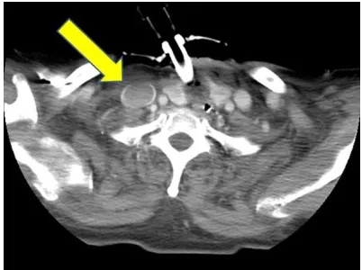
We observed the patient in our intensive care unit for 48 h postoperatively. To check the ALT flap, pinprick and Doppler tests were perfo- rmed. At 68 h postoperatively, we noticed that the right mandibular and neck areas were swol-len and red. When the lesion was found, the Doppler sound was clearly identified, and no abnormal lesion was found by the pin prick test. To evaluate the lesion, we performed trast-enhanced CT and revealed a poorly con-Figure 1. Contrast-enhanced computed
[image:2.612.88.289.71.221.2]trasted area in the right IJV, and IJVT was diag-nosed radiologically (Figure 1). Conversely, no hemorrhage (extravasation), cancerous lesion, or abscess was revealed. A vascular surgeon, pulmonologist, and cardiologist were consult-ed, and IJVT was clinically diagnosconsult-ed, with no thrombi noted in the patient’s body by the whole-body contrast-enhanced CT scan. Follow-up protocol CT scan performed 1, 2, 6, and 12 months after the surgery showed that IJVT has been resolved. Until 1.5 years after the resec-tion, she was conservatively treated using med-icines. Intravenous heparin for one month fol-lowed by warfarin for six months and subse-quent aspirin for one year was administered to the patient. After follow-up of 1 year and 11 months, the patient was doing well with no evi-dence of cancer recurrence or metastatic dis-ease, and the translated flap remained healthy to date. There was no abnormal event related to IJVT. The clot gradually became smaller, and no other event has occurred to date.
Written informed consent was obtained from the patient for publication of this case report and all accompanying images. The Ethics Co- mmittee of the University of the Ryukyus waived the requirement for review per institutional pro-tocol, as the study did not contain content that requires ethical approval, and approved the submission and publication of this case report. Discussion
We found two important issues in this case: (I) To our knowledge, this is the first case report of IJVT after combined SOHND and ALT flap recon-struction using the IJV, and (II) CT scan clearly detected IJVT within 1 week postoperatively. We performed a literature search using PubMed and Google Scholar articles published from 1906 [16] to 2018 using the following termino-logical combinations: “internal jugular vein; in- ternal jugular venous; jugular vein”, AND “thr- ombosis; stenosis; occlusion; patency”, AND, “neck dissection; supraomohyoid neck dissec-tion; upper neck dissecdissec-tion; selective neck dis-section; elective neck dissection”. We excluded non-English literature or English conference proceedings. We found two cases of IJVT asso-ciated with SOHND [17, 18]. However, the cur-rent combination (IJVT after SOHND and ALT using the IJV) was not found. For patients with H&N cancer, ND is a standard method for
radi-cal treatment [2]. Of the several types of NDs, SND is a conservative method and has been replacing radical ND [19]. SOHND is a type of SND that is selected in cases of limited local invasive H&N cancer to preserve function and minimize the esthetic deformity [20]. Recon- struction of the defect is also important to restore function and esthetics, as well as to control the cancer [6]. Therefore, flap failure of reconstruction surgery should be avoided [21]. In H&N reconstruction, many types of flaps have been reported [22]. Of those, the combi-nation of ALT and IJV is used frequently in ND for three reasons: (I) ALT has a high flap survival rate (97.8%) compared with other flaps used for H&N reconstruction [23]. (II) The IJV can be used freely for end-to-end or end-to-side anas-tomosis even when there are no branches [7]. Many reports described use of the IJV [3, 24-26]. (III) The IJV is used mostly during ND (including SOHND) and reconstruction because it has a small risk of venous twist and a large diameter with reliable venous blood flow [7, 20, 27]. Use of the ALT was first described by Song and colleagues [28], and the flap has been used worldwide because of its reliability and less donor site morbidity [5, 23]. The ALT flap is versatile and ideal for H&N reconstruction; however, flap compromise and failure cannot be avoided even in the hands of experienced surgeons [5, 29]. Moreover, Wong and col-leagues [30] performed a multivariate analysis and reported that flap failure occurs significant-ly more often at the H&N site than at other sites in the body. One reason for flap failure is vascu-lar thrombosis [6].
cancer-associ-ated thrombosis by increasing coagulability) [26, 36, 37]. Therefore, treatment of H&N can-cer using techniques such as ND and/or recon-struction surgery has many risks for IJVT. As described above, IJVT leads not only to flap fail-ure but also to life-threatening PE. For example, in a study of 300 patients with upper-extremity deep venous thrombosis, approximately 2% resulted in PE [38]. In cases of IJVT, the patient should be continuously and closely monitored postoperatively owing to a significant risk of developing PE [17]. Actually, PE is one of the reasons of sudden death after ND [34].
Our second important finding was that CT clear-ly detected IJVT. After any type of ND and/or reconstruction surgery for H&N cancer patients, the clinician should detect IJVT for three re- asons.
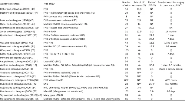
First, IJVT can occur regardless of the type of ND. Overall, the IJVT rate after ND (radical, modified, or functional neck dissection [FND]) is well described [21, 39, 40]. However, the IJVT rate after SND was less reported. To date, some reports described the rate of the IJV occlusion or IJVT; however, the occlusion and thrombosis are confused [15, 41, 42]. Basically, an occlusion is defined as the result of a throm-bosis [29]. Therefore, to distinguish “occlusion” and “thrombosis”, we accurately report them in Table 1 with a literature review as defined above [3, 10, 12-15, 21, 35, 39-53]. IJVT can occur at various rates after ND [54]. Table 1 indicates that 0%-30.4% of IJVT occurred after ND. In regards to SND, Hudgins and colleagues [14] reported a 20% rate of IJVT after SND; however, the sample size in the study was small (8 of 26 cases). Quraishi and colleagues [13] described the IJVT rate after FND or SND, but the rate was not distinguished between FND and SND. Cappiello and colleagues [43] report-ed lateral ND as SND [43]. However, those reports are small and limited to describing the association between SND and IJVT. Abovem- entioned reports did not define the IJVT rate after SND [13, 14, 43]. Notably, Harada and colleagues [12] reported that the caliber of the IJV reduces but gradually increases after 1 and 3 months of any other type of ND (including SOHND). Small caliber (vein narrowing) is a risk for IJVT [35, 44]. Therefore, the results of Harada and colleagues [12] can be postulated to indicate that IJVT can occur regardless of the type of ND. So, as described above, the
clini-cian should prevent or detect IJVT after any type of ND.
Second, IJVT possibly may be asymptomatic. In our patient, cervical swelling was the key symp-tom for detecting IJVT, and subsequent con-trast-enhanced CT confirmed it. Cervical swell-ing (edema), pain, and flap congestion are the main clinical manifestations of IJVT [9, 45]. However, the symptoms are not specific for IJVT but are commonly observed after ND or recon-struction surgery [21, 46]. By contrast, edema is not always associated with IJVT [55], and clinical manifestations are not observed in all IJVT patients [8]. However, IJVT has serious risks as indicated above. Therefore, the clini-cians should notice IJVT whether the clinical symptom manifests or not.
Thirdly, IJVT is difficult to prevent. In a system-atic review and meta-analysis, Lee and Mun [11] found little evidence that use of antithrom-botics reduces the risk of thrombosis or flap failure. Moreover, the authors also reported that the risk of using routine antithrombotics may exceed their benefits [11]. For example, after reconstruction surgery, hematoma, bleed-ing, and need for a blood transfusion are com-mon complications [5, 34, 56]. The antithrom-botics for prevention of thrombosis conversely cause other complications. Therefore, like in our case, antithrombotics are not routinely used [57]. Further, preoperative evaluation of the IJV could not predict IJVT [58], so there is a possibility that IJVT is inevitable [59].
Table 1. Reported studies of IJVT or IJV occlusion after ND
Author/References Type of ND Number of veins occlusion (%)Rate of IJV IJVT (%)Rate of Time between the surgery to occurrence of IJVT Fisher and colleagues (1988) [40] FND 14 14.3 NA (-)
Docherty and colleagues (1993) [44] FND + radiotherapy (16 cases also underwent RS) 20 40 NA (-) FND (2 cases also underwent RS) 8 0 NA (-) Lake and colleagues (1994) [47] FND (some cases underwent RS) 35 2.9 NA (-) Cotter and colleagues (1994) [48] Modified RND (4 cases also underwent RS) 79 14 NA (-) Leontsinis and colleagues (1995) [39] FND (4 cases also underwent RS) 27 NA 29.6 NA Zohar and colleagues (1995) [49] FND or RND 31 12.9 3.2 14 months Quraishi and colleagues (1997) [13] FND or SND (some cases underwent RS) 81 NA 24.7 1 day
FND or SND (some cases underwent RS) 72 NA 26.4 7 days Wax and colleagues (1997) [50] FND + RS 43 NA 18.6 1 and 7 days Brown and colleagues (1998) [21] Modified ND (16 cases also underwent RS) 29 NA 13.8 1-2 weeks Güney and colleagues (1998) [51] FND 133 NA 0 (-) Katou and colleagues (1998) [45] FND + RS or FND + RND + RS 38 0 2.6 7 days Prim and colleagues (2000) [41] FND 54 NA 0 (-) Cappiello and colleagues (2002) [43] Lateral ND (SND) 50 4 0 (-) de Bree and colleagues (2002) [15] Modified RND or SOHND or Anterolateral ND (all cases underwent RS) 23 NA 30.4 1 day-12.5 months Kubo and colleagues (2002) [3] FND + RS 58 6.9 3.4 2 and 8 days (2 cases) Yucel and colleagues (2003) [52] FND or modified radical ND type III 36 NAa 0 (-)
Harada and colleagues (2003) [12] Modified RND or SOHND (39 necks also underwent RS) 76 NAb 0 (-)
Miyasaka and colleagues (2005) [10] FND followed by RS 632 NAc 0.6 4-25 hours
Hudgins and colleagues (2005) [42] SND 28 NAd 21.4d 4-50 months
Nagata and colleagues (2006) [14] RND or modified RND or SOHND (21 necks also underwent RS) 29 3.4 NA (-) Fukuiwa and colleagues (2008) [53] ND + RS (ND type was not mentioned.) 102 NA 2.9 2-7 days Teymoortash and colleagues (2010) [46] Many types of SND 98 NA 0 (-) Makiguchi and colleagues (2015) [35] Modified RND or Extended-SOHND (Level I-IV). 37 necks also underwent RS 81 7.4 0 (-)
CT can assess intrathoracic lesions (such as those in the lungs) more accurately than ultra-sonography [60]. (II): CT can detect thromboses for a wide body range, because IJVT has the possibility for causing other site thromboses [8]. (III): CT can clearly evaluate the diameter of the vessel [36, 40]. In contrast, detection using ultrasonography depends on various parame-ters such as patient’s obesity, edema, wounds, overlying bandages, as well as the clinician’s technical experience [65]. As described above, CT is a convenient tool for detection of IJVT. However, there is no accurate protocol to de- tect IJVT after ND or reconstruction surgery for H&N cancer. By contrast, a defined protocol to detect the cancer exists [2]. IJVT can occur ear-lier because the immediate complication or reaction (such as wound infection, compres-sion of the IJV by flap or hematoma, or fistula, which are the causes of IJVT) occurs immedi-ately postoperatively [3, 54, 66]. Further, it takes a long time (4 days) from the first clinical symptom to diagnosis of IJVT [9]. Sometimes, the thrombosis does not affect the vein paten-cy or flap failure [50, 61]; however, IJVT can cause sudden death after ND [50]. Therefore, IJVT should be detected at the earliest time point with CT.
This study has several limitations. First, current conclusions are based on only one case report, which limits the generalizability of findings. Second, this is a retrospective study as most studies on thrombosis following the flap opera-tion of head and neck surgery are retrospective in nature [11]. Therefore, we believe that fur-ther prospective studies and accumulation of reported cases is warranted to establish a new protocol for detecting IJVT after cancer sur- gery.
In conclusion, we describe an extremely rare case of IJVT, and based on the observations, we suggest that CT should be performed within a week after ND and/or reconstruction surgery for H&N cancer to detect IJVT at the earliest time point. Because routine CT performed for patients with H&N cancer is very late for detect-ing IJVT safely, patient prognosis becomes worse due to pulmonary embolism or flap fa- ilure.
Acknowledgements
No funding support was received for the con-duct of this study and writing of this case
report. We thank Dr. Shoshin Yamazato, De- partment of Infectious Diseases, Respiratory and Digestive Medicine (The First Department of Internal Medicine), Graduate School of Me- dicine, University of the Ryukyus (Japan), and Dr. Shoichiro Yamazato, Department of Cardi- ovascular Medicine, Nephrology and Neurology (The Third Department of Internal Medicine), Graduate School of Medicine, University of the Ryukyus (Japan), who contributed to the pat- ient’s care. In addition, the authors would like to thank Enago (www.enago.jp) for the English language review of this study.
Disclosure of conflict of interest
None.
Address correspondence to: Tessho Maruyama, Department of Oral and Maxillofacial Surgery, Un- iversity Hospital of The Ryukyus, 207 Uehara, Ni- shihara, Okinawa 903-0215, Japan; Molecular Microbiology Group, Tropical Biosphere Research Center, University of The Ryukyus, Nishihara, Okinawa, Japan. Tel: +81 98 895 1192; Fax: +81 98 895 1431; E-mail: [email protected]
References
[1] Ferlay J, Soerjomataram I, Dikshit R, Eser S, Mathers C, Rebelo M, Parkin DM, Forman D and Bray F. Cancer incidence and mortality worldwide: sources, methods and major pat-terns in GLOBOCAN 2012. Int J Cancer 2015; 136: E359-386.
[2] National Comprehensive Cancer Network: He- ad and Neck Cancers Version 1. 2018. https:// www.nccn.org/professionals/physician_gls/ pdf/head-and-neck.pdf Accessed April 18, 2018.
[3] Kubo T, Haramoto U, Yano K, Kakibuchi M, Ta- kagi S, Nakai K, Sakai Y, Inohara H and Hos- okawa K. Internal jugular vein occlusion in head and neck microsurgical reconstruction. Ann Plast Surg 2002; 49: 490-494.
[4] Kerawala CJ and Heliotos M. Prevention of complications in neck dissection. Head Neck Oncol 2009; 1: 35.
[5] Wu CC, Lin PY, Chew KY and Kuo YR. Free tis-sue transfers in head and neck reconstruction: complications, outcomes and strategies for management of flap failure: analysis of 2019 flaps in single institute. Microsurgery 2014; 34: 339-344.
[7] Kiya K, Kubo T, Seike S and Hosokawa K. Free flap survival despite internal jugular vein th- rombosis in head and neck reconstruction. Plast Reconstr Surg Glob Open 2018; 6: e1647.
[8] Gbaguidi X, Janvresse A, Benichou J, Cailleux N, Levesque H and Marie I. Internal jugular vein thrombosis: outcome and risk factors. QJM 2011; 104: 209-219.
[9] Leci-Tahiri L, Zherka-Saracini H, Tahiri A and Koshi A. Bilateral internal jugular vein throm-bosis due to malignant tumor. J Med Case Rep 2018; 12: 42.
[10] Miyasaka M, Ichikawa K, Nishimura M, Yama- zaki A, Taira H, Imagawa K and Tanino R. Sal- vage operations of free tissue transfer follow-ing internal jugular venous thrombosis: a re-view of 4 cases. Microsurgery 2005; 25: 191-195.
[11] Lee KT and Mun GH. The efficacy of postopera-tive antithrombotics in free flap surgery: a sys-tematic review and meta-analysis. Plast Reconstr Surg 2015; 135: 1124-1139.
[12] Harada H, Omura K and Takeuchi Y. Patency and caliber of the internal jugular vein after neck dissection. Auris Nasus Larynx 2003; 30: 269-272.
[13] Quraishi HA, Wax MK, Granke K and Rodman SM. Internal jugular vein thrombosis after functional and selective neck dissection. Arch Otolaryngol Head Neck Surg 1997; 123: 969-973.
[14] Hudgins PA, Kingdom TT, Weissler MC and Mukherji SK. Selective neck dissection: CT and MR imaging findings. AJNR Am J Neuroradiol 2005; 26: 1174-1177.
[15] de Bree R, van den Berg FG, van Schaik C, Beerens AJ, Manoliu RA, Castelijns JA, Snow GB and Leemans CR. Assessment of patency of the internal jugular vein following neck dis-section and microvascular flap reconstruction by power Doppler ultrasound. J Laryngol Otol 2002; 116: 622-626.
[16] Crile G. Excision of cancer of the head and neck. With special reference to the plan of dis-section based on one hundred and thirty-two operations. JAMA 1906; 47: 1780-1786. [17] Castling B, Fowell C and Bhatia S. Intra-op-
erative internal jugular vein thrombosis compli-cating microvascular free flap transfer. Int J Oral Maxillofac Surg 2012; 41: 1229-1231. [18] Segna E, Bolzoni AR, Baserga C and Baj A. Free
flap loss caused by heparin-induced thrombo-cytopenia and thrombosis (HITT): a case report and literature review. Acta Otorhinolaryngol Ital 2016; 36: 527-533.
[19] Khafif A. Lateral neck dissection. Operative Te- chniques in Otolaryngology 2004; 15: 160-167.
[20] Yagi S, Suyama Y, Fukuoka K, Takeuchi H and Kitano H. Recipient vessel selection in head and neck reconstruction based on the type of neck dissection. Yonago Acta Med 2016; 59: 159-162.
[21] Brown DH, Mulholland S, Yoo JH, Gullane PJ, Irish JC, Neligan P and Keller A. Internal jugular vein thrombosis following modified neck dis-section: implications for head and neck flap reconstruction. Head Neck 1998; 20: 169-174.
[22] Corbitt C, Skoracki RJ, Yu P and Hanasono MM. Free flap failure in head and neck reconstruc-tion. Head Neck 2014; 36: 1440-1445. [23] Bianchi B, Ferri A, Ferrari S, Copelli C, Boni P,
Ferri T and Sesenna E. The free anterolateral thigh musculocutaneous flap for head and neck reconstruction: one surgeon’s experi-ence in 92 cases. Microsurgery 2012; 32: 87-95.
[24] Chalian AA, Anderson TD, Weinstein GS and Weber RS. Internal jugular vein versus external jugular vein anastamosis: implications for suc-cessful free tissue transfer. Head Neck 2001; 23: 475-478.
[25] Yazar S. Selection of recipient vessels in micro-surgical free tissue reconstruction of head and neck defects. Microsurgery 2007; 27: 588-594.
[26] Felstead AM and Perkins CS. Thrombosis of the internal jugular vein: a rare but important operative finding. Br J Oral Maxillofac Surg 2010; 48: 195-196.
[27] Gong ZJ, Chen YR, Wang K, Zhang S, Ren ZH and Wu HJ. Longitudinal contraction venoplas-ty in prevention of internal jugular vein throm-bosis after free flap vascular anastomosis. J Oral Maxillofac Surg 2016; 74: 1277-1283. [28] Song YG, Chen GZ and Song YL. The free thigh
flap: a new free flap concept based on the sep-tocutaneous artery. Br J Plast Surg 1984; 37: 149-159.
[29] Yang Q, Ren ZH, Chickooree D, Wu HJ, Tan HY, Wang K, He ZJ, Gong CJ, Ram V and Zhang S. The effect of early detection of anterolateral thigh free flap crisis on the salvage success rate, based on 10 years of experience and 1072 flaps. Int J Oral Maxillofac Surg 2014; 43: 1059-1063.
[30] Wong AK, Joanna Nguyen T, Peric M, Shahabi A, Vidar EN, Hwang BH, Niknam Leilabadi S, Chan LS and Urata MM. Analysis of risk factors associated with microvascular free flap failure using a multi-institutional database. Micros- urgery 2015; 35: 6-12.
[32] Ahmed N. Thrombosis after central venous cannulation. Med J Aust 1976; 1: 217-220. [33] Togashi Y, Kim YH, Masago K, Tamai K, Sa-
kamori Y, Mio T and Mishima M. Pulmonary embolism due to internal jugular vein thrombo-sis in a patient with non-small cell lung cancer receiving bevacizumab. Int J Clin Oncol 2011; 16: 444-446.
[34] Gueret G, Cosset MF, McGee K, Luboinski FB and Bourgain JL. Sudden death after neck dis-section for cancer. Ann Otol Rhinol Laryngol 2002; 111: 115-119.
[35] Makiguchi T, Yokoo S, Ogawa M and Miyazaki H. Factors influencing internal jugular vein pa-tency after neck dissection in oral cancer. Int J Oral Maxillofac Surg 2015; 44: 1218-1224. [36] Sharma P, Kumar R, Singh H, Jeph S, Patnecha
M, Reddy RM, Naswa N, Bal C and Malhotra A. Imaging thrombus in cancer patients with FDG PET-CT. Jpn J Radiol 2012; 30: 95-104. [37] Ikushima S, Ono R, Fukuda K, Sakayori M, Aw-
ano N and Kondo K. Trousseau’s syndrome: cancer-associated thrombosis. Jpn J Clin Oncol 2016; 46: 204-208.
[38] Levy MM, Albuquerque F and Pfeifer JD. Low incidence of pulmonary embolism associated with upper-extremity deep venous thrombosis. Ann Vasc Surg 2012; 26: 964-972.
[39] Leontsinis TG, Currie AR and Mannell A. In- ternal jugular vein thrombosis following func-tional neck dissection. Laryngoscope 1995; 105: 169-174.
[40] Fisher CB, Mattox DE and Zinreich JS. Patency of the internal jugular vein after functional neck dissection. Laryngoscope 1988; 98: 923-927.
[41] Prim MP, de Diego JI, Fernández-Zubillaga A, García-Raya P, Madero R and Gavilán J. Pa- tency and flow of the internal jugular vein aft- er functional neck dissection. Laryngoscope 2000; 110: 47-50.
[42] Nagata T, Matsunaga K, Kawazu T, Kawano S, Oobu K and Ohishi M. Patency assessment of the internal jugular vein after neck dissection. Int J Oral Maxillofac Surg 2006; 35: 416-420. [43] Cappiello J, Piazza C, Berlucchi M, Peretti G,
De Zinis LO, Maroldi R and Nicolai P. Internal jugular vein patency after lateral neck dissec-tion: a prospective study. Eur Arch Otorhino- laryngol 2002; 259: 409-412.
[44] Docherty JG, Carter R, Sheldon CD, Falconer JS, Bainbridge LC, Robertson AG and Soutar DS. Relative effect of surgery and radiotherapy on the internal jugular vein following functional neck dissection. Head Neck 1993; 15: 553-556.
[45] Katou F, Echigo S, Ito M, Shirai N, Ohtani H and Motegi K. Reliability of internal jugular vein in oral microvascular reconstruction. Oral Surg
Oral Med Oral Pathol Oral Radiol Endod 1998; 86: 529-533.
[46] Teymoortash A, Hoch S, Eivazi B and Werner JA. Postoperative morbidity after different ty- pes of selective neck dissection. Laryngoscope 2010; 120: 924-929.
[47] Lake GM 3rd, DiNardo LJ and Demeo JH. Performance of the internal jugular vein after functional neck dissection. Otolaryngol Head Neck Surg 1994; 111: 201-204.
[48] Cotter CS, Stringer SP, Landau S, Mancuso AA and Cassisi NJ. Patency of the internal jugular vein following modified radical neck dissec-tion. Laryngoscope 1994; 104: 841-845. [49] Zohar Y, Strauss M, Sabo R, Sadov R, Sabo G
and Lehman J. Internal jugular vein patency after functional neck dissection: venous du-plex imaging. Ann Otol Rhinol Laryngol 1995; 104: 532-536.
[50] Wax MK, Quraishi H, Rodman S and Granke K. Internal jugular vein patency in patients under-going microvascular reconstruction. Laryngo- scope 1997; 107: 1245-1248.
[51] Güney E, Yiğitbaşi OG, Canöz K, Oztürk M and Ersoy A. Functional neck dissection: cure and functional results. J Laryngol Otol 1998; 112: 1176-1178.
[52] Yucel EA, Orhan KS, Guldiken Y, Aydin K, Si- msek T, Erdamar B and Deger K. Evaluation of factors concerning the patency of the internal jugular vein after functional neck dissection. Eur Arch Otorhinolaryngol 2003; 260: 35-38. [53] Fukuiwa T, Nishimoto K, Hayashi T and Kurono
Y. Venous thrombosis after microvascular free-tissue transfer in head and neck cancer recon-struction. Auris Nasus Larynx 2008; 35: 390-396.
[54] Yu P, Chang DW, Miller MJ, Reece G and Robb GL. Analysis of 49 cases of flap compromise in 1310 free flaps for head and neck reconstruc-tion. Head Neck 2009; 31: 45-51.
[55] Chen MH, Chang PM, Chen PM, Tzeng CH, Chu PY, Chang SY and Yang MH. Prolonged facial edema is an indicator of poor prognosis in pa-tients with head and neck squamous cell carci-noma. Support Care Cancer 2010; 18: 1313-1319.
[56] Perisanidis C, Herberger B, Papadogeorgakis N, Seemann R, Eder-Czembirek C, Tamandl D, Heinze G, Kyzas PA, Kanatas A, Mitchell D, Wolff KD and Ewers R. Complications after free flap surgery: do we need a standardized classification of surgical complications? Br J Oral Maxillofac Surg 2012; 50: 113-118. [57] Chang EI, Zhang H, Liu J, Yu P, Skoracki RJ and
[58] Prim MP, De Diego JI, Moreno P, Madero R and Gavilán J. Status of internal jugular veins in pa-tients with carcinomas of the head and neck area. Otolaryngol Head Neck Surg 2004; 131: 494-496.
[59] Okazaki M, Asato H, Okochi M and Suga H. One-segment double vascular pedicled free je-junum transfer for the reconstruction of pha-ryngoesophageal defects. J Reconstr Microsurg 2007; 23: 213-218.
[60] Pata YS, Unal M and Gulhan S. Internal jugular vein thrombosis due to distant malignancies: two case reports and literature review. J La- ryngol Otol 2008; 122: 318-320.
[61] Amato MM, Rodriguez LR and Lineaweaver WC. Survival of free tissue transfer following internal jugular venous thrombosis. Plast Re- constr Surg 1999; 104: 1406-1408.
[62] Evans GR. Indirect documentation of the nega-tive effects of irradiation and neck dissection on the internal jugular vein. Ann Plast Surg 1995; 35: 670.
[63] Thomas S, Sawhney S and Kapur BM. Case re-port: bilateral massive internal jugular vein thrombosis in carcinoma of the thyroid: CT evaluation. Clin Radiol 1991; 43: 433-434. [64] Lin D, Reeck JB and Murr AH. Internal jugular
vein thrombosis and deep neck infection from intravenous drug use: management strategy. Laryngoscope 2004; 114: 56-60.
[65] Rondina MT, Lam UT, Pendleton RC, Kraiss LW, Wanner N, Zimmerman GA, Hoffman JM, Han- rahan C, Boucher K, Christian PE, Butterfield RI and Morton KA. 18F-FDG PET in the evaluation of acuity of deep vein thrombosis. Clin Nucl Med 2012; 37: 1139-1145.