REVIEW ARTICLE
Gadolinium Contrast Agents for CNS Imaging: Current
Concepts and Clinical Evidence
E. Kanal, K. Maravilla, and H.A. Rowley
SUMMARY: The aim of this article was to review the properties of the various gadolinium-based contrast agents used for CNS imaging along with the clinical evidence and published data that highlight the impact these different properties can have on diagnostic perfor-mance. In addition, approaches to optimizing image acquisition that take into account the different properties of specific gadolinium-based contrast agents and an extensive review of the safety profiles of the various agents are presented.
ABBREVIATIONS:CE⫽contrast-enhanced; CNR⫽contrast-to-noise ratio; GBCA⫽gadolinium-based contrast agent; Gd⫽gadolinium; NSF⫽nephrogenic systemic fibrosis; rCBV⫽relative cerebral blood volume; rCBF⫽relative cerebral blood flow
O
f the 9 gadolinium-based contrast agents (GBCAs) ap-proved by the United States FDA for contrast-enhanced (CE) MR imaging, 7 (gadoterate meglumine, Dotarem, Guerbet, Aulnay-sous-Bois, France; gadobutrol, Gadavist, Bayer Health-Care Pharmaceuticals, Wayne, New Jersey; gadopentetate dime-glumine, Magnevist, Bayer HealthCare; gadobenate dimeglu-mine, MultiHance, Bracco Diagnostics, Princeton, New Jersey; gadodiamide, Omniscan, GE Healthcare, Milwaukee, Wisconsin; gadoversetamide, OptiMARK, Covidien, Dublin, Ireland; gad-oteridol, ProHance, Bracco Diagnostics) are approved specifically for CE-MR imaging of the CNS.1-7The 2 agents not approved for CE-MR imaging of the CNS (gadofosveset trisodium, Ablavar, Lantheus Medical Imaging, North Billerica, Massachusetts; and gadoxetic acid, Bayer HealthCare Pharmaceuticals [Eovist or Pri-movist]) have distinct properties that render them unsuitable for this indication; Ablavar8is an intravascular “blood-pool” agent approved for MR angiography of the aortoiliac vessels, whose strong binding to serum albumin (and resulting large effective molecular size) restricts permeability across the open blood-brain barrier, which limits suitability for CNS applications, while Eo-vist9is an approved liver-specific agent inappropriate for CNSapplications because 50% of the injected dose is taken up and eliminated by hepatocytes.
Although numerous studies published in high-ranking peer-reviewed journals have confirmed the safety and efficacy of the 7 GBCAs approved for CNS imaging, differences among these agents and the impact these differences may have on diagnostic sensitivity and clinical decision-making remain underappreciated and sometimes misunderstood.
The aim of this article was to review the properties of the var-ious GBCAs used for CNS imaging together with the clinical evi-dence and published data that highlight the impact these different properties can have on diagnostic performance. In addition, ap-proaches to optimizing image acquisition that take into account the different properties of specific GBCAs and an extensive review of the safety profiles of the various agents will be presented.
For the purposes of the present review, brand names rather than chemical names have been used throughout to refer to the various GBCAs. Although chemical names would ordinarily be used in a review article such as this, it was thought that because the actual practitioners of MR imaging are typically more familiar with brand names than with chemical names, the use of brand names would help avoid the possibility of obfuscation and thereby enhance clarity in this field. Moreover, because generic formula-tions of these agents are not available in the United States, these brand names are clinically relevant designations. We have there-fore elected to specifically use the same brand names for the var-ious agents with which the reader will be most familiar.
GBCAS: WHAT THEY ARE AND WHAT THEY DO
As a heavy metal in the lanthanide series, elemental free gadolin-ium (Gd) is toxic to humans. GBCAs are formed by chelation of gadolinium to organic ligands to decrease its toxicity and render it From Magnetic Resonance Services (E.K.), Department of Radiology, University of
Pittsburgh Medical Center, Pittsburgh, Pennsylvania; Research Laboratory (K.M.), University of Washington, Seattle, Washington; and Departments of Radiology, Neurology, and Neurosurgery (H.A.R.), University of Wisconsin, Madison, Wisconsin.
Please address correspondence to Howard A. Rowley, MD, Department of Radiol-ogy, University of Wisconsin, 600 Highland Ave, Mail code 3252, Madison, WI 53792; e-mail: [email protected]
Indicates open access to non-subscribers at www.ajnr.org
Indicates article with supplemental on-line tables.
safe for human use. Structurally, there are 2 distinct categories of GBCAs: 1) macrocyclic molecules in which the Gd3⫹ion is caged
within the cavity of the ligand, and 2) open-chain or “linear” molecules (On-line Table 1). Within each category, there are both ionic and nonionic GBCAs. Despite their different molecular structures and physicochemical properties, all GBCAs used for CE-MR imaging of the CNS have an identical mechanism of ac-tion involving shortening of the T1, T2, and T2* relaxaac-tion time constants of adjacent water protons in tissues10and similar bio-distribution profiles because all are extracellular agents that do not cross the intact blood-brain barrier. The fundamental ability of GBCAs to selectively shorten T1 relaxation within a lesion com-pared with normal tissue is the basis for the most commonly ap-plied usage of contrast agents in clinical practice today.
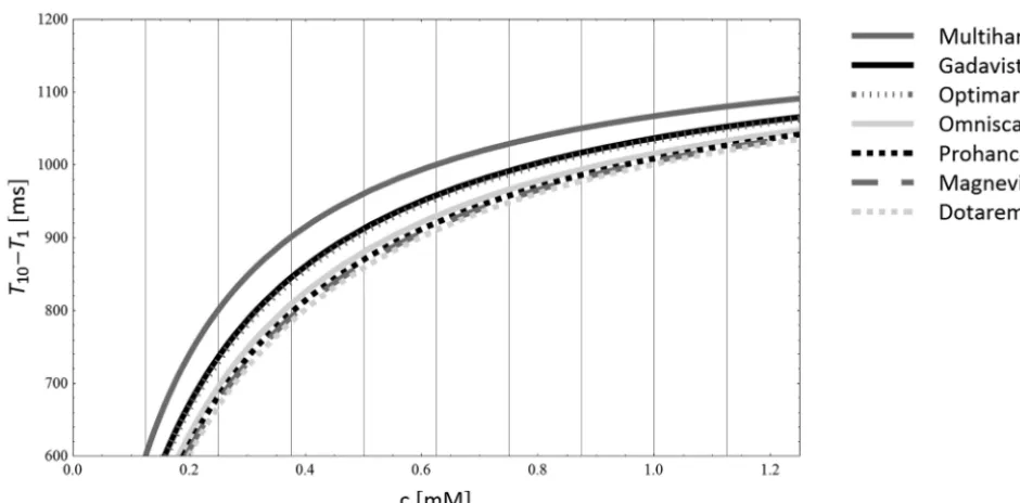
The extent to which the time constants are shortened, and hence the effectiveness of a specific GBCA in clinical practice, depends on the local concentration of the GBCA (Fig 1) and on the relaxivity valuesri(with i⫽1, 2, 2*) of the agent. Relaxivities
are defined as the slope of the linear regression generated from a plot of the measured relaxation rate (1/Ti) versus the concentra-tion of the GBCA:
1 Ti⫽
1
Ti共0兲⫹ri⫻ [GBCA]
whereTidenotes the longitudinal (T1) or transverse (T2, T2*)
relaxation times of a solution containing GBCA andTi(0), the
relaxation time of the solvent without GBCA.11 Agents with higherr1, r2,andr2*values will shorten the T1, T2, and T2* relaxation times of tissues to a greater extent, resulting in greater signal-intensity enhancement on T1-weighted images or greater signal-intensity loss on T2- and T2*-weighted im-ages, respectively.
Molecules normally tumble or rotate in space at a rate that is dependent on many factors, not the least of which is their
molec-ular mass. This tumbling rate can also be represented as the in-verse of the so-called molecular correlation time. The closer the tumbling rate of the gadolinium-containing chelate molecule to the resonant frequency of water molecules, the greater is the re-laxivity or relaxation enhancement. The molecules of each of the 7 FDA-approved CNS gadolinium chelates are all relatively small and therefore have exceedingly high tumbling rates that exceed the resonant frequencies of water molecules at the clinically used MR imaging field strengths. However, as the molecular tumbling rates between the paramagnetic contrast agent and the target wa-ter-based molecules become closer, energy exchange between the 2 molecules is facilitated and longitudinal magnetization is en-hanced (ie, the target tissue T1 shortens).
One of the approved CNS agents, MultiHance, interacts weakly and transiently with serum albumin in vivo. This interac-tion slows the molecular tumbling rate of the complex, rendering it closer to the resonant frequency of water molecules at the field strength at which they are being imaged. This increases the (en-hanced) tissue rotational correlation time (ie, slows its tumbling rate) and therefore brings it closer to matching that of water. The result is greater relaxivities/r1values and stronger relaxation en-hancement effects.3,12,13Thus, ther1of MultiHance measured in human plasma at 37°C has been reported to be as high as 7.9 L⫻ mmol⫺1 ⫻ s⫺1 at 1.5T,14 whereas the reported r1 values of GBCAs that do not significantly interact with serum albumin range from approximately 3.6 L⫻mmol⫺1⫻s⫺1at 1.5T for Dotarem to 5.2 L⫻mmol⫺1⫻s⫺1at 1.5T for Gadavist.11,14 However, reported relaxivity values may vary somewhat between investigating groups due to measurements being made under dif-ferent conditions. For example, ther1andr2values reported by Rohrer et al11were obtained by using nonsterile bovine blood plasma, while those reported by Pintaske et al14were obtained by using human blood plasma.
Six of the 7 GBCAs approved for neuroradiologic imaging are
[image:2.594.54.524.46.278.2]formulated as 0.5 mol/L concentrations, while 1 agent, Gadavist, is formulated at 1.0 mol/L. The physicochemical properties of this latter GBCA resemble those of the other conventional GBCAs, and its molecular structure differs from that of ProHance in that a hydroxy-propyl group on the ProHance molecule is replaced by a trihydroxy-butyl group on the Gadavist molecule.15Thus, the slightly higher relaxivity of Gadavist can be ascribed mainly to the slightly larger size and hence slightly slower molecular tumbling rate of the Gadavist molecule itself. Notably, the magnitude of the slowing of the molec-ular tumbling rate is smaller than that observed for GBCAs approved for neuroradiologic applications that interact with serum albumin as well as non-neuroradiologic agents such as Ablavar for which pow-erful protein binding is observed.
The degree of tissue T1 (and T2 and T2*) shortening by any GBCA is determined by the local concentration of the agent within the tissue being imaged in combination with the relative relaxivity of the agent. Varied concentrations of administered GBCAs can be theorized to result in varied rates of uptake of the agent into the extracellular fluid of the tissues being imaged, with faster tissue uptake expected with a GBCA formulated at a greater concentration. Theoretically, albeit not demonstrated clinically, in routine T1-weighted steady-state imaging, the higher concen-tration of Gadavist might impact the “ideal” postcontrast imaging time for this agent versus others with lower molarity. Recalling, however, that intravenous administration requires that all admin-istered boluses pass through the heart and lungs before being distributed to the rest of the body, it would seem that any concen-tration difference per time among the various neuroradiologic GBCAs, other than possibly first-pass effects, would be quite small and likely not clinically discernable in steady-state. In addition, one should recognize that even if the contrast agent may theoret-ically accumulate faster in the target lesion, enhancing back-ground tissues would also be expected to similarly accumulate that same amount more rapidly. Notwithstanding these theoretic considerations, in daily clinical practice, it appears that the ulti-mate steady-state concentrations in the tissues being imaged are sufficiently similar for all GBCAs regardless of the administered concentration and that the final concentration in tissues in steady-state is most likely determined predominantly by the total administered dose, based on the similarity of observed contrast enhancement when corrected for differences in relative relaxivity of the different agents as discussed later in this review. Thus, for routine, equidose, postcontrast T1-weighted steady-state imag-ing, enhanced signal intensities are dominated predominantly by the relative relaxivity values of the agents being compared. There-fore, the higher ther1value, the greater will be the contrast be-tween the lesion and nonenhancing background tissues.
Dynamic phase imaging (as is used in perfusion-weighted im-aging and in contrast-enhanced MR angiographic studies) differs from the above in that the target tissue of interest is the blood— the same “tissue” into which the agent is directly injected. Thus, per the equation above, higher GBCA concentration and/or higher relaxivity could each produce increases in signal intensity of the intravascular blood for T1-weighted imaging, or greater signal loss for T2- or T2*-weighted imaging. For dynamic bolus CE-MRA examinations, a principal objective is to time the bolus arrival within the vessels of interest to coincide with acquisition of
the lowest order phase-encoding steps ink-space to obtain max-imum vessel enhancement. Because all 7 GBCAs approved for neuroradiologic imaging are approved at identical administered total doses of 0.1 mmol/kg body weight, this means that the higher the concentration of the administered agent, the smaller the vol-ume injected. Thus, a standard administered dose to a 70-kg pa-tient would be 14 mL of any of the 6 GBCAs formulated at 0.5 mol/L, while it would be 7 mL for Gadavist formulated at 1.0 mol/L. However, the result of a smaller injected volume is that the timing window of opportunity during which to catch the higher concentration Gadavist in the vessels of interest is shortened by as much as one-half, making bolus timing more difficult and poten-tially more error-prone. This can possibly be offset by halving the injection rate when administering higher concentration agents or by diluting them with normal saline before administration. How-ever, these approaches would negate any theoretic advantage of using a higher concentration agent.
The relatively recent FDA approval of GBCAs formulated at dif-ferent concentrations but approved at identical administered doses provides opportunities for comparison among such agents. How-ever, such attempts at comparison should use identical FDA-ap-proved doses; comparisons should not be performed by using arbi-trarily defined equivalent volumes because this would result in “double-dosing” of the higher concentration agent. Such studies evaluating agents of different concentrations would be useful because the ideal time to image is dependent on many factors, which include blood vessel size, flow rate and transit time in the case of CE-MRA, perfusion dynamics (vascularity and permeability) of the anatomy/ pathology to be imaged in the case of perfusion MR, the concentra-tion and relaxivities of the administered agent, and the design of the MR imaging pulse sequence to be used to acquire the postcontrast images. Furthermore, “ideal” timing will also be highly dependent on whether first-pass, dynamic contrast information is sought as op-posed to steady-state images. “Ideal” timing tends to be spread over far longer windows of opportunity for steady-state acquisitions, while the window for first-pass, dynamic contrast information is quite short and is far more significantly impacted by such factors as the manner in whichk-space is filled and the temporal resolution of the acquired images. Imaging parameters and timing that might be ideal for one agent might prove to be quite different for another GBCA with different concentration or relaxivity.
CLINICAL EVIDENCE FOR THE ROLE OF RELAXIVITY
ON DIAGNOSTIC PERFORMANCE
Methodologic Considerations
Direct comparison among different agents is accomplished more objectively with an intraindividual crossover study in which each patient receives both GBCAs in random order in 2 identical MR imaging examinations separated by just a few days. Such studies are better designed to isolate the GBCA as the only variable being assessed. Thus, variations due to patient-, disease-, or ex-amination-related factors are eliminated or, at least, minimized. In such studies, any differences in findings can be attributed solely to the GBCA because all other variables are identical for the 2 examinations.
Summarized below are the findings of several such intraindi-vidual crossover studies performed in human subjects for CNS evaluation of various GBCAs. In organizing this discussion, we realized that there is no standardized definition for use of the term, “high relaxivity.” It is the opinion of these authors that “high relaxivity” should be defined not numerically alone, but rather by an objectively proved ability of an agent to deliver in-creased clinical utility as measured by clinically relevant increases in signal enhancement (rather than merely small but statistically significant signal increases) or, preferably, an objectively mea-sured increase in lesion number or lesion extent compared with other “standard” GBCAs.
Steady-State Imaging. To analyze the possible differential roles of relaxivity and concentration in imaging performance, findings are presented for studies that compared the following: 1) Dota-rem, Magnevist, Omniscan, OptiMARK, and ProHance (which all possess similar [standard] relaxivity values and are formulated at standard [0.5 mol/L] concentrations), 2) Gadavist (the GBCA with slightly higher relaxivity and the highest concentration [1 mol/L]), and 3) MultiHance (the GBCA with the highest relaxiv-ity and formulated at standard concentration).
1) Intraindividual Crossover Studies Comparing GBCAs with Stan-dard Relaxivity/StanStan-dard Concentration (Dotarem, Magnevist, Omniscan, OptiMARK, ProHance). To the authors’ knowledge, Greco et al26performed the only intraindividual crossover study that compared GBCAs with standard relaxivity/standard concen-tration. In that study, 2 blinded readers intraindividually com-pared Magnevist (r1-relaxivity at 1.5T: 3.9 – 4.1 L⫻mmol⫺1⫻ s⫺1)11,14with ProHance (r1-relaxivity at 1.5T: 4.1 L⫻mmol⫺1⫻ s⫺1measured in bovine plasma at 37°C)11in 80 subjects for the presence of disease, degree of enhancement, location and number of lesions, and additional information gained (definition of lesion borders, improved visualization, distinction of edema, disease classification, determination of recurrent tumor, and so forth). Neither reader noted any significant differences in terms of GBCA preference (readers 1 and 2 preferred ProHance over Magnevist in 2 and 4 cases, respectively, and Magnevist over ProHance in 1 and 2 cases, respectively), and no differences were noted between agents in terms of the additional information provided on post-contrast images (On-line Table 2).
2) Intraindividual Crossover Studies Comparing a GBCA with Slightly Higher Relaxivity and High Concentration versus Standard Relaxiv-ity/Standard Concentration Agents (Gadavist versus Either Dota-rem, Magnevist, Omniscan, OptiMARK, or ProHance). To date, 5 published reports have described intraindividual crossover com-parisons of Gadavist with standard relaxivity GBCAs.27-31Three
of these looked specifically at the potential benefit of Gadavist versus Magnevist27,28or ProHance29for the detection and visu-alization of cerebral metastases and concluded, in each case, that Gadavist is advantageous for lesion detection primarily because of improved lesion conspicuity (On-line Table 2). However, the conclusions in a study by Anzalone et al27were based solely on subjective assessment of images from 27 patients by 2 neuroradi-ologists in consensus, but unfortunately, no quantitative assess-ment of lesion enhanceassess-ment was reported. Kim et al28reported improved quantitative enhancement (lesion/brain contrast-to-noise ratio [CNR]) with Gadavist, but this study was retrospective and only compared double (0.2 mmol/kg body weight) doses of Gadavist and Magnevist. Furthermore, GBCA administration was not random (all patients received Magnevist for the first exami-nation and Gadavist for the second). In the third study in patients with brain metastases, Katakami et al29evaluated a larger number of patients by using a prospective design and concluded that a single 0.1-mmol/kg dose of Gadavist is noninferior to a double 0.2-mmol/kg dose of ProHance for lesion detection. However, despite administering a single 0.1-mmol/kg dose of ProHance as part of the study design, no assessment of single-dose ProHance images was performed. Thus, it is not possible to say whether a single dose of ProHance would have proved noninferior to a sin-gle dose of Gadavist by using their study design, sample size, and assessment methodology.
In a more recent single-center study in 51 patients with either primary or secondary brain tumors, 2 blinded readers each pre-ferred Gadavist to ProHance in more patients in terms of subjec-tive “preference in contrast enhancement,” “overall preference,” and “preference in diagnostic quality.”30However, differences in quantitative enhancement were inconsistent and sequence-de-pendent, with a higher SNR for Gadavist noted only on a second T1-FLASH sequence at approximately 10 minutes postinjection, with no differences between agents seen on T1-spin echo or MPRAGE sequences. Indeed, Bayer HealthCare (manufacturer of gadobutrol, Gadavist) reported to the FDA that the performances of 0.1-mmol/kg doses of Gadavist and ProHance for brain tumor imaging are similar.2,31In a prospective multicenter phase III study performed in 419 patients for the FDA approval of Gadavist for CNS imaging, 3 blinded readers each reported similar contrast enhancement, lesion border delineation, and lesion internal mor-phology and a similar overall accuracy of diagnosis when these 2 agents were administered at an equivalent dose of 0.1 mmol/kg body weight. The conclusion of the study was that Gadavist is noninferior to ProHance.2,31
that following Dotarem, as expected from the differences in their respective relaxivities, but this yielded no significant difference between the 2 agents for measured CNR. Most important, no differences in the number of lesions detected with either agent were observed.
3a) Intraindividual Crossover Studies Comparing the GBCA with the Highest Relaxivity/Standard Concentration versus GBCAs with Standard Relaxivity/Standard Concentration (MultiHance versus Dotarem, Magnevist, Omniscan, OptiMARK, or ProHance). Nu-merous multicenter studies have compared MultiHance with standard GBCAs by using an intraindividual crossover study de-sign with blinded image evaluation by fully independent experi-enced neuroradiologists.33-41All of these studies were designed to demonstrate superiority rather than noninferiority, and the find-ings of all concluded that MultiHance is significantly superior in terms of both qualitative enhancement (global diagnostic prefer-ence, lesion border delineation, definition of disease extent, visualization of lesion internal morphology, lesion contrast en-hancement) and quantitative enhancement (CNR, lesion-to-background ratio) (On-line Table 2). In each of these studies, the authors concluded that the superiority of MultiHance was due to its higherr1value.
3b) Intraindividual Crossover Study to Compare the Highest Re-laxivity GBCA versus the GBCA Formulated at the Highest Con-centration (MultiHance versus Gadavist). A recent study by Seidl et al41directly addressed the relative merits of high relaxivity ver-sus high gadolinium concentration. In their randomized, double-blind, intraindividual crossover study, 123 patients each under-went 1 examination with 0.1-mmol/kg MultiHance and 1 examination with 0.1-mmol/kg Gadavist. Three blinded readers consistently demonstrated a highly significant (P⬍.0001) pref-erence for MultiHance for all qualitative end points with good interreader agreement for all evaluations (On-line Table 2). In addition, significant superiority was noted for all quantitative as-sessments with a mean difference of approximately 22% in per-centage lesion enhancement between MultiHance and Gadavist.
This study demonstrated that gadolinium concentration has little-to-no practical clinical impact on steady-state morphologic imaging and that at identical approved (0.1 mmol/kg) doses, the relaxivity of the GBCA is the dominant characteristic determining the degree of enhancement.
Perfusion Imaging. Cerebral perfusion assessment by dynamic susceptibility contrast MR imaging is frequently used for evalua-tion of brain tumors, stroke, and degenerative diseases such as dementia. The technique is based on rapid intravenous injection of a GBCA and subsequent bolus tracking by using a fast suscep-tibility-weighted imaging sequence that uses the T2* relaxing properties of the GBCA. Following tracer kinetic modeling, para-metric maps of mean transit time, regional cerebral blood volume (rCBV), and regional cerebral blood flow (rCBF) can be calcu-lated by unfolding tissue concentration curves and the concentra-tion curve of the feeding artery.
Compared with conventional morphologic (static) imaging, (dynamic) perfusion imaging is more dependent on the shape of the injected contrast bolus and thus on the rate at which GBCAs are injected. Additionally, higher administered GBCA
concentra-tion and higher relaxivity might each be beneficial in augmenting the signal loss associated with the first-pass contrast bolus through the tissues of interest.
Although interindividual parallel group studies have com-pared Gadavist with Magnevist at 1.5T,42,43comparatively few intraindividual crossover studies have been performed to com-pare GBCAs for perfusion imaging. Those that have been per-formed have compared Gadavist with either Magnevist at 3T44or MultiHance at 1.5T45or 3T.46,47
1) Inter- and Intraindividual Crossover Studies Comparing GBCAs with Standard Relaxivity/High Concentration versus Standard Relax-ivity/Standard Concentration (Gadavist versus Magnevist). An in-terindividual parallel group comparison of Gadavist with Magn-evist was first performed by Griffiths et al,42who compared 10-and 20-mL injections of Gadavist with 20-mL injections of Magn-evist (all at 5 mL/s, resulting in overall injection times of 2 and 4 seconds, respectively) in 6 groups of 6 patients (36 patients over-all) at 1.5T to determine whether the higher Gd concentration of Gadavist was beneficial when using thinner sections (4 mm as opposed to 7 mm) for single-shot, gradient-recalled echo-planar imaging. They compared time-intensity curves calculated at re-gions of interest in the hemispheric white matter and thalamus in terms of maximum signal reduction (ie, the difference between mean baseline and minimum value on the time-intensity curve), full width at half minimum, and signal-to-noise measurements. No significant differences were found between 20 mL of Magnev-ist and 10 mL of GadavMagnev-ist in terms of the maximum signal changes observed in either anatomic area and at either section thickness. On the other hand, the signal changes nearly doubled when 20-mL Gadavist was compared with 20-mL Magnevist (ie, when a 2-fold higher dose of Gadavist was used), indicating that the total amount (ie, dose) of Gd was the dominant factor in determining signal response rather than the administered concentration per se. A second interindividual parallel group comparison of Gada-vist and MagneGada-vist at 1.5T was subsequently performed by the same group when investigating whether 2 gadolinium perfusion studies of the whole brain could be performed during the same table occupancy without degradation of the derived data in the second study.43In this study, 12 patients each received 2 injec-tions at a fixed rate of 5 mL/s of either 20-mL Magnevist (6 pa-tients) or 10-mL Gadavist (6 papa-tients), with each administration separated by 8 minutes. Although the study was not designed specifically to compare the 2 agents directly, the authors never-theless showed no significant differences in either the maximum signal change or full width at half maximum with 10-mL Gadavist compared with 20-mL Magnevist.
Magnevist in most of the 6 intra-axial tumors. The authors con-cluded that the higher concentration of Gadavist offers advan-tages over standard-concentration Magnevist for delineation of gray and white matter and for the demarcation of highly vascu-larized tumor tissue and that these advantages are due to an im-proved bolus effect with increased intravascular concentration during the first pass.
2) Intraindividual Crossover Studies Comparing High Relaxivity/ Standard Concentration versus Standard Relaxivity/High Con-centration (MultiHance versus Gadavist). Early intraindividual crossover studies to compare Gadavist and MultiHance were per-formed independently by Essig et al45and Thilmann et al46in healthy volunteers at 1.5T and 3T, respectively. In the study by Thilmann et al,46 16 healthy volunteers each underwent 3 DSC-MR imaging examinations separated by at least 3 days, re-ceiving a single (0.1-mmol/kg; 7-mL) dose of Gadavist, a double (14-mL) dose of Gadavist, and a single (14-mL) dose of Multi-Hance, each at an injection rate of 5 mL/s (ie, resulting in injection times of 1.4, 2.8, and 2.8 seconds, respectively). Quantitative de-terminations based on signal intensity/time curves were made at regions of interest on gray and white matter and specific arteries selected for perfusion analysis. Additionally, gray-scale and color-coded maps of regional cerebral blood volume and regional cere-bral blood flow were calculated and compared.
Quantitative analysis revealed nearly identical signal intensity/ time curves for the 2 single-dose examinations. No differences were noted in terms of maximal relative signal drop, full width at half maximum, or signal-to-noise ratio of the concentration curve at maximum concentration. Likewise, qualitative evalua-tion of rCBV and rCBF maps by 2 experienced blinded radiolo-gists revealed no differences between the 2 single-dose examina-tions with no advantage noted for either of the 2 GBCAs. More pronounced signal drops (52% versus 32%) and better quality perfusion maps (rCBV and rCBF) were obtained with double-dose Gadavist compared with either single-double-dose examination, though both single-dose examinations were considered suitable for diagnostic purposes.
More recently, Wirestam et al47performed further evaluations of data acquired by Thilmann et al46and confirmed that double-dose Gadavist results in higher absolute CBV, CBF, and mean transit time than single-dose Gadavist and that no significant dif-ferences are apparent between dose Gadavist and single-dose MultiHance.
Similar findings and conclusions to those of Thilmann et al46 were made by Essig et al45in a study comprising 12 healthy male volunteers who each underwent 4 highly standardized perfusion MR imaging examinations with 0.1- and 0.2-mmol/kg doses of Gadavist and MultiHance, each administered at 5 mL/s. As in the study by Thilmann et al,46a single dose of both agents was shown to be sufficient to achieve high-quality, diagnostically valid perfu-sion maps. Again, no differences were noted between single doses of the 2 agents for any quantitative parameter (susceptibility effect [percentage signal drops of approximately 30%], rCBV, and rCBF values) apart from full width at half maximum, which was signif-icantly greater for MultiHance. Likewise, 2 off-site blinded read-ers found no significant differences between Gadavist and MultiHance in terms of image quality, adequacy of white-to-gray
matter differentiation, or subjective preference for 1 agent or the other in terms of CBV and CBF image sets. Better overall image quality was noted with double (0.2 mmol/kg) doses of the 2 agents, for which a slightly higher susceptibility effect was seen with Gadavist. Nevertheless, the authors considered that double doses of the 2 agents provided no clinical benefit over single-dose examinations. The authors also concluded that single doses of both agents were effective at inducing sufficient signal drop on T2* EPI sequences to permit robust and reproducible quantifica-tion of perfusion parameters. Moreover, they concluded that the greater volume of injection of MultiHance had no disadvantage and gave comparable perfusion values to those obtained with the more highly concentrated Gadavist.
Contrast-Enhanced MR Angiography. Similar to perfusion imag-ing, dynamic bolus contrast-enhanced MR angiography is a rapid imaging technique in which images are acquired during the first pass of a GBCA through the vessels of interest. However, unlike DSC perfusion imaging, the level of enhancement is dependent on ther1of the agent rather than ther2*value. Accordingly, image quality and diagnostic performance are dependent not only on the image acquisition parameters but also on the contrast-injec-tion protocol. Thus, while advances in sequence design can lead to marked improvements in the spatial and temporal resolution of vessel images, it remains fundamental that bolus timing and the peak concentration of intraluminal contrast coincide with the ac-quisition of the lower order phase-encoding steps ofk-space im-age acquisition. To achieve MR angiograms with adequate homo-geneous arterial contrast and without image artifacts, contrast bolus timing must achieve high concentration and a relatively constant plateau during acquisition of the central part ofk-space, which contributes most of the image contrast. In addition, evi-dence appears to support the need to maintain a high level of Gd in the vessels during much of the higher order phase-encoding acquisition to minimize vessel edge blurring that can reduce vessel detail and visualization of smaller vessels.48
For the purposes of the present article, the focus will mainly be on studies comparing GBCAs for CE-MRA of the intracranial and supra-aortic vessels. However, the underlying principles of GBCA administration and image acquisition are common to all CE-MRA examinations across all vascular territories.
Intraindividual Crossover Studies Comparing GBCAs for CE-MRA of the Supra-Aortic Vessels
Magn-evist dose from the central part of thek-space during the MRA acquisition. On the other hand, the extended injection time for double-dose Magnevist would have provided double the window of opportunity to correctly “catch” the highest intraluminal GBCA concentration and may have contributed to better vessel wall sharpness, though this was not evaluated in the study. Em-piric adjustments to optimize signal by using a specific pulse se-quence, gadolinium agent, acquisition timing, and injection pa-rameters are, therefore, critical in achieving best image quality.
A recent study by Li et al50in 46 patients compared single-dose MultiHance and double-dose Magnevist. In this study, the 2-fold greater volume of Magnevist required to achieve a double dose was injected at a 2-fold faster rate to achieve comparable bolus geometry for the 2 examinations in each patient. Three blinded readers in the study found no differences between single-dose MultiHance and double-dose Magnevist for any qualitative rameter (vessel anatomic delineation, detection/exclusion of pa-thology, and global preference) or for quantitative measures of contrast enhancement (SNR, CNR). Indirect support for the find-ings of Li et al50comes from a study by Bu¨ltmann et al,51who compared single 0.1-mmol/kg doses of MultiHance and Magnev-ist across 19 arteries/arterial segments (comprising the internal carotid arteries; anterior, middle, and posterior cerebral arteries; vertebral arteries; and basilar artery) in 12 healthy volunteers at 3T. Maximum-intensity-projection images acquired with MultiHance were found to be markedly superior in terms of mean technical quality and vessel delineation to those acquired with Magnevist. Likewise the relative CNR was significantly greater with MultiHance, with overall increases of 23.3%, 26.7%, and 28.5% noted for the internal carotid, middle cerebral, and basilar arteries, respectively.
More recently, Kramer et al52compared Gadavist with both MultiHance and Dotarem in 20 healthy volunteers at 3T. Al-though the total dose of each GBCA administered was 0.1 mmol/kg body weight, at variance with previous studies, the au-thors acquired both static CE-MRA and dynamic (time-resolved) CE-MRA images with 0.07 mmol/kg injected initially for the ac-quisition of static images followed by a further 0.03 mmol/kg for the acquisition of dynamic MRA. A fixed injection rate of 2 mL/s was used for both injections with all 3 GBCAs, and determina-tions were made of both quantitative and qualitative end points. Qualitative assessment of static images by 3 blinded readers found Gadavist to be not significantly different from MultiHance but superior to Dotarem, while few differences were noted between MultiHance and Dotarem. In terms of quantitative assessment of static images, a higher SNR with Gadavist was noted in the prox-imal ICA but not in the distal ICA, while the CNR with Gadavist was not significantly different from that with MultiHance but significantly superior to that with Dotarem. Similar findings were obtained for dynamic MRA. Finally, no differences were noted between the different GBCAs in terms of vessel sharpness.
The manner in which the contrast agents were administered for this study is not one routinely used in clinical practice. Never-theless, it supports the advantages potentially gained with in-creased GBCA concentration if data acquisition can be appropri-ately timed to the shortened first pass of contrast bolus.
SAFETY
With recognition of the association between Gd and nephrogenic systemic fibrosis (NSF) in 2006, there has been a sharply increased focus on GBCA safety. The 7 GBCAs currently approved by the FDA for CNS imaging have nearly identical pharmacokinetic (biodistribution and blood half-lives) profiles and mechanisms of action, and 6 of the 7 have similar clearance pathways almost exclusively through the kidneys. MultiHance differs in that a small fraction of the injected dose (approximately 3%–5%) is taken up by normally functioning hepatocytes and excreted into the bile.
Nephrogenic Systemic Fibrosis
NSF is a rare, potentially life-threatening disease that has been linked to the administration of some GBCAs in patients with se-vere renal impairment. In the peer-reviewed literature, approxi-mately 78% of all unconfounded, single-agent cases of NSF have been associated with Omniscan, while a further 20% have been associated with Magnevist, and⬍2%, with OptiMARK.53Very few single-agent cases (0.5%) have been associated with the mac-rocyclic agent Gadavist, while no unconfounded cases have been reported for Dotarem, MultiHance, or ProHance.53Similarly, no unconfounded cases have been reported for the 2 GBCAs not approved for CNS imaging (Ablavar, Eovist). Of note, GBCAs associated with the lowest number of putative cases of NSF, if any, are characterized either by a macrocyclic structure with high kinetic stability (Dotarem, Gadavist, ProHance) or a unique ability to interact with or bind to plasma proteins (Ab-lavar, Eovist, MultiHance). With the latter group, it has been proposed that the aromatic moiety on these complexes, in ad-dition to being responsible for the protein-binding character-istics, may also increase the stability of the molecule by im-proving their kinetic inertia. This is possibly due to the steric effect of the bulky substituents that slightly hinders unwrap-ping of the ligand around the gadolinium.54
The introduction of specific recommendations such as patient prescreening for renal disease, the contraindication of less stable so-called high-risk GBCAs (Magnevist, Omniscan, and Opti-MARK) in patients at risk of NSF, restricting the use of GBCAs to the lowest dose needed to provide the required diagnostic infor-mation, and a drastic reduction in the number of cases in which the recommended single dose is exceeded, has resulted in a mark-edly reduced incidence of NSF. Indeed, these measures have re-sulted in the number of new NSF cases dropping close to zero.55
Adverse Reactions
Although the tolerability of GBCAs is very good, adverse reactions are observed after the administration of all agents as reported in the prescribing information approved by regulatory authorities. On the basis of published data, rates of adverse reactions follow-ing GBCA administration range between 0.03% and 2.4%.56-65Of these reactions,⬎74% are transient and mild in intensity. Reac-tions of moderate or severe intensity have been reported to range from⬍1% to 19% of cases.56,58,61-66Life-threatening and fatal reactions are very rare, with 40 deaths per 51 million administered GBCA doses reported between 2004 and 2009.65
includes reactions that can occur in any patient, such as drug overdose and drug interactions; the second includes reactions that are restricted to susceptible patients, such as drug toxicity/aug-mented effects (ie, drug idiosyncrasy or reduced tolerance) and hypersensitivity reactions that are either allergic or pseudoallergic in nature.67These latter 2 reactions are clinically indistinguish-able because their symptomatic presentations are very similar; however, they differ vastly mechanistically. Although true allergic reactions to GBCAs have been reported,68,69the available evi-dence suggests that most adverse reactions to GBCAs are pseudo-allergic in nature and are not associated with immunologic specificity.57
Acute reactions typically affect susceptible patients and occur within the first hour after GBCA injection. Mild reactions, such as nausea and vomiting, are usually self-limiting and do not require treatment.57,70Patients who experience mild reactions should be monitored for at least 30 minutes post-GBCA exposure to en-sure that there is no progression of signs and symptoms.57 Mod-erate reactions consist of more severe symptoms than mild reac-tions, or they can appear as other clinically significant reactions that require specific treatment. The vital signs of patients sus-pected of experiencing a moderate reaction should be monitored closely to make certain that progression to a severe event is avoided. Severe reactions, though quite rare, are significant in that they can present life-threatening situations; thus, immediate rec-ognition and treatment are warranted. In many cases, life-threatening events begin with mild signs and symptoms, and then evolve rapidly. As a result, it is imperative that practitio-ners monitor all patients receiving a GBCA during their MR imaging procedure, with particular attention paid to asthmatic and atopic patients and patients with allergic respiratory phe-nomena because these patients are considered more “at risk” of an adverse event. Recent legal action and findings confirm the seriousness with which the Centers for Medicare and Medicaid Services approaches the mandate that GBCA administration be under direct physician supervision.71
Regulatory authorities have not differentiated among com-mercially available GBCAs in terms of acute adverse-reaction pro-files. Most published reports consider GBCAs to have comparable adverse event profiles for mild reactions such as nausea and for severe anaphylactoid reactions.72To remain objective and scien-tifically sound, comparisons of adverse event rates across agents should ensure that the mode of data collection (study design, prospective versus retrospective) and population (sex, clinical profile, body area) are taken into account. Variations in these factors alone can result in up to 100-fold differences in apparent event rates.63-65,73-75When differences in incidence rates among GBCAs are encountered, such differences are sometimes attribut-able to methodologic bias or to the so-called Weber or Lalli effects.76,77
The Weber effect occurs when a new medication introduced to the clinical market leads to heightened safety vigilance on the part of practitioners and, as a consequence, more frequent reporting of adverse reactions.56,76,78After experience is accumulated by using a given product, previously noteworthy reactions are perceived as being predictable and less noteworthy and, therefore, may be re-ported less frequently.56,76,78Thus, the Weber effect is
character-ized by a transient increase in adverse event reporting that tends to peak in the second year following introduction of a new agent, with a subsequent return to the original baseline reaction rate observed before the introduction of the new agent.76Such an effect was recently reported with GBCAs by Davenport et al,79 who described the impact of an abrupt switch from Magnevist to MultiHance on the incidence of immediate allergic-type adverse events. After MultiHance was substituted for Magnevist, a signif-icant transient increase in the frequency of reported allergic-like reactions occurred that peaked in the second year postswitch and then declined, suggestive of the Weber effect. The subsequent reaction rate during last 3 calendar quarters of the monitoring period did not differ significantly from the original baseline reac-tion rate with Magnevist.79However, the authors did note that the study was underpowered to confirm noninferiority among differ-ent GBCAs with respect to severe reactions.
Another confounder in the comparison of adverse events among various GBCAs is the Lalli effect, which occurs when prac-titioners unintentionally project their feelings about a new prod-uct onto patients, inducing fear and anxiety. This may result in unpredictable, idiosyncratic reactions of varying severity.77
Both of these effects have been observed with contrast agents56,77,79; however, true differences between agents in terms of severe acute reaction rates have not been demonstrated in well-controlled, statistically powered, prospective, randomized, blinded studies.78The main reason for the lack of such studies is their complexity, duration, and cost due to the low incidence of adverse events. For an idea about the estimated sample size, a randomized, parallel-group study aimed at comparing the safety of agent A versus agent B with expected rates of allergic-type se-vere adverse reactions of 1/20,000 and 1/100,000, respectively, would require 350,000 patients per agent—that is, 700,000 pa-tients for 85% statistical power by using 2 group Fisher exact tests with an␣of .05 and a 2-sided significance level. Such a sample size would be required for a 5-fold difference in the rate of adverse reactions. If you assume that agent A has a reaction rate of 1/50,000 and agent B has a reaction rate of 1/100,000, the number of patients per group for the same 85% power would rise to 2,890,000.
Several reports have claimed to show differences across GBCAs.65,66However, these reports have largely been retrospec-tive surveys or observational studies with major methodologic flaws, such as reliance on the accuracy of historic recordkeeping by busy health care providers, lack of randomization, presence of unbalanced study groups, possible selection bias, potential for confounding data, and inappropriate analysis. In contrast, more reliable data are derived from prospective development studies. Unfortunately, most development studies are noncomparative and, when comparative, are typically too small to allow robust and unequivocal conclusions.78
DISCUSSION
Morphologic Imaging
factor for contrast efficacy. Thus, large-scale, well-controlled, prospective clinical studies have found no differences between GBCAs with similarr1values (Magnevist versus ProHance,26 Ga-davist versus ProHance,31), while differences have been observed between GBCAs with higher r1-relaxivity versus GBCAs with “standard” r1-relaxivity (Gadavist versus Dotarem32; MultiHance versus Dotarem, Magnevist, Omniscan, Gadavist33-41). Moreover, the studies performed so far suggest that the magni-tude of the superiority reflects the magnimagni-tude of the difference in r1-relaxivity. Thus, the difference in percentage lesion enhance-ment of approximately 9% for Gadavist compared with Dota-rem32reflects only a slightly greaterr1-relaxivity of Gadavist com-pared with Dotarem, while the larger differences of 22%–27% for MultiHance compared with Gadavist41and Magnevist,37 respec-tively, reflect the larger differences inr1-relaxivity between these agents. Notably, small differences in percentage lesion enhance-ment have not been demonstrated to add significant clinical effi-cacy,31,32while larger differences have been shown to result in significantly superior depiction of tumor morphologic features, lesion extent, and border delineation.33-41The previously cited studies also support our earlier hypothesis that in steady-state postcontrast T1-weighted imaging typically used for conven-tional CNS imaging, in which postcontrast acquisition times are delayed at least 2–5 minutes following contrast agent injection, the administered Gd concentration of the agent does not play a significant clinical role when the different GBCAs are delivered at equal FDA-approved doses.
Perfusion Imaging
The magnitude of signal drop in DSC perfusion imaging is largely, if not solely, dependent on ther2*value of the GBCA together with the local concentration of Gd. Ther1andr2values of GBCAs play no detectable role in DSC perfusion. Thus, at least theoreti-cally, the higher concentration of Gadavist should be advanta-geous for perfusion imaging compared with standard-concentra-tion GBCAs when administered at equivalent doses at a fixed injection rate. Hence, while a 75-kg patient administered a stan-dard Gadavist dose of 0.1 mmol/kg body weight at a rate of 5 mL/s would receive a total volume of 7.5 mL in 1.5 seconds, the same patient administered an equivalent gadolinium dose of standard concentration GBCA at the same rate would receive a total vol-ume of 15 mL during 3 seconds. In this setting the possibility of injecting an equivalent number of Gd molecules in a shorter time, giving a sharper contrast bolus, might be expected to result in more Gd molecules in the tissue of interest during image acquisi-tion and thus a greater susceptibility effect, as suggested by Giesel et al.44However, the experimental results of Thilmann et al46and Essig et al45revealed no significant differences for similar injected doses of Gadavist and MultiHance (identical signal intensity/time curves obtained for single-dose examinations with both GBCAs). These findings suggest that bolus dispersion (ie, physiologic wid-ening [dilution] of the contrast bolus during the first pass through the lungs and heart) leads to normalization of the resulting bolus shape to a point where the local intravascular concentration of Gd is similar for injections of equal total dose. Dilution of the con-trast bolus in this manner appears to obviate any benefit of higher Gd concentration at the short injection times (typically
⬍5 seconds) required for perfusion imaging.46In this case, the susceptibility perfusion effect is determined primarily by the administered GBCA dose, rather than by administered GBCA molarity.46There is support for this possibility in a study by van Osch et al,80which reported the altered results of perfusion measurements obtained when varying parameters, including injection rate, injection volume, and injection time, even when using the same total Gd dose.
Contrast-Enhanced MR Angiography
When injected under comparable conditions (ie, with identical bolus geometry), the greaterr1-relaxivity of MultiHance has been shown to be beneficial for CE-MRA of the supra-aortic arteries, giving superior quantitative and qualitative enhancement when administered at an equivalent dose to Magnevist51and equivalent enhancement and diagnostic performance, when a single dose of MultiHance is compared with a double dose of Magnevist.50 These findings in the supra-aortic arteries are supported by the findings of numerous intraindividual crossover studies in other vascular territories.81-86
It is reasonable to conclude that increased GBCA concentra-tion (as seen in Gadavist) and significant increases inr1 -relaxivi-ties (to the magnitude seen with MultiHance) would each be ben-eficial for increased vascular signal intensity for CE-MRA, though the benefit obtained from higher concentrations may be accom-panied by a concomitant shortening of the optimal image-acqui-sition timing window when both agents are injected at similar rates. Attempting to increase the likelihood of successfully match-ing the timmatch-ing window of the lowest order phase-encodmatch-ing steps with the first passage of the contrast agent by slower injection rates or by dilution of the administered agent may help offset any such timing problems but may simultaneously serve to negate any po-tential benefit that the higher concentration agent might have introduced.
CONCLUSIONS
The use of GBCAs in neurorardiologic applications has revolu-tionized the field since their introduction 25 years ago. Clinical applications in steady-state parenchymal imaging, MR angiogra-phy, and perfusion are developing rapidly. Radiologists should be aware in detail of the characteristics of the various agents includ-ing relaxivity, concentration, and chelate stability along with how these affect diagnostic efficacy and patient safety. In this review, we have attempted to present these in a manner that will, hope-fully, enable the physician user to optimize agent selection and imaging parameters to achieve the best results for any given clin-ical situation.
Gore, Eli Lilly, Genentech, Abbott Labs, Lundbeck, Paion, Forest Labs, National Insti-tutes of Health,Comments: clinical trial safety monitoring boards, consulting for protocols which incorporated contrast use in some cases,Payment for Lectures (including service onSpeakers Bureaus): Bracco, GE Healthcare, Continuing Medical Education organizations,Patents (planned, pending or issued): GE Healthcare,* Com-ments: MRI pulse sequence, joint patent pending,Royalties: Wisconsin Alumni Re-search Foundation,Comments: CT patent that incorporates contrast.*Money paid to the institution.
REFERENCES
1. Dotarem product label. http://www.accessdata.fda.gov/drugsatfda_ docs/label/2013/204781s000lbl.pdf. Accessed May 10, 2013 2. Gadavist product label. http://www.accessdata.fda.gov/drugsatfda_
docs/label/2011/201277s000lbl.pdf. Accessed May 10, 2013 3. Magnevist product label. http://www.accessdata.fda.gov/drugsatfda_
docs/label/2012/019596s051lbl.pdf. Accessed May 10, 2013 4. MultiHance product label. http://www.accessdata.fda.gov/drugs
atfda_docs/label/2010/021357s009lbl.pdf. Accessed May 10, 2013
5. Omniscan product label. http://www.accessdata.fda.gov/drugs
atfda_docs/label/2010/020123s037lbl.pdf. Accessed May 10, 2013 6. OptiMARK product label. http://www.accessdata.fda.gov/drugs
atfda_docs/label/2010/020937s016lbl.pdf. Accessed 10 May 10, 2013
7. ProHance product label. http://www.accessdata.fda.gov/drugs
atfda_docs/label/2010/020131s024lbl.pdf. Accessed May 10, 2013
8. Ablavar product label. http://www.accessdata.fda.gov/drugs
atfda_docs/label/2010/021711s003lbl.pdf. Accessed May 10, 2013 9. Eovist product label. http://www.accessdata.fda.gov/drugsatfda_
docs/label/2010/022090s004lbl.pdf. Accessed May 10, 2013
10. Bellin MF, Van Der Molen AJ.Extracellular gadolinium-based
con-trast media: an overview.Eur J Radiol2008;66:160 – 67
11. Rohrer M, Bauer H, Mintorovitch J, et al.Comparison of magnetic
properties of MRI contrast media solutions at different magnetic field strengths.Invest Radiol2005;40:715–24
12. Cavagna FM, Maggioni F, Castelli PM, et al.Gadolinium chelates
with weak binding to serum proteins: a new class of high-efficiency, general purpose contrast agents for magnetic resonance imaging.
Invest Radiol1997;32:780 –96
13. Giesel FL, von Tengg-Kobligk H, Wilkinson ID, et al.Influence of
human serum albumin on longitudinal and transverse relaxation rates (R1 and R2) of magnetic resonance contrast agents.Invest
Ra-diol2006;41:222–28
14. Pintaske J, Martirosian P, Graf H, et al.Relaxivity of gadopentetate
dimeglumine (Magnevist), gadobutrol (Gadavist), and gadobenate dimeglumine (MultiHance) in human blood plasma at 0.2, 1.5, and 3 Tesla. Invest Radiol 2006;41:213–21, Erratum in Invest Radiol
2006;41:859
15. Laurent S, Elst LV, Muller RN.Comparative study of the
physico-chemical properties of six clinical low molecular weight gadolin-ium contrast agents.Contrast Media Mol Imaging2006;1:128 –37
16. Yuh WT, Fisher DJ, Engelken JD, et al.MR evaluation of CNS
tumors: dose comparison study with gadopentetate dimeglumine and gadoteridol.Radiology1991;180:485–91
17. Myhr G, Rinck PA, Børseth A.Gadodiamide injection and
gadopen-tetate dimeglumine: a double-blind study in MR imaging of the CNS.Acta Radiol1992;33:405– 09
18. Bale´riaux D, Matos C, De Greef D.Gadodiamide injection as a
con-trast medium for MRI of the central nervous system: a comparison with gadolinium-DOTA.Neuroradiology1993;35:490 –94
19. Valk J, Algra PR, Hazenberg CJ, et al.A double-blind, comparative
study of gadodiamide injection and gadopentetate dimeglumine in MRI of the central nervous system.Neuroradiology1993;35:173–77
20. Brugie`res P, Gaston A, Degryse HR, et al.Randomised double blind
trial of the safety and efficacy of two gadolinium complexes (Gd-DTPA and Gd-DOTA).Neuroradiology1994;36:27–30
21. Akeson P, Jonsson E, Haugen I, et al.Contrast-enhanced MRI of the
central nervous system: comparison between gadodiamide injec-tion and gadolinium-DTPA.Neuroradiology1995;37:229 –33
22. Oudkerk M, Sijens PE, van Beek EJ, et al.Safety and efficacy of
Dota-rem (Gd-DOTA) versus Magnevist (Gd-DTPA) in magnetic
reso-nance imaging of the central nervous system. Invest Radiol
1995;30:75–78
23. Grossman RI, Rubin DI, Hunter G, et al.Magnetic resonance
imag-ing in patients with central nervous system pathology: q compari-son of OptiMARK (Gd-DTPA-BMEA) and Magnevist (Gd-DTPA).
Invest Radiol2000;35:412–19
24. Runge VM, Armstrong MR, Barr RG, et al.A clinical comparison of
the safety and efficacy of MultiHance (gadobenate dimeglumine) and Omniscan (gadodiamide) in magnetic resonance imaging in patients with central nervous system pathology. Invest Radiol
2001;36:65–71
25. Runge VM, Parker JR, Donovan M.Double-blind, efficacy
evalua-tion of gadobenate dimeglumine, a gadolinium chelate with en-hanced relaxivity, in malignant lesions of the brain.Invest Radiol
2002;37:269 – 80
26. Greco A, Parker JR, Ratcliffe CG, et al.Phase III, randomized, double
blind, crossover comparison of gadoteridol and gadopentetate dimeglumine in magnetic resonance imaging of patients with intra-cranial lesions.Australas Radiol2001;45:457– 63
27. Anzalone N, Gerevini S, Scotti R, et al.Detection of cerebral
metas-tases on magnetic resonance imaging: intraindividual comparison of gadobutrol with gadopentetate dimeglumine. Acta Radiol
2009;50:933– 40
28. Kim ES, Chang JH, Choi HS, et al.Diagnostic yield of double-dose
gadobutrol in the detection of brain metastasis: intraindividual comparison with double-dose gadopentetate dimeglumine.AJNR Am J Neuroradiol2010;31:1055–58
29. Katakami N, Inaba Y, Sugata S, et al.Magnetic resonance evaluation
of brain metastases from systemic malignances with two doses of gadobutrol 1.0M compared with gadoteridol: a multicenter, phase II/III study in patients with known or suspected brain metastases.
Invest Radiol2011;46:411–18
30. Koenig M, Schulte-Altedorneburg G, Piontek M, et al.
Intra-individ-ual, randomised comparison of the MRI contrast agents gadobutrol versus gadoteridol in patients with primary and secondary brain tumours, evaluated in a blinded read.Eur Radiol2013;23:3287–95 31. Center for Drug Evaluation and Research. FDA Advisory Committee
Briefing Document. New Drug Application 201–277. http:// www.fda.gov/downloads/drugs/developmentapprovalprocess/ developmentresources/ucm255169.pdf. Accessed February 6, 2014
32. Anzalone N, Scarabino T, Venturi C, et al.Cerebral neoplastic
en-hancing lesions: multicenter, randomized, crossover intraindi-vidual comparison between gadobutrol (1.0M) and gadoterate me-glumine (0.5M) at 0.1 mmol Gd/kg body weight in a clinical setting.
Eur J Radiol2013;82:139 – 45
33. Colosimo C, Ruscalleda J, Korves M, et al.Detection of intracranial
metastases: a multicenter, intrapatient comparison of gadobenate dimeglumine-enhanced MRI with routinely used contrast agents at equal dosage.Invest Radiol2001;36:72– 81
34. Knopp MV, Runge VM, Essig M, et al.Primary and secondary brain
tumors at MR imaging: bicentric intraindividual crossover com-parison of gadobenate dimeglumine and gadopentetate dimeglu-mine.Radiology2004;230:55– 64
35. Colosimo C, Knopp MV, Barreau X, et al.A comparison of
Gd-BOPTA and Gd-DOTA for contrast-enhanced MRI of intracranial tumours.Neuroradiology2004;46:655– 65
36. Essig M, Tartaro A, Tartaglione T, et al.Enhancing lesions of the
brain: intra-individual crossover comparison of contrast enhance-ment after gadobenate dimeglumine versus established gadolin-ium comparators.Academic Radiology2006;13:744 –51
37. Maravilla KR, Maldjian JA, Schmalfuss IM, et al.Contrast
enhance-ment of central nervous system lesions: multicenter intraindi-vidual crossover comparative study of two MR contrast agents. Ra-diology2006;240:389 – 400
38. Kuhn MJ, Picozzi P, Maldjian JA, et al.Evaluation of intraaxial
gadopentetate dimeglumine for visualization and assessment, and implications for surgical intervention.J Neurosurg2007;106:557– 66
39. Rowley HA, Scialfa G, Gao PY, et al.Contrast-enhanced MR imaging
of brain lesions: a large-scale intraindividual crossover comparison of gadobenate dimeglumine versus gadodiamide.AJNR Am J Neu-roradiol2008;29:1684 –91
40. Rumboldt Z, Rowley HA, Steinberg F, et al.Multicenter,
double-blind, randomized, intra-individual crossover comparison of gadobenate dimeglumine and gadopentetate dimeglumine in MRI of brain tumors at 3 Tesla.J Magn Reson Imaging2009;29:760 – 67
41. Seidl Z, Vymazal J, Mechl M, et al.Does higher gadolinium
concen-tration play a role in the morphologic assessment of brain tumors? Results of a multicenter intraindividual crossover comparison of gadobutrol versus gadobenate dimeglumine (the MERIT Study).
AJNR Am J Neuroradiol2012;33:1050 –58
42. Griffiths PD, Wilkinson ID, Wels T, et al.Brain MR perfusion
imag-ing in humans.Acta Radiol2001;42:555–59
43. Griffiths PD, Pandya H, Wilkinson ID, et al.Sequential dynamic
gadolinium magnetic resonance perfusion-weighted imaging: ef-fects on transit time and cerebral blood volume measurements.
Acta Radiol2006;47:1079 – 84
44. Giesel FL, Mehndiratta A, Risse F, et al.Intraindividual comparison
between gadopentetate dimeglumine and gadobutrol for magnetic resonance perfusion in normal brain and intracranial tumors at 3 Tesla.Acta Radiol2009;50:521–30
45. Essig M, Lodemann KP, Le-Huu M, et al.Intraindividual
compari-son of gadobenate dimeglumine and gadobutrol for cerebral mag-netic resonance perfusion imaging at 1.5 T. Invest Radiol
2006;41:256 – 63
46. Thilmann O, Larsson EM, Bjo¨rkman-Burtscher IM, et al.
Compari-son of contrast agents with high molarity and with weak protein binding in cerebral perfusion imaging at 3 T.J Magn Reson Imaging
2005;22:597– 604
47. Wirestam R, Thilmann O, Knutsson L, et al.Comparison of
quanti-tative dynamic susceptibility-contrast MRI perfusion estimates ob-tained using different contrast-agent administration schemes at 3T.Eur J Radiol2010;75:e86 –91
48. Beranek-Chiu J, Froehlich JM, Wentz KU, et al.Improved vessel
delineation in keyhole time-resolved contrast-enhanced MR an-giography using a gadolinium doped flush.J Magn Reson Imaging
2009;29:1147–53
49. Pediconi F, Fraioli F, Catalano C, et al.Gadobenate dimeglumine
(Gd-BOPTA) vs gadopentetate dimeglumine (Gd-DTPA) for con-trast-enhanced magnetic resonance angiography (MRA): improve-ment in intravascular signal intensity and contrast to noise ratio.
Radiol Med2003;106:87–93
50. Li Y, Li X, Li D, et al.Multicenter, intraindividual comparison of
single-dose gadobenate dimeglumine and double-dose gadopen-tetate dimeglumine for MR angiography of the supra-aortic arter-ies (the Supra-Aortic Value Study). AJNR Am J Neuroradiol
2013;34:847–54
51. Bu¨ltmann E, Erb G, Kirchin MA, et al.Intra-individual crossover
comparison of gadobenate dimeglumine and gadopentetate dime-glumine for contrast-enhanced MR angiography of the supraaortic vessels at 3 Tesla.Invest Radiol2008;43:695–702
52. Kramer JH, Arnoldi E, Franc¸ois CJ, et al.Dynamic and static
mag-netic resonance angiography of the supra-aortic vessels at 3.0 T: intraindividual comparison of gadobutrol, gadobenate dimeglu-mine, and gadoterate meglumine at equimolar dose.Invest Radiol
2013;48:121– 8
53. Pirovano G, Munley J, Schultz C, et al.Nephrogenic systemic
fibrosis: a review of published cases and results from three prospec-tive observational studies.Insights Imaging2012;3(suppl 1):S293
54. Ide´e JM, Port M, Robic C, et al.Role of thermodynamic and kinetic
parameters in gadolinium chelate stability.J Magn Reson Imaging
2009;30:1249 –58
55. Bennett CL, Qureshi ZP, Sartor AO, et al.Gadolinium-induced
nephrogenic systemic fibrosis: the rise and fall of an iatrogenic dis-ease.Clin Kidney J2012;5:82– 88
56. Abujudeh HH, Kosaraju VK, Kaewlai R.Acute adverse reactions to
gadopentetate dimeglumine and gadobenate dimeglumine: experi-ence with 21,659 injections.AJR Am J Roentgentol2010;194:430 –34 57. American College of Radiology. Manual on Contrast Media. Version
9. 2013. http://www.acr.org. Accessed January 4, 2013
58. Bruder O, Schneider S, Nothnagel D, et al.Acute adverse reactions to
gadolinium-based contrast agents in CMR: multicenter experience with 17,767 patients from the EuroCMR Registry.JACC Cardiovasc Imaging2011;4:1171–76
59. Cochran ST, Bomyea K, Sayre JW.Trends in adverse events after IV
administration of contrast media. AJR Am J Roentgenol
2001;176:1385– 88
60. Forsting M, Palkowitsch P.Prevalence of acute adverse reactions to
gadobutrol: a highly concentrated macrocyclic gadolinium chelate: review of 14,299 patients from observational trials.Eur J Radiol
2010;74:e186 –92
61. Murphy KP, Szopinski KT, Cohal RH, et al.Occurrence of adverse
reactions to gadolinium-based contrast material and management of patients at increased risk: a survey of the American Society of Neuroradiology Fellowship Directors.Acad Radiol1999;6:656 – 64
62. Li A, Wong CS, Wong MK, et al.Acute adverse reactions to magnetic
resonance contrast media: gadolinium chelates. Br J Radiol
2006;79:368 –71
63. Dillman JR, Ellis JH, Cohan RH, et al.Frequency and severity of
acute allergic-like reactions to gadolinium-containing I.V. contrast
media in children and adults. AJR Am J Roentgenol
2007;189:1533–38
64. Hunt CH, Hartman RP, Hesley GK.Frequency and severity of
ad-verse effects of iodinated and gadolinium contrast materials:
retro-spective review of 456,930 doses. AJR Am J Roentgenol
2009;193:1124 –27
65. Prince MR, Zhang H, Zou Z, et al.Incidence of immediate
gadolin-ium contrast media reactions. AJR Am J Roentgenol
2011;196:W138 – 43
66. Jung JW, Kang HR, Kim MH, et al.Immediate hypersensitivity
re-action to gadolinium-based MR contrast media. Radiology
2012;264:414 –22
67. Vervloet D, Durham S. Adverse reactions to drugs. BMJ
1998;316:1511–14
68. Hasdenteufel F, Luyasu S, Renaudin JM, et al.Anaphylactic shock
after first exposure to gadoterate meglumine: two case reports doc-umented by positive allergy assessment.J Allergy Clin Immunol
2008;121:527–28
69. Schiavino D, Murzilli F, Del Ninno M, et al.Demonstration of an
IgE-mediated immunological pathogenesis of a severe reaction to gadopentetate dimeglumine. J Invest Allergol Clin Immunol
2003;13:140 – 42
70. European Society on Urogenital Radiology (ESUR). ESUR Guidelines on Contrast Media. Version 8.1. http://www.esur.org/esur-guidelines/. Ac-cessed January 4, 2013
71. United States of America ex rel. Lynch vs. Imagimed LLC, et al. (N.D. N.Y.); as reported on http://www.justice.gov/opa/pr/2013/ August/13-civ-958.html. Accessed September 14, 2013
72. Runge V.Safety of approved MR contrast media for intravenous
injection.J Magn Reson Imaging2000;12:205–13
73. Bleicher AG, Kanal E.Assessment of adverse reaction rates to a
newly approved MRI contrast agent: review of 23,553 adminis-trations of gadobenate dimeglumine. AJR Am J Roentgenol
2008;191:W307–11
74. Shellock FG, Parker JR, Venetianer C, et al.Safety of gadobenate
dime-glumine (MultiHance): summary of findings from clinical studies and postmarketing surveillance.Invest Radiol2006;41:500 – 09
75. Shellock FG, Parker JR, Pirovano G, et al.Safety characteristics of
dimeglu-mine.J Magn Reson Imaging2006;24:1378 – 85, Erratum inJ Magn Reson Imaging2007;26:217
76. Weber JC.Epidemiology of adverse reactions to nonsteroidal
anti-inflammatory drug.In: Rainsford KD, Velo GP, eds.Side Effects of Antiinflammatory/Analgesic Drugs Advances in Inflammation Re-search. Vol. 6. New York: Raven Press; 1984:1–7
77. Lalli AF.Urographic contrast media reactions and anxiety.
Radiol-ogy1974;112:267–71
78. Semelka RC, Hernandes Mde A, Stallings CG, et al.Objective
eval-uation of acute adverse events and image quality of gadolinium-based contrast agents (gadobutrol and gadobenate dimeglu-mine) by blinded evaluation: pilot study.Magn Reson Imaging
2013;31:96 –101
79. Davenport MS, Dillman JR, Cohan RH, et al. Effect of abrupt
substi-tution of gadobenate dimeglumine for gadopentetate dimeglumine on rate of allergic-like reactions.Radiology2013;266:773– 82
80. van Osch MJ, Vonken EJ, Wu O, et al.Model of the human
vascula-ture for studying the influence of contrast injection speed on cere-bral perfusion MRI.Magn Reson Med2003;50:614 –22
81. Prokop M, Schneider G, Vanzulli A, et al.Contrast-enhanced MR
angiography of the renal arteries: blinded multicenter crossover comparison of gadobenate dimeglumine and gadopentetate dime-glumine.Radiology2005;234:399 – 408
82. Gerretsen SC, le Maire TF, Miller S, et al.Multicenter, double-blind,
randomized, intraindividual crossover comparison of gadobenate dimeglumine and gadopentetate dimeglumine for MR angiography of peripheral arteries.Radiology2010;255:988 –1000
83. Knopp MV, Giesel FL, von Tengg-Kobligk H, et al.
Contrast-en-hanced MR angiography of the run-off vasculature: intraindividual comparison of gadobenate dimeglumine with gadopentetate dime-glumine.J Magn Reson Imaging2003;17:694 –702
84. Stein PD, Chenevert TL, Fowler SE, et al.Gadolinium-enhanced
magnetic resonance angiography for pulmonary embolism: a mul-ticenter prospective study (PIOPED III).Ann Intern Med2010;152: 434 – 43, W142– 43
85. Woodard PK, Chenevert TL, Sostman HD, et al.Signal quality of
single dose gadobenate dimeglumine pulmonary MRA examina-tions exceeds quality of MRA performed with double dose
gadopentetate dimeglumine. Int J Cardiovasc Imaging
2012;28:295–301
86. Wang J, Yan F, Liu J, et al.Multicenter, intra-individual
compar-ison of single dose gadobenate dimeglumine and double dose gadopentetate dimeglumine for MR angiography of the periph-eral arteries (the periphperiph-eral VALUE study). J Magn Reson