ORIGINAL RESEARCH
EXTRACRANIAL VASCULAR
Intraplaque Hemorrhage and the Plaque Surface in Carotid
Atherosclerosis: The Plaque At RISK Study (PARISK)
A.C. van Dijk, M.T.B. Truijman, B. Hussain, T. Zadi, G. Saiedie, A.A.J. de Rotte, M.I. Liem, A.F.W. van der Steen, M.J.A.P. Daemen, P.J. Koudstaal, P.J. Nederkoorn, J. Hendrikse, XM.E. Kooi, and A. van der Lugt
ABSTRACT
BACKGROUND AND PURPOSE: An important characteristic of vulnerable plaque, intraplaque hemorrhage, may predict plaque rupture. Plaque rupture can be visible on noninvasive imaging as a disruption of the plaque surface. We investigated the association between intraplaque hemorrhage and disruption of the plaque surface.
MATERIALS AND METHODS: We selected the first 100 patients of the Plaque At RISK study, an ongoing prospective noninvasive plaque imaging study in patients with mild-to-moderate atherosclerotic lesions in the carotid artery. In carotid artery plaques, disruption of the plaque surface (defined as ulcerated plaques and/or fissured fibrous cap) and intraplaque hemorrhage were assessed by using MDCTA and 3T MR imaging, respectively. We used a2 test and multivariable logistic regression to assess the association between intraplaque hemorrhage and disrupted plaque surface.
RESULTS:One hundred forty-nine carotid arteries in 78 patients could be used for the current analyses. Intraplaque hemorrhage and plaque ulcerations were more prevalent in symptomatic compared with contralateral vessels (hemorrhage, 38% versus 11%;P⬍.001; and ulcerations, 27% versus 7%;P⫽.001). Fissured fibrous cap was more prevalent in symptomatic compared with contralateral vessels (13% versus 4%;P⫽.06). After adjustment for age, sex, diabetes mellitus, and degree of stenosis, intraplaque hemorrhage was associated with disrupted plaque surface (OR, 3.13; 95% CI, 1.25–7.84) in all vessels.
CONCLUSIONS: Intraplaque hemorrhage is associated with disruption of the plaque surface in patients with a carotid artery stenosis of ⬍70%. Serial studies are needed to investigate whether intraplaque hemorrhage indeed increases the risk of plaque rupture and subse-quent ischemic stroke during follow-up.
ABBREVIATIONS:ECST⫽European Carotid Surgery Trial; PARISK⫽Plaque At RISK
T
he need to identify patients with mild-to-moderate carotid artery stenosis and an increased stroke risk who might benefit from surgical treatment has shifted research interest fromassess-ment of the degree of carotid stenosis to assessassess-ment of vulnerable plaque characteristics.1 Vulnerable plaques are atherosclerotic
plaques more prone to rupture and are associated with a higher risk for thromboembolism and ischemic stroke.2,3Intraplaque
hemorrhage is an important characteristic of the vulnerable plaque.4Prevalence of intraplaque hemorrhage has been shown to
be higher in symptomatic than in asymptomatic lesions.5
More-over, the presence of intraplaque hemorrhage in carotid artery disease is associated with an increased risk of cerebral ischemic events.6-8
The pathophysiologic mechanism leading to intraplaque hem-orrhage is a topic of debate. However, a common viewpoint is that small leaky neovessels in the atherosclerotic plaques are a likely Received December 23, 2014; accepted after revision March 14, 2015.
From the Departments of Radiology (A.C.v.D., B.H., T.Z.,G.S., A.v.d.L.), Neurology (A.C.v.D., P.J.K.), and Biomedical Engineering (A.F.W.v.d.S.), Erasmus Medical Center, Rotterdam, the Netherlands; Departments of Radiology (M.T.B.T., M.E.K.) and Clini-cal Neurophysiology (M.T.B.T.) and Cardiovascular Research Institute Maastricht School for Cardiovascular Diseases (M.T.B.T., M.E.K.), Maastricht University Medical Center, Maastricht, the Netherlands; Department of Radiology (A.A.J.d.R., J.H.), Uni-versity Medical Center Utrecht, Utrecht, the Netherlands; and Departments of Neurology (M.I.L., P.J.N.) and Pathology (M.J.A.P.D.), Amsterdam Medical Center, Amsterdam, the Netherlands.
This work was performed within the framework of the Center for Translational Molecular Medicine (www.ctmm.nl), project PARISK (grant 01C-202), and was sup-ported by the Dutch Heart Foundation.
Paper previously presented in part at: Annual Meetings of the European Congress of Radiology, May 28 –31, 2013, Vienna, Austria; the European Stroke Conference, May 28 –31, 2013, London, UK; and the European Society for Magnetic Resonance in Medicine and Biology, October 3–5, 2013, Toulouse, France.
Please address correspondence to Aad van der Lugt, MD, PhD, Erasmus MC–Uni-versity Medical Center Rotterdam, Department of Radiology, ’s Gravendijkwal 230, 3015 CE Rotterdam, the Netherlands; e-mail: a.vanderlugt@erasmusmc.nl
source of intraplaque hemorrhage.5,9,10 The presence of
intra-plaque hemorrhage is thought to initiate several biologic pro-cesses like phagocytosis and local inflammation, leading to the release of proteolytic enzymes, deposition of free cholesterol and subsequently plaque growth, plaque destabilization, and possible plaque rupture.5,9-12Plaque rupture can be visible on imaging as
a disruption of the atherosclerotic plaque surface (plaque ulcer-ation and/or a fissured fibrous cap).13,14A previous study
re-ported that plaque ulceration on CTA was useful for the predic-tion of intraplaque hemorrhage on MR imaging in a broad group of symptomatic patients referred for carotid artery imaging.15
Ul-cerated plaques themselves are independently associated with an increased risk of ipsilateral ischemic events as well.16,17
The aim of the current study was to investigate the association between intraplaque hemorrhage, as assessed on MR imaging, and disruption of the plaque surface, assessed on MDCTA, in symptomatic patients with a carotid artery stenosis of⬍70%.
MATERIALS AND METHODS
Study PopulationPatients were derived from the Plaque At RISK (PARISK) study (clinical trials.gov, NCT01208025). Details of the PARISK study are previously described.18The PARISK study is an ongoing
pro-spective multicenter cohort study focusing on the identification of patients with mild-to-moderate carotid artery stenosis with an increased risk of recurrent stroke by using noninvasive plaque imaging. Eligible for inclusion are patients with a TIA, including amaurosis fugax, or minor stroke in the carotid artery territory and a mild-to-moderate stenosis (30%– 69%) of the ipsilateral internal carotid artery. “TIA” was defined as an episode of tem-porary and focal cerebral dysfunction of vascular origin, lasting for a maximum 24 hours, leaving no persistent neurologic defi-cits. “Minor stroke” was defined as an episode of temporary and focal cerebral dysfunction of vascular origin, lasting for ⬎24 hours or a nondisabling stroke with a modified Rankin Scale score ofⱕ3. “Amaurosis fugax” was defined as a sudden loss of vision of presumed vascular origin and confined to 1 eye. The degree of stenosis was determined with clinically obtained Doppler sonog-raphy or MDCTA. The upper cutoff value of 70% was based on the NASCET criteria. The lower cutoff value was an atheroscle-rotic plaque with a thickness of at least 2–3 mm, which corre-sponds to a European Carotid Surgery Trial (ECST) stenosis of 30%.19Exclusion criteria were a probable cardiac source of
em-bolism, a clotting disorder, severe comorbidity, standard contra-indications for MR imaging, a documented allergy to MR imaging or CT contrast agents, or a renal clearance of⬍30 mL/min. Insti-tutional review board approval was obtained in all university hos-pitals, and all patients gave written informed consent. For the current analyses, we selected the first 100 included patients.
Cardiovascular Risk Factors
“Hypercholesterolemia” was defined as fasting total cholesterol of
⬎5 mmol/L or the use of cholesterol-lowering medication at the time of the TIA or ischemic stroke. We defined “hypertension” as systolic blood pressure of⬎140 mm Hg or a diastolic blood pres-sure of⬎90 mm Hg during 2 episodes of at least 15 minutes of continuous noninvasive blood pressure measurement or
treat-ment with antihypertensive medication. “Diabetes mellitus” was defined as a fasting serum glucose level of⬎6.9 mmol/L, 2-hour postload glucose level of⬎11.0 mmol/L, or the use of antidiabetic medication. We assessed smoking status at the time of the TIA or ischemic stroke and dichotomized it into current smoker or no current smoker. In addition, we recorded body mass index, the use of cardiovascular medications, and medical history.
MDCTA Data Acquisition and Analysis
We performed image acquisition by using a standardized proto-col, as discussed in the study design article.18All MDCTA studies
were evaluated by trained readers blinded to clinical data and other imaging tests. The MDCTA images were transferred to a workstation equipped with dedicated 3D analysis software (Leon-ardo and syngo.via; Siemens, Erlangen, Germany). The multipla-nar reformatting application allowed analysis of both carotid ar-teries in oblique, coronal, and sagittal planes. Image quality was rated on a 3-point scale: 1) poor, defined as low contrast and major artifacts and not eligible for analysis; 2) moderate, defined as moderate artifacts and eligible for analysis; and 3) good, de-fined as few or no artifacts and eligible for analysis.
First, we evaluated the presence of an atherosclerotic plaque in both carotid arteries, defined as the presence of calcifications and/or thickening of the vessel wall (ⱖ⬃1 mm). If present, dis-ruption of the plaque surface was assessed in both arteries at the same time. “Disruption of the plaque surface,” defined as the presence of plaque ulceration and/or a fissured fibrous cap, was assessed by 2 independent observers (B.H., 12 months, and A.C.v.D., 4 years of experience); discrepancies were solved by consensus and/or an experienced third observer (A.v.d.L, 10 years of experience). We defined “plaque ulceration” as an extension of contrast material of⬎1 mm into the atherosclerotic plaque on at least 2 orthogonal planes (Fig 1).13,20We defined “fissured fibrous
cap” according to the criteria of Saba and Mallarini,21extension of
contrast material of⬍1 mm into the atherosclerotic plaque and an angle ofⱖ230° with the lumen (Fig 2).
[image:2.594.301.534.48.164.2]stenosis in the carotid bifurcations and internal carotid arteries was measured according to the ECST and NASCET criteria, per-pendicular to the central lumen line.19,22A custom-made plug-in
for the freely available ImageJ software (National Institutes of Health, Bethesda, Maryland) was used to quantify calcifications in both carotid arteries within 3 cm proximal and distal to the bifur-cation. We used a threshold of 600 HU to differentiate calcifica-tions from contrast material in the lumen; calcification volume was expressed in cubic millimeters. A detailed description of the measurements is provided elsewhere.23
MR Imaging Data Acquisition and Analysis
Image acquisition was performed on a 3T MR imaging system (Achieva; Philips Healthcare, Best, the Netherlands; or Discovery MR 750; GE Healthcare, Milwaukee, Wisconsin). A multi-sequence contrast-enhanced protocol was used; a detailed de-scription of this protocol is provided in the study design article.18
For this study, we used the 3D-T1W fat suppressed spoiled gradi-ent echo sequence (GE Healthcare) or the 2D-T1W inversion re-covery turbo field echo sequence (Philips Healthcare). A 3D vol-ume of the extracranial carotid artery (GE Healthcare) or 15 transverse adjoining sections of 2 mm each covering the entire plaque (Philips Healthcare) were imaged.
All MR imaging studies were evaluated by a trained reader (M.T.B.T., 4 years of experience) blinded to clinical data and other imaging tests. MR images were reviewed by using a standard DICOM viewer. Image quality was rated on a 5-point scale: 1) low SNR, limits use, arterial wall and vessel margins unidentifiable; 2) marginal SNR, arterial wall visible, with the substructure, lumen, and outer boundaries indistinct; 3) marginal SNR, wall structures identifiable with the lumen and outer boundaries partially ob-scured; 4) high SNR with minimal artifacts; vessel wall, lumen, and adventitial margins well-defined; and 5) high SNR without artifacts, wall architecture depicted in detail, lumen and adventi-tial boundary clearly defined.24 Intraplaque hemorrhage was
scored in both arteries at the same time and was defined as a hyperintense signal in the plaque compared with the adjacent sternocleidomastoid muscle (Fig 3).24In 47 vessels, the presence
of intraplaque hemorrhage was assessed by a second independent observer (A.C.v.D, 4 years of experience; the minimum interval between MDCTA and MR imaging scores was 8 months) to assess
interobserver variability, and an excellent agreement was found (⫽0.95; 95% CI, 0.86 –1.00).
Statistical Analysis
Baseline characteristics are shown for all patients; vessel charac-teristics are shown for all vessels and for symptomatic and con-tralateral vessels separately. Data are presented as mean⫾SD, median (25th–75th percentile), or number of patients (percent-age). Differences between symptomatic and contralateral vessels were evaluated by using a2test for categoric data and a Student
ttest or Mann-WhitneyUtest for continuous data. For the anal-ysis of calcification volume, we used natural log-transformed val-ues and added 1.0 mm3to the nontransformed values to deal with
participants with a calcification volume of zero. First, we used a2
test to assess the association between intraplaque hemorrhage and disrupted plaque surface (ulcerated plaques and/or a fissured fi-brous cap) in all vessels. We used all vessels because we assumed that the underlying pathophysiologic mechanism would be simi-lar in symptomatic and contralateral vessels. Additionally, a logis-tic regression was used to further investigate the association be-tween intraplaque hemorrhage, other plaque characteristics, and cardiovascular risk factors on the one hand and disrupted plaque surface on the other. We used a generalized estimation equation approach with an unstructured correlation matrix to adjust for the correlation between both carotid arteries in each patient. Ad-justments were made for age and sex (model 1) supplemented with all variables with aPvalue⬍.10 in model 1 (model 2, in-cluded the degree of stenosis according to the ECST criteria to correct for differences in stenosis).
Analyses were repeated to assess the association between intra-plaque hemorrhage and ulcerated intra-plaques alone. In addition, analyses were repeated for the symptomatic artery. The location of intraplaque hemorrhage and disrupted plaque surface was vi-sually correlated by 2 independent observers after manual align-ment of the MR imaging and MDCTA scans based on vessel ge-ometry and the location of the plaque. Statistical analyses were performed by using STATA software (Version 13.1; StataCorp, College Station, Texas). P ⬍ .05 was considered statistically significant.
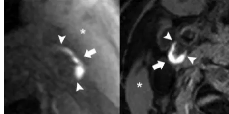
FIG 2. Fissured fibrous cap on MDCTA. Transversal (left) and longitu-dinal (right) MDCTA images of the carotid bifurcation. A “fissured fibrous cap,” defined as an extension of contrast material of⬍1 mm into the atherosclerotic plaque and an angle ofⱖ230° with the lumen, is visible only on the transversal plane (arrows).
[image:3.594.301.534.48.164.2] [image:3.594.53.285.48.165.2]RESULTS
Patient Characteristics
In 20 of the 100 patients, MDCTA (n⫽18) or MR imaging (n⫽ 2) of the carotid arteries had not been performed due to contra-indications. Two patients were excluded due to inferior image quality of the MDCTA. Of the remaining 78 patients, 149 vessels could be used for the current analyses. Seven contralateral vessels were excluded from analysis due to the absence of plaque (n⫽5) or occlusion of the carotid artery (n⫽2). Baseline characteristics are shown inTable 1. Forty-one of the 78 patients (53%) had an ischemic stroke; 37 (47%) patients had a TIA, including 7 with amaurosis fugax.
Vessel Characteristics
The median interval between the neurologic event and MDCTA was 32 days (25th to 75th percentile, 14 –56 days); the median interval between the event and MR imaging was 44 days (25th to 75th percentile, 27– 62 days). Characteristics of all vessels and symptomatic and contralateral vessels separately are shown in Table 2. The mean severity of stenosis was 51%⫾17% (ECST). The prevalence of intraplaque hemorrhage in the symptomatic and contralateral vessels was 38% and 11%, respectively.
Twenty-six of the 149 vessels (17%) showed plaque ulceration; the preva-lence in the symptomatic vessels and the contralateral vessels was 27% and 7%, respectively. In 13 of the 149 vessels (9%), a fissured fibrous cap was present; most were found in the symptomatic vessels (n⫽10), but the difference was not significant (P⫽.06).
[image:4.594.53.284.334.579.2]Intraplaque Hemorrhage and Disrupted Plaque Surface InTable 3, the association between the presence of intraplaque hemorrhage and disrupted plaque surface (ulcerated plaque and/or fissured fibrous cap) is shown. In vessels with intraplaque hemorrhage, a disrupted plaque surface was significantly more prevalent compared with vessels without intraplaque hemorrhage (45% versus 15%;P⬍ .001).Table 4 shows the results of the multivariable logistic regression. After correction for age and sex, the presence of intraplaque hemorrhage was associated with a disrupted plaque surface (OR, 3.98; 95% CI, 1.73–9.16;P⫽.001). Additionally, we found an association between the degree of ste-nosis and disrupted plaque surface (OR per 10% increase 1.42; 95% CI, 1.09 –1.84;P⫽.009). After correction for age, sex, and all variables withP⬍.10, intraplaque hemorrhage was still signifi-cantly associated with disrupted plaque surface (OR, 3.13; 95% CI, 1.25–7.84;P⫽.02). Diabetes mellitus was inversely associated with disrupted plaque surface (OR, 0.31; 95% CI, 0.11– 0.94;P⫽ .04). Similar results were found when the analyses were repeated to assess the association between intraplaque hemorrhage and ulcerated plaque alone. The association between intraplaque hemorrhage and disrupted plaque surface was attenuated when the analyses were repeated in only the symptomatic arteries (On-line Tables 1–3). The location of the plaque ulceration and/or fissured fibrous cap was the same as that of the intraplaque hemorrhage in 16 of the 21 lesions (76%); an example is shown inFig 4.
Table 1: Clinical characteristics of patients (nⴝ78)a Clinical Characteristic
Age (yr) 67⫾9
Male sex 57 (73%)
Classification event
TIA 30 (38%)
Stroke 41 (53%)
Amaurosis fugax 7 (9%)
Hypercholesterolemia 43 (55%)
Hypertension 56 (72%)
Diabetes mellitus 19 (24%)
Current smoking
No 58 (74%)
Yes 20 (26%)
Body mass index 25.2 (24.3–28.4)
Current use
Antiplatelet therapy 36 (46%)
Oral anticoagulants 0 (0%)
Statins 36 (46%)
Antihypertensive medication 50 (64%)
Antidiabetic medication 13 (17%)
History
Ischemic stroke or TIA 13 (17%)
Ischemic heart disease 16 (21%)
Peripheral arterial disease 15 (19%)
a
[image:4.594.302.533.496.582.2]Data are mean⫾SD, absolute numbers of patients (%), or median (25th–75th per-centile).
Table 2: Vessel characteristicsa
Vessel Characteristic All Vessels
Symptomatic Vessels
Contralateral Vessels
PValue, Symptomatic versus Contralateral Vessels
No. 149 78 71
Plaque ulceration 26 (17%) 21 (27%) 5 (7%) .001b
Fissured fibrous cap 13 (9%) 10 (13%) 3 (4%) .06
Calcium volume (mm3) 21.0 (3.6–56.2) 24.2 (6.2–71.8) 17.2 (1.9–53.1) .09
Degree of stenosis (ECST) (%) 51⫾17 55⫾17 47⫾16 .004b
Degree of stenosis (NASCET) (%) 8 (0–32) 14 (0–35) 2 (0–26) .03b
Intraplaque hemorrhage 38 (26%) 30 (38%) 8 (11%) ⬍.001b
a
Data are absolute numbers of vessels (%), median (25th–75th percentile), or mean⫾SD. bP⬍.05.
Table 3: Association of intraplaque hemorrhage and disrupted plaque surface in all vesselsa
Disrupted Plaqueb
Intact Plaque Surface Total Intraplaque hemorrhage present
(n⫽38)
17 (45%) 21 (55%) 38
Intraplaque hemorrhage absent (n⫽111)
17 (15%) 94 (85%) 111
Total 34 115 149
a
P⬍.001. b
[image:4.594.54.540.618.714.2]DISCUSSION
This study shows that intraplaque hemorrhage is associated with a disrupted plaque surface (plaque ulceration and/or fissured fi-brous cap) and plaque ulceration alone. In 76% of the vessels with both intraplaque hemorrhage and a disrupted plaque surface, in-traplaque hemorrhage and plaque ulceration or fissured fibrous cap shared the same location. The association between these 2 vulnerable plaque characteristics may support the notion that in-traplaque hemorrhage increases the risk of plaque rupture. How-ever, because of the cross-sectional study design, serial studies are needed to further investigate the role of intraplaque hemorrhage in plaque rupture.
Some methodologic issues need to be discussed first. A strength of our prospective study is that most vessels showed a mild stenosis. Most previous studies focused on patients with a moderate or severe carotid artery stenosis. However, plaque im-aging in this patient group is less relevant for clinical decision-making because patients with a moderate (male patients) or se-vere (all patients) symptomatic carotid artery stenosis are already eligible for carotid endarterectomy.16Moreover, the amount of
calcification will probably be higher in patients with a moderate or severe carotid artery stenosis, complicating the detection of plaque surface morphology. Second, we reviewed MDCTA and MR images of all patients with a disrupted plaque surface and intraplaque hemorrhage to investigate whether the intraplaque hemorrhage and plaque disruption were at the same location.
The finding that the location was indeed similar in 76% strengthens the association between the 2 vulnerable plaque characteristics.
A limitation of our multicenter study is that we used different MDCTA and MR imaging scanners. MDCTA scanning protocols, however, were similar. We used the 3D-T1W fat suppresssed spoiled gradient echo or the 2D-T1W inversion recovery turbo field echo MR imaging sequence to detect intraplaque hemor-rhage. Bitar et al25and Cappendijk et al26used sequences like ours
and found a good sensitivity, specificity, and interobserver agree-ment for the detection of intraplaque hemorrhage in both se-quences. They also found good agreement between MR imaging– depicted intraplaque hemorrhage and histologic sections. In addition, our overall prevalence of intraplaque hemorrhage of 26% is in accordance with that in the literature.4,5,15Correction
for MR imaging protocol used in the multivariate model did not change our results. A second limitation is the lack of histology. We used MDCTA instead of MR imaging for the evaluation of the plaque surface, because MDCTA has been shown to have a high sensitivity and specificity for the detection of plaque ulceration and has fewer limitations such as partial volume effects and flow artifacts that mimic plaques.27,28In addition, when we reviewed
the MR images and MDCTA images side by side, plaque ulcer-ations were hardly visible on the MR images. Saba et al27showed
[image:5.594.54.535.65.193.2]a good sensitivity and specificity for the detection of plaque ulcer-ation by using MDCTA and multiplanar reconstruction (sensitivity, FIG 4. Correlated intraplaque hemorrhage on MR imaging and plaque ulceration on MDCTA. An example of MR imaging and MDCTA images of the left carotid bifurcation. On the 3D-T1W fat suppressed echo-spoiled gradient echo MR imaging sequence, intraplaque hemorrhage is visible in the atherosclerotic plaque (A,arrow). MDCTA images show a small ulceration on both the transversal and longitudinal plane at the site of the intraplaque hemorrhage (BandC,arrows;arrowheadsindicate the edges of the plaque ulceration).
Table 4: Multivariable OR for the association among clinical characteristics, vessel characteristics, and disrupted plaque surface in all vessels
Characteristic
Multivariable (Age, Sex)
Multivariablea(Age, Sex,
FactorsP< .10)
OR (95% CI) PValue OR (95% CI) PValue
Age 1.05 (1.00–1.10) .07 1.05 (1.00–1.10) .07
Sex 0.42 (0.15–1.12) .08 0.50 (0.17–1.42) .19
Hypertension 1.98 (0.73–5.35) .18
Diabetes mellitus 0.40 (0.14–1.10) .08 0.31 (0.11–0.94) .04b
Hypercholesterolemia 1.41 (0.63–3.15) .40
Current smoking 0.48 (0.16–1.46) .20
Intraplaque hemorrhage 3.98 (1.73–9.16) .001b 3.13 (1.25–7.84) .02b
Degree of stenosis (ECST, per 10%) 1.42 (1.09–1.84) .009b 1.30 (1.00–1.69) .07
Calcification volume 0.86 (0.69–1.08) .21
aWe corrected for age, sex, diabetes mellitus, and degree of stenosis (ECST).
b
[image:5.594.78.511.223.364.2]75.8%; specificity, 90.8%). We found an overall prevalence of plaque ulceration of 17%. Similar prevalence of plaque ulceration was found in other studies, ranging from 13% to 22%.15,20,29,30
Detection of the presence of a fissured fibrous cap has been stud-ied less intensively. We found an overall prevalence of the pres-ence of a fissured fibrous cap of 9%, in agreement with the 11% found by Saba and Mallarini.20Rupture of the fissured fibrous cap
was significantly associated with cerebrovascular symptoms (P⫽ .003).20Based on the good sensitivity and specificity of MDCTA
for plaque ulcerations and the good agreement between MR im-aging– depicted intraplaque hemorrhage and histology, we there-fore conclude that it is legitimate to use imaging techniques such as MDCTA and MR imaging to study the relationship between intraplaque hemorrhage and the disruption of the plaque sur-face.25,26Moreover, the use of MDCTA and MR imaging provides
the opportunity to investigate the relationship in a specific cate-gory of patients—that is, those with a mild-to-moderate carotid artery stenosis, patients who are treated medically and thus have no available histology. The final limitation of our study is that it is cross-sectional. Our results should therefore be confirmed in a serial study.
Rupture of an atherosclerotic carotid artery plaque is an im-portant cause of ischemic stroke. Therefore, understanding the pathophysiology of plaque rupture is very important. Infiltration of macrophages in pathologic intima thickening plays a key role in the development of atherosclerotic plaques. The combination of macrophage infiltration, apoptosis, and hypoxia-induced necro-sis leads to the development of more advanced atherosclerotic plaques with a lipid-rich necrotic core. Previous studies already showed an association between lipid-rich necrotic core volumes and plaque ulceration.29-31In these larger plaques, hypoxia and
the inflammatory response are assumed to promote neovascular-ization. As stated previously, the common viewpoint is that these small leaky neovessels are responsible for the occurrence of intra-plaque hemorrhage and subsequently the development of an un-stable rupture-prone plaque.5,9-12,32Our results—if confirmed in
serial studies— can support the pathophysiologic relation be-tween intraplaque hemorrhage and disrupted plaque surface. An alternative viewpoint to explain this relationship is that repeated fissuring of the plaque and associated formation of nonocclusive luminal thrombus incorporated in the plaque could be the cause of intraplaque hemorrhage.5Nevertheless, observations that
in-traplaque hemorrhage is related to high attenuation of plaque neovessels, in the absence of plaque fissuring, supports the more common view of small leaky neovessels as the cause of intraplaque hemorrhage.5,10
CONCLUSIONS
We showed that intraplaque hemorrhage is associated with a dis-rupted plaque surface (plaque ulceration and/or fissured fibrous cap) in patients with a⬍70% carotid artery stenosis. Our findings suggest a strong association between these 2 vulnerable plaque characteristics. Nevertheless, because of the cross-sectional study design, additional serial studies are needed to evaluate whether intraplaque hemorrhage indeed increases the risk for plaque rup-ture and subsequent TIA or ischemic stroke.
APPENDIX
Participating centers: Academic Medical Center, Amsterdam (P.J. Nederkoorn); Atrium Medisch Centrum, Heerlen (A.H.C.M.L. Schreuder); Erasmus Medical Center, Rotterdam (A. van der Lugt, P.J. Koudstaal); Flevoziekenhuis, Almere (M. Limburg); Kennemer Gasthuis, Haarlem (M. Weisfelt); Laurentius Zieken-huis, Roermond (A.G. Korten); Maasstad ZiekenZieken-huis, Rotterdam (R. Saxena); Maastricht University Medical Center (M.E. Kooi, R.J. van Oostenbrugge, W.H. Mess); Orbis Medisch Centrum, Sittard (N.P. van Orshoven); Sint Antonius Ziekenhuis, Nieu-wegein (S.C. Tromp); Sint Franciscus Gasthuis, Rotterdam (S.L.M. Bakker); Slotervaartziekenhuis, Amsterdam (N.D. Kruyt); Tergooi Ziekenhuizen, Hilversum/Blaricum (J.R. de Kruijk); University Medical Center Utrecht (J. Hendrikse, G.J. de Borst); Viecuri Medisch Centrum, Venlo (B.J. Meems); Vlietland Ziekenhuis, Schiedam (J.C.B. Verhey); IJsselland Ziekenhuis, Capelle a/d IJsel (A.D. Wijnhoud).
Disclosures: Anouk C. van Dijk—RELATED:Grant: Center for Translational Molecular Medicine and supported by the Dutch Heart Foundation,*Comments: Project PARISK (grant 01C-202). Antonius F.W. van der Steen—RELATED:Grant: Center for Translational Molecular Medicine, project PARISK,*Comments: This work is part of a public-private partnership by the Center for Translational Molecular Medicine and the Dutch Heart Foundation; I am Co-Principal Investigator of this grant. Mat J.A.P. Daemen—RELATED:Grant: Center for Translational Molecular Medicine,* Com-ments: The Center for Translational Molecular Medicine provided the grant for the PARISK research program. Peter J. Koudstaal—UNRELATED: editor and author, Com-ments:Textbook of Neurology, Reed Elsevier. Paul J. Nederkoorn—UNRELATED: Grants/Grants Pending: 1) Preventive Antibiotics in Stroke Study, the Netherlands Organization for Health Research and Development (no. 171002302) and the Nether-lands Heart Foundation (no. 2009B095); 2) The ThRombolysis in UnconTrolled Hypertension study: Fonds NutsOhra, grant No. 1302– 026. Marianne Eline Kooi— RELATED:Grant: Center for Translational Molecular Medicine.* Aad van der Lugt— RELATED:Grant: Center for Translational Molecular Medicine,* Dutch Heart Foun-dation*;UNRELATED:Grants/Grants Pending: GE Healthcare,*Comments: MRI vendor;Payment for Lectures (including service on Speakers Bureaus): GE Health-care,*Comments: MRI vendor. *Money paid to the institution.
REFERENCES
1. Yuan C, Mitsumori LM, Beach KW, et al.Carotid atherosclerotic plaque: noninvasive MR characterization and identification of vul-nerable lesions.Radiology2001;221:285–99
2. Naghavi M, Libby P, Falk E, et al.From vulnerable plaque to vulner-able patient: a call for new definitions and risk assessment strate-gies, part II.Circulation2003;108:1772–78
3. Naghavi M, Libby P, Falk E, et al.From vulnerable plaque to vulner-able patient: a call for new definitions and risk assessment strate-gies, part I.Circulation2003;108:1664 –72
4. Saam T, Hetterich H, Hoffmann V, et al.Meta-analysis and system-atic review of the predictive value of carotid plaque hemorrhage on cerebrovascular events by magnetic resonance imaging.J Am Coll Cardiol2013;62:1081–91
5. Teng Z, Sadat U, Brown AJ, et al.Plaque hemorrhage in carotid artery disease: pathogenesis, clinical and biomechanical consider-ations.J Biomech2014;47:847–58
6. Altaf N, Daniels L, Morgan PS, et al.Detection of intraplaque hem-orrhage by magnetic resonance imaging in symptomatic patients with mild to moderate carotid stenosis predicts recurrent neuro-logical events.J Vasc Surg2008;47:337– 42
7. Kwee RM, van Oostenbrugge RJ, Mess WH, et al.MRI of carotid atherosclerosis to identify TIA and stroke patients who are at risk of a recurrence.J Magn Reson Imaging2013;37:1189 –94
9. Michel JB, Virmani R, Arbustini E, et al.Intraplaque haemorrhages as the trigger of plaque vulnerability.Eur Heart J2011;32:1977– 85, 85a, 85b, 85c
10. Virmani R, Kolodgie FD, Burke AP, et al.Atherosclerotic plaque progression and vulnerability to rupture: angiogenesis as a source of intraplaque hemorrhage. Arterioscler Thromb Vasc Biol2005; 25:2054 – 61
11. Michel JB, Delbosc S, Ho-Tin-Noe´ B, et al.From intraplaque haem-orrhages to plaque vulnerability: biological consequences of intra-plaque haemorrhages. J Cardiovasc Med (Hagerstown) 2012;13: 628 –34
12. Takaya N, Yuan C, Chu B, et al.Presence of intraplaque hemorrhage stimulates progression of carotid atherosclerotic plaques: a high-resolution magnetic resonance imaging study.Circulation2005; 111:2768 –75
13. Lovett JK, Gallagher PJ, Hands LJ, et al.Histological correlates of carotid plaque surface morphology on lumen contrast imaging.
Circulation2004;110:2190 –97
14. Saba L, Sanfilippo R, Pirisi R, et al.Multidetector-row CT angiogra-phy in the study of atherosclerotic carotid arteries.Neuroradiology
2007;49:623–37
15. U-King-Im JM, Fox AJ, Aviv RI, et al.Characterization of carotid plaque hemorrhage: a CT angiography and MR intraplaque hemor-rhage study.Stroke2010;41:1623–29
16. Rothwell PM, Eliasziw M, Gutnikov SA, et al; Carotid Endarterec-tomy Trialists Collaboration.Endarterectomy for symptomatic ca-rotid stenosis in relation to clinical subgroups and timing of sur-gery.Lancet2004;363:915–24
17. Rothwell PM, Gibson R, Warlow CP; European Carotid Surgery Tri-alists’ Collaborative Group.Interrelation between plaque surface morphology and degree of stenosis on carotid angiograms and the risk of ischemic stroke in patients with symptomatic carotid steno-sis.Stroke2000;31:615–21
18. Truijman MT, Kooi ME, van Dijk AC, et al. Plaque At RISK (PARISK): prospective multicenter study to improve diagnosis of high-risk carotid plaques.Int J Stroke2014;9:747–54
19. Randomised trial of endarterectomy for recently symptomatic ca-rotid stenosis: final results of the MRC European Caca-rotid Surgery Trial (ECST).Lancet1998;351:1379 – 87
20. de Weert TT, Cretier S, Groen HC, et al.Atherosclerotic plaque surface morphology in the carotid bifurcation assessed with multidetector computed tomography angiography.Stroke2009;40:1334 – 40
21. Saba L, Mallarini G.Fissured fibrous cap of vulnerable carotid plaques and symptomaticity: are they correlated? Preliminary re-sults by using multi-detector-row CT angiography.Cerebrovasc Dis
2009;27:322–27
22. North American Symptomatic Carotid Endarterectomy Trial Col-laborators.Beneficial effect of carotid endarterectomy in symp-tomatic patients with high-grade carotid stenosis.N Engl J Med
1991;325:445–53
23. de Weert TT, Cakir H, Rozie S, et al.Intracranial internal carotid artery calcifications: association with vascular risk factors and ischemic cerebrovascular disease. AJNR Am J Neuroradiol
2009;30:177– 84
24. Yuan C, Mitsumori LM, Ferguson MS, et al.In vivo accuracy of mul-tispectral magnetic resonance imaging for identifying lipid-rich necrotic cores and intraplaque hemorrhage in advanced human ca-rotid plaques.Circulation2001;104:2051–56
25. Bitar R, Moody AR, Leung G, et al.In vivo 3D high-spatial-resolution MR imaging of intraplaque hemorrhage.Radiology2008;249:259 – 67 26. Cappendijk VC, Cleutjens KB, Heeneman S, et al.In vivo detection of
hemorrhage in human atherosclerotic plaques with magnetic reso-nance imaging.J Magn Reson Imaging2004;20:105–10
27. Saba L, Caddeo G, Sanfilippo R, et al.Efficacy and sensitivity of axial scans and different reconstruction methods in the study of the ul-cerated carotid plaque using multidetector-row CT angiography: comparison with surgical results.AJNR Am J Neuroradiol2007; 28:716 –23
28. Yu W, Underhill HR, Ferguson MS, et al.The added value of longi-tudinal black-blood cardiovascular magnetic resonance angiogra-phy in the cross sectional identification of carotid atherosclerotic ulceration.J Cardiovasc Magn Reson2009;11:31
29. Homburg PJ, Rozie S, van Gils MJ, et al.Association between carotid artery plaque ulceration and plaque composition evaluated with multidetector CT angiography.Stroke2011;42:367–72
30. Saba L, Sanfilippo R, Sannia S, et al.Association between carotid artery plaque volume, composition, and ulceration: a retrospective assessment with MDCT.AJR Am J Roentgenol2012;199:151–56 31. Underhill HR, Yuan C, Yarnykh VL, et al.Predictors of surface
dis-ruption with MR imaging in asymptomatic carotid artery stenosis.
AJNR Am J Neuroradiol2010;31:487–93