ORIGINAL RESEARCH
INTERVENTIONAL
Long-Term Outcomes of Patients with Stent Tips Embedded
into Internal Carotid Artery Branches during Aneurysm Coiling
XS.P. Ban,XO.-K. Kwon,XS.U. Lee,XJ.S. Bang,XC.W. Oh,XH.J. Jeong,X M.J. Cho,XE.-A. Jeong, andXT. Kim
ABSTRACT
BACKGROUND AND PURPOSE: During stent-assisted coiling of ICA aneurysms, stent tips are sometimes unintentionally embedded into ICA branches. Stent tips can be visualized because they have radiopaque markers. Concerns regarding stent tip misplacement include risks of artery perforation and occlusion. The aim of this study was to evaluate the long-term outcomes of ICA branches with embedded stent tips.
MATERIALS AND METHODS: ICA branches with embedded stent tips were identified among 35 patients with unruptured ICA aneurysms treated with stent-assisted coiling between November 2003 and November 2014. Patient clinical and angiographic outcomes associated with the embedded stent tip were analyzed.
RESULTS:Most of the 35 studied aneurysms were paraclinoid ICA aneurysms (n⫽30). The most commonly involved ICA branch was the posterior communicating artery (26 patients, 74.3%), followed by the anterior choroidal artery (8 patients, 22.9%) and ophthalmic artery (1 patient, 2.9%). During the follow-up period (38.6⫾17.9 months), no new neurologic deficits developed. Neither hemorrhagic nor throm-boembolic events occurred. Angiography was performed during the final follow-up evaluation at a mean of 32.7⫾18.0 months, and all ICA branches with embedded stent tips showed patent blood flow without severe luminal narrowing.
CONCLUSIONS: In our experience, placement of a stent tip into ICA branches during stent-assisted coiling was not associated with any major adverse events.
ABBREVIATIONS:AchoA⫽anterior choroidal artery; OphA⫽ophthalmic artery; PcomA⫽posterior communicating artery; SAC⫽stent-assisted coiling
S
elf-expandable stents have provided considerable assis-tance in increasing the indications for aneurysm coiling. Complex and wide-neck aneurysms, which are considered challenging or impossible to treat with simple coiling, can now be treated.1Generally, self-expandable stents for cerebral aneurysm treatment have proximal and distal radiopaque markers to identify the end of the stent. These markers are made from coiled tantalum wire or platinum bands, which are larger than the stent strut itself. The markers are attached to the triangular pointed stent tip wire (strut) part. During stent-assisted coil-ing (SAC), the stent tip can sometimes be unintentionally
em-bedded into a small arterial branch. We have occasionally en-countered this event, particularly during SAC performed for ICA aneurysms, which is well-visualized due to the radiopaque markers. This phenomenon raises concerns regarding the po-tential risks of arterial perforation due to the sharp end and constant arterial pulsating motion of the stent, as well as the chances of vessel occlusion. Stent tip markers may also disrupt blood flow by themselves or by intimal injury (thrombus for-mation or intimal hyperplasia), which may then lead to infarc-tion. Despite these concerns, we were unable to find a report regarding the clinical or radiologic outcomes of embedded stent tips within ICA branches. Thus, we reviewed our series of patients who had undergone SAC for unruptured ICA aneu-rysms and in whom stent tips were embedded within the ICA branch orifice. We analyzed clinical and angiographic data to identify the outcomes of these cases.
MATERIALS AND METHODS
PatientsThis study was approved by the institutional review board at our institution. Informed consent from enrolled patients was waived. Received November 7, 2017; accepted after revision January 3, 2018.
From the Department of Neurosurgery, Seoul National University Bundang Hospi-tal, Seongnam, Gyeonggi-do, Korea.
Please address correspondence to Tackeun Kim, MD, Department of Neurosur-gery, Seoul National University Bundang Hospital, 82 Gumi-ro 173 beon-gil, Bun-dang-gu, Seongnam-si, Gyeonggi-do, 13620, Korea; e-mail: midabo@naver.com
Indicates article with supplemental on-line table.
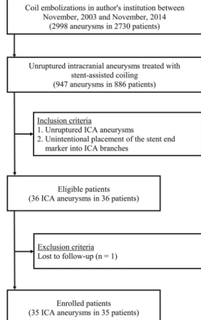
We retrospectively reviewed patient medical records, including pre- and posttreatment radiologic studies. Our data base con-tained a series of 2730 patients with 2998 intracranial aneurysms treated with endovascular techniques between November 2003 and November 2014. Among these patients, 886 with 947 unrup-tured intracranial aneurysms were treated with SAC. In this series, data from 664 patients with 706 ICA aneurysms who were treated with SAC were collected and reviewed. Among these patients, the placement of a stent tip into an ICA branch was identified in 36 aneurysms of 36 patients. In our subsequent analysis, 1 patient who was lost to follow-up was excluded. The patient selection criteria are outlined inFig 1.
Antiplatelet Therapy Protocol and Endovascular Procedures
Patients with unruptured aneurysms received dual antiplatelet agents (100 mg of aspirin and 75 mg of clopidogrel) for at least 5 days before embolization. One day before coil embolization, P2Y12 reaction units were measured using VerifyNow (Accumet-rics, San Diego, California). Patients with clopidogrel resistance (⬎220 P2Y12 reaction units) received a modified antiplatelet reg-imen. Details regarding our antiplatelet agent protocol have been previously described.2A 3000-IU bolus dose of intravenous hep-arin was administered after the placement of the femoral artery sheath, and heparin was later infused at an hourly booster dose of 1000 IU, with monitoring of the activated clotting time. After the procedure, dual antiplatelet treatment continued for 1 year; after
1 year, this therapy was exchanged for daily oral treatment with 100 mg of aspirin for an additional year.
All endovascular procedures were performed with the patient under general anesthesia using an Integris Allura scanner (Philips Healthcare, Best, the Netherlands) before 2014 and an IFNX-8000V scanner (Toshiba, Tokyo, Japan) from 2014. The jailing technique was mainly used for SAC. First, a target aneurysm was selected using a microcatheter (Excelsior SL-10; Stryker Neuro-vascular, Kalamazoo, Michigan). Then, the parent artery was nav-igated with another microcatheter (Prowler Select Plus, Codman & Shurtleff, Raynham, Massachusetts; or Neuro Renegade, Stryker Neurovascular) for stent delivery. A stent was deployed into the parent artery across the aneurysm neck. Then, the aneu-rysm was coiled using platinum detachable coils until complete occlusion was achieved or further coiling was deemed unsafe. Stent type, length, and placement were determined by the neuro-vascular team and were based on aneurysm location and neck diameter. Among 706 ICA aneurysms treated with SAC, Enter-prise stents (Codman & Shurtleff) were most commonly used (n⫽607, 86.0%), followed by Neuroform stents (Stryker Neuro-vascular) in 71 cases (10.1%). The other stents used included Low-Profile Visualized Intraluminal Support stents (MicroVention, Tustin, California) in 19 cases (2.7%) and Solitaire AB stents (Covidien, Irvine, California) in 9 cases (1.3%). Of the 35 cases, 32 were treated with a single Enterprise stent and 3 were treated with a single Neuroform stent.
Stent Tips of Enterprise and Neuroform Stents
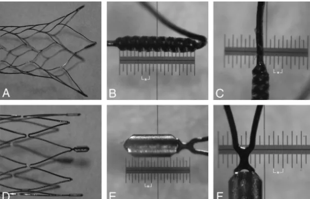
The Enterprise stent is a laser-cut stent with a closed-cell design. This stent has flared proximal and distal ends, consisting of 4 pointed parts. Each end has 4 radiopaque tantalum coil markers for increased visibility, which flare out for fixation of the stent onto the vascular wall when fully deployed (Fig 2A). Due to the sharp, pointed, flaring parts, these stents can be embedded into the small ICA branches. The radiopaque markers of the tips allow them to be clearly visualized. The diameter of a radiopaque marker was thicker than that of the stent strut. Stent strut width and thickness were approximately 0.0015 inches (0.0381 mm) and 0.0031 inches (0.0787 mm), respectively. The thick-ness and length of the radiopaque marker were approximately 0.008 inches (0.2032 mm) and 0.043 inches (1.0922 mm), re-spectively (Fig 2B, -C).
The Neuroform stent is a laser-cut stent with an open-cell or half-open-cell design. This stent has 4 distal and 4 proximal ra-diopaque platinum markers (Fig 2D). Each open-cell segment may serve as a separate fixing device to enhance the apposition of the stent to the vessel wall like the flared ends of the Enterprise stent. Like the Enterprise stent, the stent tip may be placed and embedded into the branch orifice. Stent strut width and thickness were approximately 0.0027 inches (0.0686 mm). The thickness and length of the radiopaque marker were approximately 0.0116 inches (0.2946 mm) and 0.030 inches (0.762 mm), respectively (Fig 2E, -F).
Evaluation of Clinical and Radiologic Outcomes
The clinical outcomes of all patients were evaluated using the mRS score on admission and at the final outpatient follow-up visit.
[image:2.594.68.268.50.368.2]Newly developed neurologic deficits as well as hemorrhagic and thromboembolic complications during follow-up periods were also reviewed. The placement and embedding of a stent tip with a marker into a branching artery was defined as a stent tip that was placed at the level of branching arteries, with the tip marker clearly visualized within the artery orifice via angiographic views, in-cluding rotational angiograms. According to our institutional protocol, patients underwent imaging follow-up at 3, 6, 12, 24, and 36 months with skull plain radiography and at 1, 2, and 3 years with 3D-TOF MRA, including source imaging. Any time a major recanalization was found, DSA was performed. DSA follow-up studies were routinely conducted between 3 and 5 years after coil embolization if no major recanalization of an aneurysm was suspected. In this study, each patient underwent a minimum of 12 months of radiologic follow-up. Vessel pa-tency was documented by follow-up DSA. If follow-up DSA was not performed, vessel patency was confirmed using the follow-up axial MRA source images.
Data Analysis
All statistical analyses were performed using SPSS software (Version 22.0; IBM, Armonk, New York). Continuous vari-ables are presented as mean⫾SD. Binary variables were com-pared using the Fisher exact test. Statistical significance was set atP⬍.05.
RESULTS
The overview of patients with stent tips embedded into the ICA branches is summarized in the On-line Table. Eight (22.9%) pa-tients were men, and 27 (77.1%) were women. The mean age was 49.3⫾12.8 years (range, 21–76 years). The cases included 30 (85.7%) paraclinoid ICA aneurysms, 3 (8.6%) ophthalmic artery (OphA) aneurysms, 1 (2.9%) cavernous ICA aneurysm, and 1 (2.9%) posterior communicating artery (PcomA) aneurysm. The most common ICA branching artery with an embedded stent tip
was the PcomA (26 patients, 74.3%). Other involved branching arteries in-cluded the anterior choroidal artery (AchoA) in 8 (22.9%) patients and the OphA in 1 (2.9%) patient. The incidence of stent tips embedded within ICA branches was not associated with the type of stent (closed-cell-design stent, 5.3%, versus open-cell-design stent, 4.2%;P⫽.424). Most of these events occurred during deployment of the dis-tal end of the stent at the upward ICA curvature near the ICA branches (32 cases, 91.4%), followed by unintentional stent advancement during microcath-eter manipulation (3 cases, 8.6%).
The mean clinical follow-up dura-tion was 38.6 ⫾ 17.9 months (range, 12–91 months). The preoperative mRS score was 0 in 30 (85.7%) patients and 1 in 5 (14.3%) patients. The final fol-low-up mRS score was 0 in 32 (91.4%) patients and 1 in 3 (8.6%) patients. No patient experienced mRS score deterioration. No newly devel-oped neurologic deficits were observed. Neither hemorrhagic nor thromboembolic (or ischemic) complications occurred during the follow-up period.
The mean angiographic follow-up duration was 32.7⫾18.0 months (range, 12–91 months). The mean initial diameter of the ICA branches with embedded stent tips was 1.79⫾ 0.69 mm (range, 0.51–2.81 mm). On final follow-up angiography (15 cases were measured by MRA with source imaging and 20 cases were measured by DSA), all ICA branches with an embedded stent tip had persistent blood flow without severe luminal nar-rowing. In 1 case, the radiopaque stent marker appeared to penetrate the PcomA. In the angiogram, the marker appeared to be located outside the arterial wall. However, no clinical event was observed (case 11).
Illustrative Cases
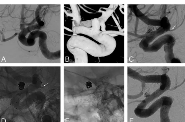
Case 11. A 64-year-old patient presented with an unruptured left paraclinoid ICA aneurysm (Fig 3A, -B). The aneurysm was treated with coil embolization using a 4.5⫻20 mm Neuroform stent. Postembolization angiography showed the placement of the stent marker in the PcomA (Fig 3C, -D). Although a radiopaque stent marker appeared to penetrate the PcomA in the 5-year-follow-up angiograms, the PcomA was patent and no hemorrhagic compli-cation occurred (Fig 3E, -F).
Case 13. A 23-year-old patient presented with an unruptured left paraclinoid ICA aneurysm (Fig 4A, -B). The aneurysm was treated with coil embolization using a 4.5⫻ 22 mm Enterprise stent. Postembolization angiography showed the placement of the stent marker into the AchoA (Fig 4C, -D). Follow-up angiography at 3 years showed patent blood flow to the AchoA without severe lu-minal narrowing (Fig 4E, -F).
[image:3.594.56.375.47.252.2]DISCUSSION
This study demonstrates that stent tip embedding into the ICA branches during SAC of cerebral aneurysms was not asso-ciated with any major adverse events. No arterial rupture or occlusion was observed during at least 1 year (12–91 months) of follow-up.
During SAC of ICA aneurysms, stent tips can be deployed and unintentionally embedded into ICA branching arteries due to their characteristics. In our study, the incidence of this event was 5.1%, and the PcomA was the most commonly involved ICA branch, followed by the AchoA. Although we attempted to deploy stents so as avoid embedding the stent tip into the orifice of these
ICA branches, the distal part of a stent may be deployed flat around the PcomA and AchoA due to the ICA curvature, resulting in stent tips embedding into these ICA branches. In this study, this issue was the most common cause of an embedded stent tip within ICA branches (91.4%). Because the ICA usu-ally starts to curve upward near the PcomA and the diameter of the PcomA is commonly larger than that of the AchoA, it is likely that the stent tips are more easily embedded into the PcomA than into the AchoA.
Regarding the structure of the stents, because a closed-cell-design stent im-mediately transmits a force from one end to the other end, embedding of a stent tip into ICA branches due to stent advancement during microcath-eter manipulation may be more com-monly observed in patients receiving a closed-cell-design stent. Concerning this possible mechanism, in this study, all 3 cases (8.6%) in which the distal part of a stent was initially deployed proxi-mally near the PcomA and then ad-vanced and embedded into the PcomA due to microcatheter manipulation were treated with the Enterprise stent (closed-cell design). However, because the most common cause of embedding of a stent tip into ICA branches was the ICA cur-vature, no association was observed among the types of stents used in this study.
This event raises the following con-cerns: vessel rupture (perforation) and occlusion. Vessel rupture due to the forceful advancement of stents has been reported. The sharp ends of the stent may contribute to arterial rupture.3,4 The lack of this complication in our study suggests that stent ends were in-serted into the branching arteries with-out excessive force. The presence of an embedded stent tip in small arteries can be a risk factor for further arterial injury with persistent arterial pulsation, at least theoreti-cally. However, this study shows that this risk is not likely in practice.
Vessel occlusion or significant flow disruption may develop through intimal injury by the stent tip in a delayed manner as well as mechanical occlusion by stent tips and large markers them-selves during the acute period. Therefore, we can consider several plausible mechanisms of vessel occlusion or flow disruption. A stent covering the small arterial ostium does not usually lead to arterial occlusion.5-7Unlike a simple stent covering the arterial FIG 3. A, An unruptured left paraclinoid ICA aneurysm on pre-embolization DSA.B, 3D image of
the aneurysm.CandD, A distal stent marker of the Neuroform stent (arrow) is deployed into the posterior communicating artery, as shown on postembolization angiography.EandF, The blood flow to the PcomA with malposition of the stent marker is patent on 5-year-follow-up cerebral angiography. A radiopaque stent marker appears to penetrate the PcomA (arrow).
[image:4.594.55.376.46.254.2] [image:4.594.56.375.317.527.2]orifice, embedded stent tips have greater luminal occupancy due to the sharp triangular stent tip labeled with a radiopaque marker. Radiopaque markers at the stent tip have a larger area and volume than the stent. In a coronary artery study of a swine model, 2-fold thicker stents were 49% more thrombogenic and increased flow stagnation and disruption.8Stent markers are 5.3-fold thicker than the stent strut in the Enterprise stent and 4.3-fold thicker than the stent strut in the Neuroform stent. In addition, persistent arterial pulsation with stent tips at the arterial orifice may also lead to further repeat intimal injury and subsequent thrombus forma-tion and neointimal hyperplasia.4,9However, this issue was not detected in our series of patients. The triangular shape of the stent tip may reduce such risks by stopping further advancement of the stent strut into the arterial lumen. The persistent need for blood flow to the ICA branches may also help prevent occlusion or se-vere stenosis.10
Furthermore, luminal narrowing by intimal hyperplasia may be spontaneously reversed.11The proliferated neointima reaches a maximal thickness by 2 months, and this neointimal layer grad-ually becomes thin, more sclerotic, and less cellular by 8 months. Kim et al9reported that most in-stent stenoses would spontane-ously improve to 91% of the initial mean diameter after 24 months. In our series, we did not perform angiographic follow-up until 12 months after SAC. Thus, luminal narrowing before this time was not confirmed. However, neither ischemic symptoms nor infarction developed in our study. One case (case 11) showed a radiopaque stent marker that appeared to be outside the PcomA lumen on 5-year-follow-up angiographic images. It is uncertain whether the stent tip was located outside the artery. However, no clinical symptoms or radiologic findings associated with arterial rupture were observed immediately after coiling, which suggests that the marker was more likely inside the artery and encased by neointimal hyperplasia. Although the mechanism has not yet been clearly determined, this study demonstrates that vessel oc-clusion by an embedded stent tip into the lumen is not common in practice.
Our study has several limitations including its retrospective nature and small sample size from a single institution. Further-more, this study showed only the results of placement of stent markers from Enterprise and Neuroform stents, which are only 2 varieties among many expandable stents. In 15 cases, follow-up imaging was performed with TOF-MRA according to our institu-tional protocol. Although the patency of ICA branches with em-bedded stent tips was confirmed by MRA images, we did not show the changes in the diameters of ICA branches with embedded stent tips because this would be inaccurate due to the metal arti-facts of the embedded stent tip on MRA images. Further research
using computational fluid dynamics is warranted to more com-pletely understand the changes in the diameter of ICA branches with embedded stent tips and the difference in the flow distur-bance effect between stent struts and radiopaque markers.
CONCLUSIONS
In our experience, placement of a stent tip into ICA branches during SAC was not associated with any major adverse events. During 12–91 months of follow-up, vessel rupture, significant blood flow reduction, and arterial occlusion were not detected.
REFERENCES
1. Geyik S, Yavus K, Yurttutan N, et al.Stent-assisted coiling in endo-vascular treatment of 500 consecutive cerebral aneurysms with
long-term follow-up. AJNR Am J Neuroradiol 2013;34:2157– 62
CrossRef Medline
2. Hwang G, Huh W, Lee JS, et al.Standard vs modified antiplatelet preparation for preventing thromboembolic events in patients with high on-treatment platelet reactivity undergoing coil emboli-zation for an unruptured intracranial aneurysm: a randomized
clinical trial.JAMA Neurol2015;72:764 –72CrossRef Medline
3. Chalouhi N, Jabbour P, Singhal S, et al.Stent-assisted coiling of intracranial aneurysms: predictors of complications,
recanaliza-tion, and outcome in 508 cases.Stroke2013;44:1348 –53CrossRef
Medline
4. Wang CC, Li W, Feng ZZ, et al.Preliminary experience with
stent-assisted coiling of aneurysms arising from small (<2.5
mm) cerebral vessels using the Low-Profile Visualized
Intralu-minal Support Device.AJNR Am J Neuroradiol2017;38:1163– 68
CrossRef Medline
5. D’Urso PI, Lanzino G, Cloft HJ, et al.Flow diversion for intracranial
aneurysms: a review.Stroke2011;42:2363– 68CrossRef Medline
6. Masuo O, Terada T, Walker G, et al.Study of the patency of small arterial branches after stent placement with an experimental in
vivo model.AJNR Am J Neuroradiol2002;23:706 –10Medline
7. Seong J, Wakhloo AK, Lieber BB.In vitro evaluation of flow diver-tors in an elastase-induced saccular aneurysm model in rabbit. J Biomech Eng2007;129:863–72CrossRef Medline
8. Kolandaivelu K, Swaminathan R, Gibson WJ, et al.Stent thrombo-genicity early in high-risk interventional settings is driven by stent design and deployment and protected by polymer-drug coatings. Circulation2011;123:1400 – 09CrossRef Medline
9. Kim YS, Lee SW, Yeom JA, et al.Angiographic findings of in-stent intimal hyperplasia after stent-assisted coil embolization: are
they permanent findings?J Neurosurg2016;124:328 –33CrossRef
Medline
10. Iosif C, Berg P, Ponsonnard S, et al.Role of terminal and anasto-motic circulation in the patency of arteries jailed by flow-diverting stents: animal flow model evaluation and preliminary results. J Neurosurg2016;125:898 –908CrossRef Medline
11. Schatz RA, Palmaz JC, Tio FO, et al.Balloon-expandable
intracoro-nary stents in the adult dog.Circulation1987;76:450 –57CrossRef