Case Reports in Clinical Medicine, 2018, 7, 397-429 http://www.scirp.org/journal/crcm ISSN Online: 2325-7083 ISSN Print: 2325-7075
DOI: 10.4236/crcm.2018.76036 Jun. 22, 2018 397 Case Reports in Clinical Medicine
Tropical Coronary Artery Disease and
Arrhythmogenic Potentials—The Changing
Pattern towards Endomyocardial Fibrosis
—An Analysis
Ramachandran Muthiah
Zion Hospital, Azhagiamandapam, Kanyakumari District, India
Abstract
Aim: To analyse the increasing burden of coronary artery disease (CAD) in tropical and subtropical belts of the Equator since it remains blurred and car-ries a grim prognosis. Introduction: Endomyocardial fibrosis [EMF] is a tropical febrile disorder, confined to peculiar and limited geographical areas. Plaque buildup in endocardium and coronary arteries, causing ischemic in-jury and arrhythmic episodes, is a vanishing mystery in its pathogenesis and emphasizing alternative routes for understanding and treatment of this enig-matic disease. Case Report: 15 cases in various age groups were reported with potential complications of coronary artery disease and arrhythmias, associated with endocardial lesions, the characteristic feature of endomyocardial fibrosis. Conclusion: The narrowing of coronary arteries as a result of thickening of the walls, spasm, inflammation, plaques and its rupture produce ischemic ep-isodes which can occur slowly or suddenly in a devastating pattern with arr-hythmogenic potentials. The important steps to prevent and decrease the risk of CAD is to reduce the chance of getting this disorder by epidemiological measures with an advice of blood thinning medications such as small daily dose aspirin, antibiotics in susceptible individuals and revascularization in es-tablished myocardial infarction.
Keywords
Endomyocardial Fibrosis, Endocardial Plaques, Egg-Cell Calcification (Endocardial), Arrhythmias, Ischemic Injury, Newer Therapeutic Strategies, RAS Vaccine
How to cite this paper: Muthiah, R. (2018) Tropical Coronary Artery Disease and Arrhythmogenic Potentials—The Changing Pattern towards Endomyocardial Fibro-sis—An Analysis. Case Reports in Clinical Medicine, 7, 397-429.
https://doi.org/10.4236/crcm.2018.76036
Received: May 17, 2018 Accepted: June 19, 2018 Published: June 22, 2018
Copyright © 2018 by author and Scientific Research Publishing Inc. This work is licensed under the Creative Commons Attribution International License (CC BY 4.0).
R. Muthiah
DOI: 10.4236/crcm.2018.76036 398 Case Reports in Clinical Medicine
1. Introduction
Coronary artery disease (CAD) remains the most common etiology for high morbidity and mortality worldwide. The worldwide burden is set to reach 47
millionaffected individuals by the year 2020 as projected by World Health
Or-ganization (WHO) [1]. The understanding of pathophysiology of coronary
ar-tery disease had led to a decrease in the mortality towards the turn of the 20th century [2]. There has been a greater focus in research aimed at all aspects of CAD in the last decade. The INTERHEART-South Asia study identified that the risk factors like abdominal obesity, smoking, hypertension, diabetes, psychoso-cial factors and lack of physical activity are more in urban areas and contribute for 89% of acute myocardial infarction in Indians.
The rising incidence of CAD is a new phenomenon in developing countries. Several Western studies have demonstrated a significant role of various nutrients like fat, saturated fat and cholesterol in the causation of CAD [3][4]. In contrast, the traditional Indian diet is low in fat content and, therefore cannot be the sole cause for the high prevalence of CAD in Indians. The findings of genome-wide association studies provide insights on the genetic architecture of coronary ar-tery disease and the first common susceptible locus for CAD was identified at 9p21 [5]. Among the modifiable risk factors, consumption of coconut and its oil contain high amount of saturated fat, thought to be highly atherogenic, but a re-cent study states that there is no specific role of coconut in the causation of CAD
[6].
Recently, an increase in the incidence of CAD was reported from southern states of India and other etiologies, the infectious or inflammatory conditions such as Endomyocardial fibrosis may provide an insight in its analysis and so these cases had been reported.
2. Case Reports
Case 1.
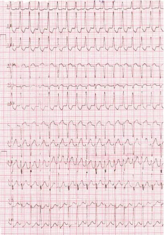
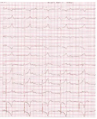
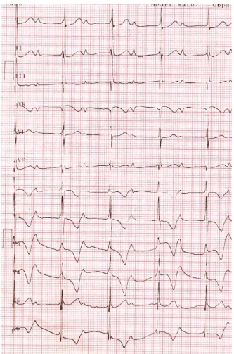
A 65 years old female was admitted with sudden onset of tachycardia as
shown in Figure 1, which revealed a narrow QRS tachycardia and responded to
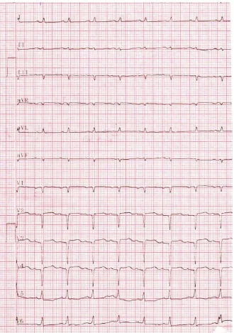
intravenous adenosine as in Figure 2. Echocardiography revealed “egg-shell”
pattern of endocardial calcification in left ventricle (LV) as shown in Figure 3, suggesting left ventricular EMF (Endomyocardial fibrosis) and M-mode LV study revealed hypokinesia of left ventricular posterior wall with moderate LV
dysfunction (EF-41%) as in Figure 4. Patient was treated with anticoagulants,
antiplatelets, nitrates, statins, amiodarone 100 mg daily with azithromycin 500 mg weekly for 6 months.
Case 2.
A 45-year-old male was admitted with sudden onset of chest discomfort and
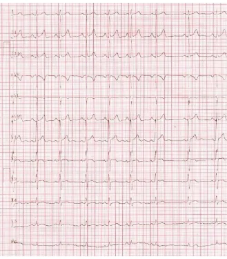
ECG revealed ST-depression in precordial leads as in Figure 5. Blood chemistry
R. Muthiah
[image:3.595.209.540.70.541.2]DOI: 10.4236/crcm.2018.76036 399 Case Reports in Clinical Medicine
Figure 1. Showing narrow QRS tachycardia.
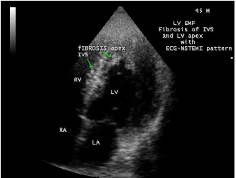
plaque rupture in CAD cases). Patient was treated with heparin, dual antiplatelet agents (aspirin, clopidogrel), nitrates and statins for 5 days and ECG reverted to normal as in Figure 6. Echocardiography revealed calcified fibrous tissue in in-terventricular septum (IVS) and left ventricular apex as in Figure 7. Patient was advised periodic follow up with the continuation of medications.
Case 3.
A 42 years old male was admitted with sudden onset of chest discomfort for 3
hours duration. ECG revealed acute anterior wall infarction as shown in Figure
8. The patient was thrombolysed with streptokinase and further treated with
R. Muthiah
[image:4.595.210.540.68.537.2]DOI: 10.4236/crcm.2018.76036 400 Case Reports in Clinical Medicine
Figure 2. Showing the reverted sinus rhythm with 6 mg intravenous adenosine and a QS pattern in V1-V4, suggesting an old anterior myocardial infarction.
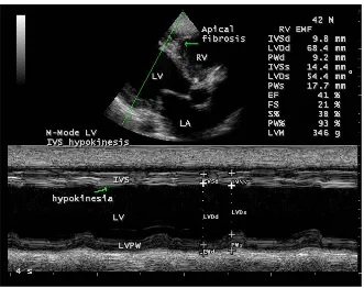
symptom free and echocardiography revealed RV (right ventricle) apical fibrosis
as in Figure 9 and hypokinesia of interventricular septum as shown in Figure
10. The patient was referred for revascularization procedures. Case 4.
A 4-year-old female child was referred for cardiac evaluation since her pulse
rate was <60 bpm. ECG revealed complete heart block as shown in Figure 11
R. Muthiah
[image:5.595.210.541.69.324.2]DOI: 10.4236/crcm.2018.76036 401 Case Reports in Clinical Medicine
Figure 3. Showing the “egg-shell” calcification of Left ventricle (LV) (arrow), suggesting LV EMF (Endomyocardial fibrosis).
Figure 4. The M-mode LV study (green line) showing the rim of IVS (interventricular septum) calcification (upper arrow) and hypokinesia of LV (left ventricle) posterior wall (lower arrow) with LV dysfunction (EF-41%).
[image:5.595.212.536.370.617.2]R. Muthiah
[image:6.595.210.542.69.421.2]DOI: 10.4236/crcm.2018.76036 402 Case Reports in Clinical Medicine
Figure 5. ECG showing ST depression in precordial leads (NSTEMI).
Case 5.
A 15-year-old female was presented with anginal episodes with ECG changes of T-inversion in V1-V3 and echocardiography revealed apical fibrosis in right
ventricle suggesting EMF as in Figure 14. She was on nitrates, antiplatelet
agents, statins and azithromycin 500mg weekly.
Case 6—showing plaque like lesions in IVS (interventricular septum) with
ST-T changes in a 78-year-old female as shown in Figure 15 and advised
ni-trates, antiplatelet agents, statins and azithromycin 500 mg weekly.
Case 7. A 60-year-old male presented with acute myocardial infarction and
echocardiography revealed IVS (interventricular) calcification as in Figure 16
and revasculaized with PCI (percutaneous coronary intervention).
Case 8. 52-year-old male presented with anginal episodes and
echocardiogra-phy revealed finger-like projections of LV myocardium as in Figure 17. The
pa-tient was advised blood-thinning medications (low dose aspirin 75 mg daily), statins and nitrates.
Case 9. A 60-year-old female was admitted with sudden onset of tachycardia and echocardiography revealed “egg-shell” calcification of right ventricular
R. Muthiah
[image:7.595.209.538.67.442.2]DOI: 10.4236/crcm.2018.76036 403 Case Reports in Clinical Medicine
Figure 6. ECG normalizing after 5 days course of heparin therapy.
three times daily and the rhythm was controlled.
Case 10. A 60-year-old male presented with sinus bradycardia revealed endo-cardial plaques in interventricular septum (IVS) as in Figure 19. The patient was treated with amoxycillin and deriphyllin for 10 days and the rhythm was res-tored to normal.
Case 11. A 28-year-old female having infertility for a period of 5 years re-vealed interatrial septal (IAS) calcification suggesting EMF as in Figure 20.
Case 12. A 25-year-old male presented with anginal episodes, having elevated CK-MB and C-reactive protein levels, but the ECG remain normal and echocar-diography revealed RV apical fibrosis as in Figure 21.
Case 13. Showing plaque like lesion in a neonate as in Figure 22, presented
with atrial fibrillation (fast response) and reverted with intravenous adenosine Case 14. Showing a thrombotic lesion in LV apex in a 55-year old male as in
Figure 23.
Case 15. A 66-year-old female presented with RV EMF and rheumatic mitral
valve involvement as shown in Figure 24 and Figure 25 and was advised
R. Muthiah
[image:8.595.208.540.70.320.2] [image:8.595.215.533.352.709.2]DOI: 10.4236/crcm.2018.76036 404 Case Reports in Clinical Medicine
Figure 7. Showing the fibrosis in IVS (interventricular septum) and LV apex (arrows).
R. Muthiah
DOI: 10.4236/crcm.2018.76036 405 Case Reports in Clinical Medicine
[image:9.595.209.537.69.326.2]Figure 9. Showing IVS (interventricular septum) calcification (lower arrow) and RV (right ventricle) apical fibrosis (upper arrow).
Figure 10. M-mode LV study (green line) showing hypokinesia of IVS (interventricular septum) (lower arrow) with RV apical fibrosis (upper arrow).
3. Discussion
3.1. Review of Literature
[image:9.595.209.540.367.629.2]R. Muthiah
[image:10.595.209.543.64.474.2]DOI: 10.4236/crcm.2018.76036 406 Case Reports in Clinical Medicine
Figure 11. ECG showing complete heart block (heart rate 50 bpm) in a 4 –year old female child.
incidence of coronary artery disease among expatriate Indians in Kenya and he believed that the ingestion of animal fat was not an important etiological factor
[7] [8]. Virchow, first proposed an association between infection and IHD
(ischemic heart disease) > 100 years ago. Cardiotropic viruses were first impli-cated in the pathogenesis of CAD in 1968 when experimental Coxsackie B4 virus
infection in mice was shown to produce acute coronary arteritis [9]. Other
in-vestigators suggest a link between Coxsackie B virus and coronary artery disease, following a report of myocardial infarction, occurring in two normolipidemic male patients due to an unknown viral illness [10].
3.2. Etiopathogenesis
R. Muthiah
[image:11.595.210.537.68.318.2]DOI: 10.4236/crcm.2018.76036 407 Case Reports in Clinical Medicine
Figure 12. Showing endocardial plaque-like lesion (apical fibrosis) in the RV (right ven-tricular) apex (arrow) in a 4-year-old female child with complete heart block.
The inflammatory nature of early cellular infiltrate suggest that either infection or autoimmune phenomena with viruses, bacteria and non-viral obligatory intracellular parasites, all being implicated as potential precipitants of the athe-rosclerotic process. Bacterial infection may lead to molecular sequelae that might have effects on the initiation and maintenance of atherosclerotic process. During infection, plasma clotting factors increases, leading to a hypercoagulable state with an increase in procoagulant activity at the level of vascular endothelium and a shift of prostaglandin metabolism towards thrombosis [11][12]. Increased addition of circulating leucocytes to the vascular endothelium also occurs. Of particular interest are the changes in lipid metabolism during acute infection as serum triglycerides and VLDL (very low density lipoproteins) levels increase with levels of LDL (low density lipoprotein)-cholesterol and HDL (high density
lipoproteins) decreasing concomitantly [13] and the endothelium is damaged
R. Muthiah
[image:12.595.208.541.65.568.2]DOI: 10.4236/crcm.2018.76036 408 Case Reports in Clinical Medicine
Figure 13. ECG showing the increase in heart rate (60 bpm) after cefotaxime therapy.
Recent febrile illness has been associated significantly with myocardial infarc-tion [16]. The individuals having seropositive for C. pnemoniae and H. pylori, shown to possess elevated levels of fibrinogen, a risk factor for CAD. C-reactive protein (CRP), an another acute-phase protein, when present at levels approaching the upper limit of normal, is an indirect evidence of CAD [17] and correlate with
poor prognosis in unstable angina [18]. However, high levels of acute-phase
R. Muthiah
[image:13.595.209.539.70.339.2] [image:13.595.209.539.370.643.2]DOI: 10.4236/crcm.2018.76036 409 Case Reports in Clinical Medicine
Figure 14. Showing apical fibrosis in the right ventricle (arrow).
Figure 15. Showing the plaque-like lesion in IVS (interventricular septum) (upper ar-row).
R. Muthiah
[image:14.595.208.539.70.325.2]DOI: 10.4236/crcm.2018.76036 410 Case Reports in Clinical Medicine
[image:14.595.209.540.368.631.2]Figure 16. Showing IVS (interventricular septum) calcification (arrow) in a 60-year-old male.
Figure 17. Showing the finger-like projections of fibrosis (left arrow).
blood vessels, serous cavities and it is immunologically mediated similar to rheumatic process, sometimes coexist in the same individual as in Figure 24 and
R. Muthiah
[image:15.595.209.540.69.320.2]DOI: 10.4236/crcm.2018.76036 411 Case Reports in Clinical Medicine
Figure 18. Showing “egg-shell” calcification of RV (right ventricle) myocardium (arrow).
Figure 19. Showing endocardial plaque like lesion in IVS (interventricular septum) (ar-row).
[image:15.595.208.540.350.615.2]R. Muthiah
[image:16.595.209.541.72.349.2]DOI: 10.4236/crcm.2018.76036 412 Case Reports in Clinical Medicine
[image:16.595.210.539.391.656.2]Figure 20. Showing the calcification of interatrial septum (IAS) (arrow) in a 28-year-old female having infertility.
Figure 21. Showing streaks of apical fibrosis in RV apex (arrow) in a 25-year-old male.
R. Muthiah
[image:17.595.208.540.69.328.2] [image:17.595.208.539.359.629.2]DOI: 10.4236/crcm.2018.76036 413 Case Reports in Clinical Medicine
Figure 22. Showing plaque-like lesion in RV apex (arrow) in a neonate.
Figure 23. Showing a thrombotic lesion at LV apex (arrow) in a 55-year-old male.
R. Muthiah
[image:18.595.208.541.71.332.2]DOI: 10.4236/crcm.2018.76036 414 Case Reports in Clinical Medicine
Figure 24. Showing fibrosis of RV apex (upper arrow) and anterior mitral leaflet (lower arrow).
Figure 25. Showing RV apical fibrosis (upper arrow) with rheumatic mitral stenosis (lower arrow).
fever in endemic areas can be challenging [26]. The active phase with recurrent
flare-up of inflammation due to repeated insults GAS (Group A β hemolytic
[image:18.595.208.541.372.634.2]R. Muthiah
DOI: 10.4236/crcm.2018.76036 415 Case Reports in Clinical Medicine
Table 1. Types of distribution of fibrosis in endomyocardial fibrosis [19][20].
Type 1 fibrosis at the apex only Type 2 fibrosis at apex extends to valvular area Type 3 fibrosis at valvular region only
Type 4 isolated lesions of fibrosis at apex and valvular region Type 5 patchy areas of fibrosis other than the apex and valve
evolving to a chronic phase of cardiac manifestations.
Endocardial thickening is the hallmark of EMF, which corresponds micro-scopically to an increased number and abnormal stimulation of cardiac fibrob-lasts in the subendocardium leading to increased collagen synthesis. Inflamma-tory cell infiltrates composed mainly of lymphocytes are prevalent along the in-terface between the endocardium and myocardium. Myocardial lesions such as interstitial fibrosis and scar formation are prominent in areas adjacent to sub-endocardial fibrosis and altered blood vessels, suggesting that they likely occur in response to ischemic injury caused by microvascular changes and there is a lack of vessels in the outer endocardium, in contrast to rheumatic heart disease.
Endomyocardial fibrosis is a multisystem disorder and it also affects the re-productive system, manifested as loss of secondary sexual characters, testicular atrophy, fibrosis of fimbrial ends of fallopian tube, leading to dysfunctional ute-rine bleeding and infertility as in Case 11, associated with IAS (interatrial sep-tum) calcification. Poor genital hygiene and genital infections play an important role in the causation of EMF in endemic areas. EMF also involve the renalsystem and causes renal failure, occasionally presents with seizures as the initial manife-station with endocardial lesions in cardiac chambers.
3.3. Echocardiographic Features
Echocardiography has become the mainstay of diagnosis of EMF [27]. It has
been used as the screening tool at the community level as the diagnosis of EMF can be confirmed at bedside.
In acute cases of EMF, the endocardial lesions are covered with a soft, spongy,
greyish-green layer of thrombus as shown in Figure 23. Large endocardial
pla-ques are unique echocardiographic feature of EMF as in Figure 12, Figure 14,
Figure 15, Figure 19 and Figure 22. EMF is marked by focal or diffuse areas of endocardial calcification, characterized by a white, smooth, and shiny
endocar-dial surface as shown in Figure 9 and Figure 16 in interventricular septum
(IVS), atrial septum as in Figure 20 and “egg-shell” calcification of myocardium as in Figure 3 and Figure 18. The restricted movement of fibrotic ventricular apex and its obliteration are accompanied by compensatory contractile mechan-ism that results in exaggerated and distinctive motion of the basal portion of the ventricle, the so called “Merlon sign” [28][29].
R. Muthiah
DOI: 10.4236/crcm.2018.76036 416 Case Reports in Clinical Medicine
10. The presence of a linear calcification, distal to the pericardium along the
in-ner border of myocardium as in Figure 21, enhanced density of moderator or
other intraventricular bands suggests EMF and the occurrence of endomyocar-dial calcification spots as in Figure 7 is usually a marker of “burnt-out” disease
[33]. The fibrotic lesions may be >1 cm thick and extend finger-like projections into the myocardium [34] as shown in Figure 17.
3.4. Management
3.4.1. Medical Therapy
It has no specific treatment for EMF and carries poor prognosis since most pa-tients present with advanced heart failure. Medical management consists of symptomatic treatment of heart failure with diuretics, angiotensin-converting enzyme inhibitors offered in combination with aspirin or anticoagulants in view of occurrence of coronary artery disease. The response to medical therapy is generally poor and unproven. When combined with rheumatic fever as evi-denced by ASO (anti-streptolysin O) positivity, penicillin prophylaxis is a po-werful modifier of the disease and causes regression when initiated early [35] in majority of cases.
3.4.2. Investigational Therapy
The fibrosis is a scarring process that over time impacts cardiac structure and function in Endomyocardial fibrosis. Therapies directed at cardiac fibrosis could reduce the progression of the disease. Medications that target the
ren-nin-angiotensin system (RAS), transforming growth factor-β and endothelin
(ET) are in various stages of development [36]. Biomarkers such as propeptides
and telopeptides, released during synthesis and degradation of collagen type 1 and III of extracellular matrix (ECM) [37], the structural component of myocar-dium, are used to identify fibrosis and to assess the efficacy of medications [38].
3.4.3. RAS Inhibitors
The renin-angiotensin (RAS) system plays a central role in fibroblast activation and it is an important target for drug therapy. It is a complex system with two counterbalancing axes. In addition to familiar ACE/Ang II/AT1 axis, an ACE2/Ang-(1-7)/Mas receptor axis has been identified, which has antifibrogenic and antiproliferative effects in heart. Administration of Ang-(1-7) and overex-pression of ACE2 can reduce cardiac fibrosis in animal models [39]. The routine use of RAS inhibiting medications, the ACE inhibitors, angiotensin II receptor blockers (ARBs) and aldosterone antagonists in hypertensive patients have shown to reduce fibrosis in humans.
Vaccines with angiotensin II effects (RAS vaccine) effectively decreased car-diac fibrosis in immunized mice, Ang II signaling was inhibited, and anti-Ang II antibodies increased.
3.4.4. TGF-β Inhibitors
R. Muthiah
DOI: 10.4236/crcm.2018.76036 417 Case Reports in Clinical Medicine
and renal dysfunction associated with these agents could impact its use in clini-cal practice [40].
3.4.5. ET Inhibitors
Bosentan has been demonstrated to inhibit ECM formation, decreased collagen synthesis and increase collagenase suppression [41] in animal models.
3.4.6. HDAC (Histone Deacetylase) Inhibitors
These agents have positive results in reducing cardiac fibrosis as well as Ang II
receptor and TGF-β levels in animal models and promising future therapeutic
targets [42].
3.4.7. Ivabradine
It is an oral medication that provides selective heart rate reduction by inhibiting the f-channels of the sinoatrial node [43]. It effectively reduces the fibrosis, cir-culating Ang II and aldosterone levels in animal models.
3.4.8. Imatinib
Inhibition of the protein, FIPILI-PDGFR alphaP, a constitutively activated tyro-sine kinase by imatinib is a potential therapeutic target for patients with early EMF [44].
3.4.9. Novel Treatment Strategies
1) Stem cell therapy
Transplantation of a variety of stem cells following myocardial infarction has been demonstrated to decrease cardiac fibrosis and cardiac muscle apoptosis
[45]. The rationale of therapy is to improve blood supply to the ischemic areas of heart and to promote cardiac cell regeneration by a direct or paracrine factors by stem cells [46][47].
The stem cells studied in cardiovascular research ranged from bone marrow to adipose tissue to skeletal muscle stem cells. These cells could potentially face re-jection and it is possible to reprogram adult cells and transform these into plu-ripotent cells (similar properties as embryonic stem cells), termed as “induced pluripotent stem cells”, which can be auto-transplanted and therefore would not be rejected.
The bone marrow-derived monoclonal, mesenchymal stem cells are most rea-dily available for transplantation in the body. Following injection of mononuc-lear stem cells in patients with myocardial infarction, there is improvement in LV ejection fraction within months [48], improve exercise capacity, decrease in scar tissue and a reduction in mortality in a 5-year follow up [49] occurs.
recent-R. Muthiah
DOI: 10.4236/crcm.2018.76036 418 Case Reports in Clinical Medicine
ly, there has been an interest to develop and inject multiple stem cells that can communicate with each other termed as “cardiocluster”. These clusters are cocktails of cells that include cardiac progenitor cells, mesenchymal stem cells, endothelial progenitor cells and fibroblasts. They have the potential to promote cardiac cell regeneration in disease states such as CAD (coronary artery disease)
when cell function is reduced [55]. Thus, stem cell therapy continues to be a
promising treatment modality in both acute and chronic CAD. 2) Nanomedicine
Nanotechnology has led to an interesting and promising direction in the treatment of coronary artery disease. HDL (high density lipoprotein) are thought to have a protective role since they are involved in the transportation of cholesterol away from the peripheral tissues. Nanotechnology has been used in the synthesis of a dimyristoyl phosphatidyl choline, which mimics the surface characteristics of HDL by mediating the removal of cholesterol from the peri-pheral tissue and transport it to the liver and this agent showed significant re-duction in plaque volume and cholesterol content in aorta in animal models
[56].
Fumagillin is an antiangiogenic drug that has been shown to promote plaque regression in coronary vasculature, but causes neurocognitive effects at thera-peutic effect in high doses.
Several nanoparticle-based antithrombotic agents such as D-phenylalanyl- L-prolyl-Larginyl-Chloromethyl ketone, perfluorocarbon-core nanoparticle, collagen IV nanoparticles, which improve collagen formation while reducing oxidative stress by mimicking Annexin A1, a glucocorticoid regulatory protein in animal models [57].
Molecular mimicry with genetic predisposition may result in autoimmune damage to endothelial lining of the heart and blood vessels, and treatment with nanobacteria is promising with reversal of calcific deposits within the vascula-ture.
3) During PCI (percutaneous coronary interventions)
R. Muthiah
DOI: 10.4236/crcm.2018.76036 419 Case Reports in Clinical Medicine
Synthetic alternatives such as electrospunnanosized fibrous scaffolds for co-ronary artery bypass grafts have been studied in graft procedures [64][65].
4) Novel oral anticoagulants
This group consists of ximelagatran, dabigatran, rivaroxaban, darexaban and
apixaban [66]. Dabigatran is a competitive inhibitor of thrombin and the other
agents, edoxaban, rivaroxaban and apixaban are inhibitors of clotting factor Xa. Dabigatran reduces the ischemic events at higher doses (110 and 150 mg), which has a bleeding risk and low dose therapy could be used without significant in-crease in bleeding risk [67].
5) Alirocumab
It is a monoclonal antibody produced by recombinant DNA technology, known to block the LDL regulator protein (PCSK9-Proprotein Convertase Sub-tilisin/Kexin Type 9), and reduce the LDL cholesterol to 66-73% when combined with atorvastatin, whereas atorvastatin alone cause reduction by 17% only [68].
6) ARNi (Angiotensin receptor-neprilysin inhibitor)
It is a combination of Sacubitril (neprilysin inhibitor component) and valsar-tan (Angiotensin II receptor antagonist), commonly referred to as LCZ696 or ARNi [69][70] and it is more effective in the treatment of heart failure than the traditional ACE (angiotensin converting enzyme) inhibitors [71][72].
7) Role of antibiotics
Plaque formation in both blood vessels (atherosclerotic/infective) and endo-cardium (infective) is a process of complexity and infection plays an important role in its pathogenesis. Antibiotic treatment should slow its progress in and early eradication of the organism is important to prevent future cardiovascular events. The Chlamydia Pneumoniae, the most studied intracellular pathogen in vascular infections and antibiotics may eliminate the organism from epithelial cells, but difficult to clear it from the circulating monocytes. It is sensitive to macrolides, the azithromycin (approved for sexually transmited Chlamydia Trachomatis infection), roxithromycin, tetracyclines and fluroquinolones (gatif-loxacin) [73]. Azithromycin is readily taken up into atherosclerotic plaques [74], given once weekly in clinical trials since a single dose may require 10 days for elimination and generally well tolerated during long-term prophylaxis, but ga-strointestinal symptoms and superinfection by candidiasis may also occur. In
ACADEMIC trial [75], there was a reduction in markers of inflammation such
as C–reactive protein, TNF-α, IL-1, 6, but antibody titers were unchanged after 6 months of therapy with azithromycin. In ISAR-3 (Intracoronary Stenting And Antibiotic Regimen-3) investigated roxithromycin, an effective anti-chlamydial macrolide for the prevention of restenosis after coronary stent deployment {76] and showed that it was ineffective at lower titers and favoured for patients with high titers (>1:512). These results raise the intriguing possibility that antibiotics might be selectively beneficial in a subgroup of patients with active infection, a more vigorous immune response or both.
R. Muthiah
DOI: 10.4236/crcm.2018.76036 420 Case Reports in Clinical Medicine
known to be effective for the treatment of anaerobic organisms and parasites.
Additional metronidazole had improved outcome in STAMINA trial [77] when
azithromycin is ineffective for Chlamydia Pneumoniae and complete eradication may take 1-year of treatment [78] (long-term therapy) since the organism is re-sistant to antibiotics when engulfed within the monocytes [79].
Leptospirosis (Weil’s disease, first described by Adolf Weil in Heidelberg, Germany in 1886) is a zoonosis, caused by gram negative aerobic spirochete of the genus, leptospira interrogans and it is potentially pathogenic to humans (Leptospira biflexa is not), causing myocarditis and arrhythmic episodes [80], 4 to 30 days after the initial phase (fever with rigors, jaundice, conjunctival suffu-sion, pharyngitis, muscle tenderness and rigidity). It is immunologically me-diated and responds to antibiotics, the ceftriaxone, penicillin, ampicillin, azith-romycin and doxycycline 200 mg/week is recommended in high risk areas.
Previous use of sulfonamides, macrolides, penicillin, cephalosporin had no ef-fect on myocardial infarction (MI) risk and vancomycin may cause coronary ar-tery spasm (Kounis syndrome) [81].
Initiation of antibiotics with cefotaxime or amoxicillin provide better outcome in EMF patients in endemic areas. Once allergic stimulus was removed with small doses of chlorpheniramine maleate (avil) and dexamethasone (4mg), the ECG changes may become normalize at early stages of involvement and arr-hythmic episodes reverse spontaneously or by specific treatment with adenosine, verapmil, amiodarone and cardioversion in resistant cases.
8) Pinocembrin
Pinocembrin is a major flavonoid derived from propolin, plays a role in the treatment of myocardial ischemia and reperfusion injury through its antioxidant effect, reduction of calcium overload, as well as inhibition of inflammation and myocardial cell apoptosis in animal models [82]. Pinocembrin exerts its
antiarr-hythmic effect by increasing the activity of Ca2+-Mg2+-ATPase, thereby
main-taining cardiac channels and upregulating the expression of cardiomyocyte li-gament junction proteins [83]), the Cx43 (a gap junction protein, the main con-nexin of cardiomyocytes), ZO-1 (Claudin-1), Kir 2.1 and suppression of the re-distribution of ZO-1 and Cx43, regulated by GJA1 and KCNJ2 genes to maintain the synchronization of electrical activity of the body and the development of heart [84].
9) Levosimendan
R. Muthiah
DOI: 10.4236/crcm.2018.76036 421 Case Reports in Clinical Medicine
ratio, an increase in cGMP level decrease the increase in cytosolic calcium dur-ing ischemia and protects from arrhythmic episodes [85].
10) Antioxidants
Direct induction of lipid peroxidation has arrhythmogenic effect on the heart. The stress affect the Na+, K+-ATPase activity and accelerates thermodenatura-tion of this enzyme which plays a key role in maintaining the transmembrane potential and electrical stability of the heart. Antioxidants prevent cardiac fibril-lation during acute ischemia and reoxygenation of the heart [86].
11) Surgical therapy
Patients with end-stage EMF may not be suitable for surgery. Surgical man-agement is mainly for relief of complications which are responsible for poor prognosis to medical therapy. Pericardio-peritoneal shunt for massive and re-current pericardial effusion and in fewer cases, a right atrial to pulmonary ar-terial shunting and pericardial stripping may improve the outcome. In most cas-es, a clearly delineated cleavage plane allows for the removal of stiff, fibrotic en-docardium (endocardiectomy) and improves the ventricular function, however, variable rates of recurrence after surgery have been reported [87].
3.5. Outcome
EMF still causes significant morbidity in specific geographical pockets. Un-treated EMF carries very poor prognosis and it represents the second leading cause of pediatric admissions for acquired heart disease after rheumatic heart
disease [88]. The long-term outcome in medical treatment for advanced cases
carries 75% mortality at 2 years [89]. An unknown number of cases evolve
ra-pidly to heart failure and experienced sudden death caused by ventricular arr-hythmias [90].
3.6. Preventive Measures
EMF affects both indigenous and non-indigenous inhabitants, suggests that it is an environmental disease, caused by an infective agent, transmitted to the sus-ceptible individual by a vector which is confined to the tropical and subtropical belts, especially in the hot, humid coastal areas and rain-forest regions. The dis-ease affects the children and young adults in an epidemic fashion in tropical countries and it has remained poorly understood, not appearing in public health agenda of the countries [91].
Investment in research is extremely limited. Improvement in environmental sanitation and hygienic measures may bring this neglected disease under con-trol. Establishment of research stations and health care centers under the direct vision of WHO (World Health Organization) is mandatory in these tropical na-tions.
3.7. Case Analysis
R. Muthiah
[image:26.595.208.537.93.394.2]DOI: 10.4236/crcm.2018.76036 422 Case Reports in Clinical Medicine
Table 2. Sociodemographic and clinical features.
Social factors
Clinical features Age
(years) Sex Rural/urban Economic status
Case 1 65 female rural poor Chest discomfort, palpitations Case 2 45 male urban high Chest discomfort Case 3 42 male rural poor Sudden onset of chest discomfort Case 4 4 female rural poor Bradycardia
Case 5 15 female urban poor Anginal episodes Case 6 78 female rural poor ST-T changes Case 7 60 male rural poor Acute myocardial infarction Case 8 52 male rural poor Anginal episodes Case 9 60 female rural poor Sudden onset of tachycardia Case 10 60 male rural poor Bradycardia Case 11 28 female urban middle class Infertility
Case 12 25 male rural poor Anginal episodes, Elevated CKMB Normal ECG
Case 13 Neonate female rural poor Atrial fibrillation with fast response Case 14 55 male urban middle class Recent anterior wall infarction
Case 15 66 female rural RV EMF with Rheumatic mitral valve disease
The reported cases clearly showing an association between endomyocardial fibrosis and coronary artery disease with arrhythmogenic potentials. Septal cal-cification of interatrial septum (IAS) and interventricular septum (IVS) is a spe-cial feature, more prone to ischemic episodes even before the ECG changes oc-cur. Sudden onset of arrhythmias in children, young adults and older age group herald the lesions of EMF in echocardiography and similarly with infarction ep-isodes.
The patients were advised for sedentary lifestyle recruitment and under peri-odic check up in cardiology clinic.
4. Conclusion
Endomyocardial fibrosis (EMF) is characterized by extensive fibrosis with calci-fication and architectural distortion [92] of the heart. It is the disease of rural belts with a high prevalence in south India and appears to have regional
varia-tions in endemic countries [93] [94]. The changing pattern of coronary artery
disease with characteristic epidemiologica features [95] in endemic areas provide the way to create alternate guidelines in the management of acute coronary syn-drome and arrhythmogenic episodes in future.
References
Co-R. Muthiah
DOI: 10.4236/crcm.2018.76036 423 Case Reports in Clinical Medicine
ronary Artery Disease in Young. World Journal of Cardiology, 8, 728-774. https://doi.org/10.4330/wjc.v8.i12.728
[2] Ford, E.S., Ajani, U.A., Croft, J.B., Critchley, J.A., Labarthe, D.R., Kottke, T.E., Giles, W.H. and Capewell, S. (2007) Explaining the Decrease in U.S. Deaths from Coronary Disease, 1980-2000. New England Journal of Medicine,356, 2388-2398. https://doi.org/10.1056/NEJMsa053935
[3] Shekella, R.B., Shyrock, A.M., Paul, O., et al. (1981) Diet, Serum Cholesterol and
Death from Coronary Artery Disease. The Western Electric Study. New England
Journal of Medicine,304, 75-80.
[4] Shaper, A.G. (1996) Reflections on the Seven Countries Study. Lancet,347, 208. https://doi.org/10.1016/S0140-6736(96)90396-7
[5] Assimes, T.L. and Herrington, D.M. (2018) Genetic Risk Scores in Premature
Co-ronary Artery Disease. Circulation: Genomic and Precision Medicine, 11, e002006.
[6] Dileep Kumar, P. (1997) The Role of Coconut and Coconut Oil in Coronary Heart
Disease in Kerala, South India. Tropical Doctor, 27, 215-217. https://doi.org/10.1177/004947559702700409
[7] Charters, A.D. and Arya, B.P. (1960) Incidence of Ischemic Heart Disease among
Indians in Kenya. Lancet, 1, 288-289.
https://doi.org/10.1016/S0140-6736(60)90222-1
[8] Charters, A.D. (1961) Aetiology of Coronary Heart Disease in Kenya. The Indian
Practitioner,15, 663-667.
[9] Sohal, R.S., Burch, G.E, Chu, K.C., Leiderman, E. and Colcolough, H.L. (1968)
Ul-trastuctural Changes in Cardiac Capillaries of Coxsackie B4 Infected Mice.
Labora-tory Investigation, 19, 399-405.
[10] Burch, G.E. and Shewey, L.L. (1976) Viral Coronary Arteritis and Myocardial
In-farction. American Heart Journal, 92, 11-14. https://doi.org/10.1016/S0002-8703(76)80398-5
[11] Reines, H.D., Halushka, P.V., Cook, J.A., Wise, W.C. and Rambo, W. (1982) Plasma Thromboxane Concentrations Are Raised in Patients Dying with Septic Shock. Lancet, 2, 174-175.https://doi.org/10.1016/S0140-6736(82)91027-3
[12] Nieminen, M.S., Mattila, K. and Valtonen, V. (1993) Infection and Inflammation as Risk Factors for Myocardial Infarction. European Heart Journal, 14, 12-16.
[13] Alvarez, C. and Ramos, A. (1986) Lipids, Lipoproteins, and Apoproteins in Serum
during Infection. Clinical Chemistry, 32, 142-145.
[14] Lopes-Virella, M.F. (1994) Interactions between Bacterial Lipopolysaccharides and
Serum Lipoproteins and Their Possible Role in Coronary Heart Disease. European
Heart Journal, 14, 118-124.
[15] Benditt, E.P. and Benditt, J.M. (1973) Evidence for a Monoclonal Origin of Human Atherosclerotic Plaques. Proceedings of the National Academy of Sciences, USA, 70, 1753-1756.https://doi.org/10.1073/pnas.70.6.1753
[16] Spodick, D.H., Flessas, A.P. and Johnson, M.M. (1984) Association of Acute Respi-ratory Symptoms with Onset of Acute Myocardial Infarction: Prospective Investiga-tion of 150 Consecutive Patients and Matched Control Patients. American Journal of Cardiology, 53, 481-482.https://doi.org/10.1016/0002-9149(84)90016-X [17] Mendall, M.A., Patel, P., Ballam, L., Strachan, D. and Northfield, T.C. (1996)
R. Muthiah
DOI: 10.4236/crcm.2018.76036 424 Case Reports in Clinical Medicine
[18] Liuzzo, G., Biasucci, L.M., Gallimore, J.R., et al. (1994) The Prognostic Value of
C-Reactive Protein and Serum Amyloid A Protein in Severe Unstable Angina. New
England Journal of Medicine,331, 417-424. https://doi.org/10.1056/NEJM199408183310701
[19] Shaper, A.G. Hutt, M.S. and Coles, R.M. (1968) Necropsy Study of Endomyocardial
Fibrosis and Rheumatic Heart Disease in Uganda, 1950-1965. British Heart Journal, 30, 391-401.https://doi.org/10.1136/hrt.30.3.391
[20] Hutt, M.S.R. (1970) Pathology of African Cardiomyopathies. Pathologia et
Micro-biologia,35, 37.https://doi.org/10.1159/000162197
[21] Olsen, E.G.J. and Sekiguchi, M. (1990) Cardiomyopathy Update No, 3: Restrictive
Cardiomyopathy and Arrhythmias. University of Tokyo Press,Tokyo, 1-7.
[22] Nwokolo, C. (1962) The Evolution of Endomyocardial Fibrosis. West African
Med-ical Journal,11, 51-57.
[23] Mocumbi, A.O. and Falase, A.O. (2013) Recent Advances in the Epidemiology,
Di-agnosis and Treatment of Endomyocardial Fibrosis in Africa. Heart, 99, 1481-1487. https://doi.org/10.1136/heartjnl-2012-303193
[24] Davies, J., Spry, C.J., Vijayaraghavan, G. and De Souza, J.A. (1983) A Comparison of the Clinical and Cardiological Features of Endomyocardial Disease in Temperate and Tropical Regions. Postgraduate Medical Journal, 59, 179-185.
https://doi.org/10.1136/pgmj.59.689.179
[25] Tharakan, J.A. (2011) Electrocardiogram in Endomyocardial Fibrosis. Indian
Pac-ing and Electrophysiology Journal, 11, 129-133.
[26] Kennedy, N., Miller, P., Adamczick, C. and Molyneux, E. (2012) Endomyocardial
Fibrosis: The First Report from Malawi. Pediatrics and International Child Health, 32, 86-88.https://doi.org/10.1179/1465328111Y.0000000036
[27] Mocumbi, A.O., Corrilho, C., Sarathchandra, P., Ferreira, M.B., Yacoub, M. and Burke, M. (2011) Echocardiography Accurately Assesses the Pathological Abnor-malities of Chronic Endomyocardial Fibrosis. International Journal of Cardiovas-cular Imaging,27, 955-964.https://doi.org/10.1007/s10554-010-9753-6
[28] Berensztein, C.S., Pinero, G., Marcotegui, M., et al. (2000) Usefulness of
Echocardi-ography and Doppler EchocardiEchocardi-ography in Endomyocardial Fibrosis. Journal of
American Society of Echocardiography,13, 226-230. https://doi.org/10.1016/S0894-7317(00)70008-3
[29] Vijayaraghavan, G., Davies, J., Sadanandan, S., et al. (1983) Echocardiographic Fea-tures of Tropical Endomyocardial Disease in South India. British Heart Journal, 50, 450-459.https://doi.org/10.1136/hrt.50.5.450
[30] Mocumbi, A.O. (2012) Endomyocardial Fibrosis: A Form of Endemic Restrictive
Cardiomyopathy. Global Cardiology Science & Practice, 1, 11. https://doi.org/10.5339/gcsp.2012.11
[31] Okereke, Q.U.J., Chikwendu, V.C., Ihenacho, H.N.C., et al. (1991) Non-Invasive Diagnosis of Endomyocardial Fibrosis in Nigeria Using Two-Dimensional Echocar-diography. Tropical Cardiology, 17, 97-105.
[32] Mady, C., Salemi, V.M.C., Ianni, B.M., et al. (2005) Quantitative Assessment of Left Ventricular Regional Wall Motion in Endomyocardial Fibrosis. Arquivos Brasileiros De Cardiologia,84, 241-244.
[33] Vijayaraghavan, G. and Sivasankaran, S. (2012) Tropical Endomyocardial Fibrosis
in India: A Vanishing Disease! Indian Journal of Medical Research, 136, 729-738.
Fibro-R. Muthiah
DOI: 10.4236/crcm.2018.76036 425 Case Reports in Clinical Medicine
sis.
[35] Steer, A.C. and Carapetes, J.R. (2009) Prevention and Treatment of Rheumatic Heart Disease in the Developing World. Nature Reviews Cardiology, 6, 689-698. https://doi.org/10.1038/nrcardio.2009.162
[36] Leask, A. (2010) Potential Therapeutic Targets for Cardiac Fibrosis: TGF Beta, An-giotensin, Endothelin, CCN, and PDGF, Partners in Fibroblast Activation.
Circula-tion Research, 106, 1675-1680.https://doi.org/10.1161/CIRCRESAHA.110.217737 [37] Fan, D., Takawale, A., Lee, J. and Kassiri, Z. (2012) Cardiac Fibroblasts, Fibrosis and
Extracellular Matrix Remodeling in Heart Disease. Fibrogenesis Tissue Repair, 5, 15.https://doi.org/10.1186/1755-1536-5-15
[38] Roubille, F., Busseuil, D., Merlet, N., et al. (2014) Investigational Drugs Targeting Cardiac Fibrosis. Expert Review of Cardiovascular Therapy,12, 111-125.
https://doi.org/10.1586/14779072.2013.839942
[39] Simoes e Silva, A.C., Silveira, K.D., Ferreira, A.J. and Teixeira, M.M. (2013) ACE,
Angiotensin-(1-7) and Mas Receptor Axis in Inflammation and Fibrosis. British Journal of Pharmacology,169, 477-492.https://doi.org/10.1111/bph.12159 [40] Edgley, A.J., Krum, H. and Kelly, D.J. (2012) Targeting Fibrosis for the Treatment
of Heart Failure: A Role for Transforming Growth Factor-Beta. Cardiovascular
Therapy,30, e30-e40. https://doi.org/10.1111/j.1755-5922.2010.00228.x
[41] Clozel, M. and Salloukh, H. (2005) Role of Endothelin in Fibrosis and Anti-Fibrotic Potential of Bosentan. Annals of Medicine, 37, 2-12.
https://doi.org/10.1080/07853890410018925
[42] Tao, H., Shi, K.H., Yang, J.J., et al. (2014) Histone Deacetylases in Cardiac Fibrosis: Current Perspectives for Therapy. Cell Signaling, 26, 521-527.
https://doi.org/10.1016/j.cellsig.2013.11.037
[43] Amgen (2014) Ivabradine for the Treatment of Chronic Heart Failure, October, 22. [44] Cools, J., DeAngelo, D.J., Gotlib, J., Stover, E.H., Legare, R.D., Cortes, J., Kutok, J.,
Clark, J., Galinsky, I., Griffin, J.D., Cross, N.C., et al. (2003) A Tyrosine Kinase Created by Fusion of the PDGFRA and FIPILI Genes as a Therapeutic Target of
Imatinib in Idiopathic Hypereosinophilic Syndrome, New England Journal of
Med-icine,348, 1201-1214.https://doi.org/10.1056/NEJMoa025217
[45] Elnakish, M.T., Kuppusamy, P. and Khan, M. (2013) Stem Cell Transplantation as a Therapy for Cardiac Fibrosis. Journal of Pathology, 229, 347-354.
https://doi.org/10.1002/path.4111
[46] Kastrup, J. (2010) Gene Therapy and Angiogenesis in Patients with Coronary
Ar-tery Disease. Expert Review of Cardiovascular Therapy, 8, 1127-1138. https://doi.org/10.1586/erc.10.95
[47] Kastrup, J. (2011) Stem Cells Therapy for Cardiovascular Repair in Ischemic Heart
Disease. How to Predict and Secure Optimal Outcome? EPMA (European
Associa-tion for Predictive Preventive & Personalized Medicine) Journal, 2, 107-117. [48] Stamm, C., Westphal, B., Kleine, H., Petzsch, M., Kittner, C., Klinge, H.,
Schumi-chen, C., Nienaber, C., Freund, M. and Steinhoff, P.G. (2003) Autologous Bone-Marrow Stem-Cell Transplantation for Myocardial Regeneration. Lancet,361, 45-46.https://doi.org/10.1016/S0140-6736(03)12110-1
[49] Yousef, M., Schannwell, C.M., Kostering, M., Zeus, T., Brehm, M. and Strauer, B.E. (2009) The BALANCE Study: Clinical Benefit and Long-Term Outcome after Intracoronary Autologous Bone Marrow Cell Transplantation in Patients with
R. Muthiah
DOI: 10.4236/crcm.2018.76036 426 Case Reports in Clinical Medicine
2262-2269.https://doi.org/10.1016/j.jacc.2009.02.051
[50] Beltrami, A.P., Barlucchi, L., Torella, D., Baker, M., Limana, F., Chimenti, S., Kasahara, H., Rota, M., Musso, E., Urbanek, K., et al. (2003) Adult Cardiac Stem Cells Are Multipotent and Support Myocardial Regeneration. Cell,114, 763-776. https://doi.org/10.1016/S0092-8674(03)00687-1
[51] Leri, A. (2009) Human Cardiac Stem Cells: The Heart of a Truth. Circulation,120,
2515-2518.https://doi.org/10.1161/CIRCULATIONAHA.109.911107
[52] Chan, S.S.K., Shueh, Y.Z., Liu, Y.W. and Hsieh, P.C.H. (2009) Hamessing Endo-genous Intra-And Extra-Cardiac Stem Cells for Cardiac Regeneration-Hope or Hipe? Drug Discovery Today. Therapeutic Strategies,6, 127-133.
https://doi.org/10.1016/j.ddstr.2009.06.004
[53] Ferreira-Martins, J., Ogorek, B., Cappetta, D., Matsuda, A., Signore, S., et al. (2012) Cardiomyogenesis in the Developing Heart Is Regulated by c-kit-Positive Cardiac Stem Cells. Circulation Research, 110, 701-715.
https://doi.org/10.1161/CIRCRESAHA.111.259507
[54] Malliaras, K., Makkar, R.R., Smith, R.R., Cheng, K., Wu., E., Bonow, R.O., et al (2014) Intracoronary Cardiosphere-Derived Cells after Myocardial Infarction: Evi-dence of Therapeutic Regeneration in the Final 1-Year Results of the CADUCEUS Trial (Cardiosphere-Derived Autologous Stem CELLS to Reverse Ventricular Dys-function). Journal of American College of Cardiology, 63, 110-122.
https://doi.org/10.1016/j.jacc.2013.08.724
[55] Nguyen, N. and Sussman, M.A. (2015) Rejuvenating the Senescent Heart. Current
Opinion in Cardiology,30, 235-239.
https://doi.org/10.1097/HCO.0000000000000161
[56] Cho, B.H., Park, J.R., Nakamura, M.T., Odinstsov, B.M., Wallig, M.A. and Chung,
B.H. (2010) Synthetic Dimyristoylphosphatidyl Choline Liposomes Assimilating into High-Density Lipoprotein Promote Regression of Atheroslerotic Lesions in Cholesterol-Fed Rabbits. Experimental Biology and Medicine, 235, 1194-1203. https://doi.org/10.1258/ebm.2010.009320
[57] Fredman, G., Kamaly, N., Spolitu, S., Milton, J., Ghorpade, D., et al. (2015) Targeted Nanoparticles Containing the Proresolving Peptide Ac2-26 Protect against Ad-vanced Atherosclerosis in Hypercholesterolemic Mice. Science Translational Medi-cine, 7,275ra20. https://doi.org/10.1126/scitranslmed.aaa1065
[58] Rhee, J.W. and Wu, J.C. (2013) Advances in Nanotechnology for the Management
of Coronary Artery Disease. Trends in Cardiovascular Medicine, 23, 39-45. https://doi.org/10.1016/j.tcm.2012.08.009
[59] Tsukie, N., Nakano, K., Matoba, T., Masuda, S., Iwata, E., Miyagawa, M., Zhao, G., Meng, W., Kishimoto, J., Sunagawa, K., et al. (2013) Pitavastatin-Incorporated Na-noparticle-Eluting Stents Attenuate In-Stent Stenosis Without Delayed Endothelial
Healing Effects in a Porcine Coronary Artery Model. Journal of Atherosclerosis
And Thrombosis, 20, 32-45.https://doi.org/10.5551/jat.13862
[60] Danenberg, H.D., Golomb, G., Groothuis, A., Gao, J., Epstein, H., Swaminathan, R., V., Seifert, P. and Edelman, E.R. (2003) Liposomal Alendronate Inhibits Systemic Innate Immunity and Reduces In-Stent Neointimal Hyperplasia in Rabbits. Circula-tion, 108, 2798-2804.https://doi.org/10.1161/01.CIR.0000097002.69209.CD [61] Margolis, J., McDonald, J., Heuser, R., Klinke, P., Waksman, R., Virmani, R., Desai,
R. Muthiah
DOI: 10.4236/crcm.2018.76036 427 Case Reports in Clinical Medicine
[62] Joo, J.R., Nam, H.Y., Nam, S.H., Baek, I. and Park, J.S. (2009) Thermal Process for Enhancing Mechanical Strength of PLGA Nanoparticle Layers On Coronary Stents, Bulletin of the Korean Chemical Society, 30, 1985-1988.
https://doi.org/10.5012/bkcs.2009.30.9.1985
[63] Yin, R.X., Yang, D.Z. and Wu, J.Z. (2014) Nanoparticle Drug- and Gene-Eluting Stents for the Prevention and Treatment of Coronary Restenosis. Theranostics,4, 175-200.https://doi.org/10.7150/thno.7210
[64] Hashi, C.K., Zhu, Y., Yang, G.Y., Young, W.L., Hsiao, B.S., Wang, K., Chu, B. and Li, S. (2007) Antithrombogenic Property of Bone Marrow Mesenchymal Stem Cells in Nanofibrous Vascular Grafts. Proceedings of the Natlional Academy of Sciences, USA, 104, 11915-11920.https://doi.org/10.1073/pnas.0704581104
[65] Stankus, J.J., Soletti, L., Fujimoto, K., Hong, Y., Vorp, D.A. and Wagner, W.R. (2007) Fabrication of Cell Microintegrated Blood Vessel Constructs through Elec-trohydrodynamic Atomization. Biomaterials,28, 2738-2746.
https://doi.org/10.1016/j.biomaterials.2007.02.012
[66] McMahon, S.R., Brummel Ziedins, K. and Schneider, D.J. (2016) Novel Oral
Anti-coagulants in the Management of Coronary Artery Disease. Coronary Artery
Dis-ease,27, 412-419.https://doi.org/10.1097/MCA.0000000000000387
[67] Oldgren, J., Budaj, A., Granger, C.B., Khder, Y., Roberts, J., et al. (2011) Dabigatran vs Placebo in Patients with Acute Coronary Syndrome on Dual Antiplatelet Thera-py: A Randomized, Double-Blind, Phase II Trial. European Heart Journal, 32, 2781-2789.https://doi.org/10.1093/eurheartj/ehr113
[68] Della Pepa, G., Bozzetto, L., Annuzzi, G. and Rivellese, A.A. (2017) Alirocumab for the Treatment of Hypercholesterolaemia. Expert Review of Clinical Pharmacology, 10, 571-582.https://doi.org/10.1080/17512433.2017.1318063
[69] Pham, A.Q., Patel, Y. and Gallagher, B. (2015) LCZ696 (Angiotensin-Neprilysin In-hibition): The New Kid on the Heart Failure Block? Journal of Pharmacy Practica, 28, 137-145.https://doi.org/10.1177/0897190014568675
[70] Seki, T., Goto, K., Kansui, Y., Ohtsubo, T., Matsumura, K. and Kitazono, T. (2017) Angiotensin II Receptor-Neprilysin Inhibitor Sacubitril/Valsartan Improves Endo-thelial Dysfunction in Spontaneously Hypertensive Rats. Journal of American Heart Association,6, pii: e006617.https://doi.org/10.1161/JAHA.117.006617
[71] Vaduganathan, M. and Desai, A.S. (2016) Angiotensin-Neprilysin Inhibition as a Paradigm for AII? Current Cardiology Reports, 18, 115.
https://doi.org/10.1007/s11886-016-0784-z
[72] McMurray, J.J., Packer, M., Desai, A.S., Gong, J., Lefkowitz, M.P., Rizkala, A.R., et al. (2014) Angiotensin-Neprilysin Inhibition versus Enalapril in Heart Failure. New England Journal of Medicine, 371, 993-1004.
https://doi.org/10.1056/NEJMoa1409077
[73] Chirgwin, K., Roblin, P.M. and Hammerschlag, M.R. (1989) In Vitro
Susceptibili-ties of Chlamydia Pneumoniae (Chlamydia sp. Strain TWAR). Antimicrobial
Agents and Chemotherapy,33, 1634-1635.https://doi.org/10.1128/AAC.33.9.1634 [74] Schneider, C.A., Diedrichs, H., Riedel, K.D., Zimmermann, T. and Hopp, H.W.
(2000) In Vivo Uptake of Azithromycin in Human Coronary Plaques. American
Journal of Cardiology, 86, 789-791, A9.
R. Muthiah
DOI: 10.4236/crcm.2018.76036 428 Case Reports in Clinical Medicine
Infection with Chlamydia (ACADEMIC) Study. Circulation, 99, 1540-1547. https://doi.org/10.1161/01.CIR.99.12.1540
[76] Neumann, F., Kastrati, A., Miethke, T., Pogatsa-Murray, G., Mehilli, J., Valina, C., et al. (2001) Treatment of Chlamydia Pneumoniae Infection with Roxithromycin and Effect on Neointimal Proliferation after Coronary Stent Placement (ISAR-3): A Randomized, Double-Blind, Placebo-Controlled Trial. Lancet,357, 2085-2089. https://doi.org/10.1016/S0140-6736(00)05181-3
[77] Stone, A.F., Mendall, M.A., Kaski, J.C., et al. (2002) Effect of Treatment for Chla-mydia Pneumoniae and Helicobacter Pylori on Markers of Inflammation and Car-diac Events in Patients with Acute Coronary Syndromes: South Thames Trial of
Antibiotics in Myocardial Infarction and Unstable Angina (STAMINA).
Circula-tion,106, 1219-1223.https://doi.org/10.1161/01.CIR.0000027820.66786.CF [78] Grayston, J.T. (2003) Antibiotic Treatment of Atheroslerotic Cardiovascular
Dis-ease. Circulation, 107, 1228-1230.
https://doi.org/10.1161/01.CIR.0000056032.56396.89
[79] Cercek, B., Shah, P.K., Noc, M., et al. (2003) Effect of Short-Term Treatment with Azithromycin on Recurrent Ischemic Events in Patients with Acute Coronary Syn-drome in the Azithromycin in Acute Coronary SynSyn-drome (AZACS) Trial: A Ran-domised Controlled Trial. Lancet, 361, 809-813.
https://doi.org/10.1016/S0140-6736(03)12706-7
[80] Rajiv, C., Manjuran, R.J., Sudhayakumar, N. and Haneef, M. (1996) Cardiovascular Involement in Leptospirosis. Indian Heart Journal, 48, 691-694.
[81] Martinez, E., Sahni, S., Al Cheema, M. and Iftikhar, A. (2018) Vancomycin-Induced
Coronary Artery Spasm: A Case of Kounis Syndrome. British Medical Journal Case
Reports,pii: bcr-2017-222846.https://doi.org/10.1136/bcr-2017-222846
[82] Saad, M.A., Abdel Salam, R.M., Kenawy, S.A. and Attia, A.S. (2015) Pinocembrin
Attenuates Hippocampal Inflammation, Oxidative Perturbations and Apoptosis in a Rat Model of Global Cerebral Ischemia Reperfusion. Pharmacological Reports,67, 115-122.https://doi.org/10.1016/j.pharep.2014.08.014
[83] Zhang, G.-M., Zhao, Z.-Y., Hu, H.-J., Yang, J. and Gu, G.Q. (2018) Effect of Pino-cembrin Pre-Treatment on Expressions of Cx43 Protein and Claudin-1 in
Myocar-dial Ischemia Cardiomyocytes of Arrhythmic Rats. Tropical Journal of
Pharma-ceutical Research,17, 415-422.https://doi.org/10.4314/tjpr.v17i3.5
[84] Seki, A., Nishii, K. and Hagiwara, N. (2015) Gap Junctional Regulation of Pressure Fluid Force and Electrical Fields in the Epigenetics of Cardiac Morphogenesis and Remodeling. Life Sciences, 129, 27-34.https://doi.org/10.1016/j.lfs.2014.10.022 [85] Du Toit, E.F., Muller, C.A., MCCarthy, J. and Opie, L.H. (1999) Levosimendan:
Ef-fects of a Calcium Sensitizer on Function and Arrhythmias and Cyclic Nucleotide Levels during Ischemia/Reperfusion in the Langendorff-Perfused Guinea Pig Heart. The Journal of Pharmacology and Experimental Therapeutics,290, 505-514. [86] Meerson, F.Z., Belkina, L.M., Sazontova, T.G., Saltikova, V.A. and Arkhipenko, Y.V.
(1987) The Role of Lipid Peroxidation in Pathogenesis of Arrhythmias and Preven-tion of Cardiac FibrillaPreven-tion with Antioxidants. Basic Research in Cardiology, 82, 123-138.https://doi.org/10.1007/BF01907060
[87] Moraes, C.R., Buffolo, E., Moraes Neto, F., Rodrigues, J.V., Gomes, C.A., Branco, J.N. and Aguiar, L. (1996) Recurrence of Fibrosis after Endomyocardial Fibrosis Surgery. Arquivos Brasileiros De Cardiologia,67, 297-299. [In Portuguese]
R. Muthiah
DOI: 10.4236/crcm.2018.76036 429 Case Reports in Clinical Medicine
to Surgery and Outcome. Cardiovascular Journal of Africa, 25, 204-211. https://doi.org/10.5830/CVJA-2014-034
[89] D’Arbela, P.G., Mutazindwa, T., Patel, A.K. and Somers, K. (1972) Survival after First Presentation with Endomyocardial Fibrosis. British Heart Journal, 34, 403-407. https://doi.org/10.1136/hrt.34.4.403
[90] Mocumbi, A.O., Yacoub, S. and Yacoub, M.H. (2008) Neglected Tropical Cardi-omyopathies, II: Endomyocardial Fibrosis: Myocardial Disease. Heart, 94, 384-390. https://doi.org/10.1136/hrt.2007.136101
[91] Mocumbi, A.O. and Ferreira, M.B. (2010) Neglected Cardiovascular Diseases in Africa: Challenges and Opportunities. Journal of American College of Cardiology, 55, 680-687.https://doi.org/10.1016/j.jacc.2009.09.041
[92] Lachaud, M., et al. (2018) Tropical Endomyocardial Fibrosis: Perspectives, PubMed. NCBI (National Center for Biotechnology Information).
[93] Kutty, V.R., Abraham, S. and Kartha, C.C. (1996) Geographical Distribution of En-domyocardial Fibrosis in South Kerala. International Journal Epidemiology, 25, 1202.https://doi.org/10.1093/ije/25.6.1202
[94] Radhakumary, C., Kumari, T.V. and Kartha, C.C. (2001) Endomyocardial Fibrosis
Is Associated with Selective Deposition of Type I Collagen. Indian Heart Journal, 53, 486.