1-Bromoadamantane
Richard Betz, Peter Klu¨fers* and Peter Mayer
Ludwig-Maximilians Universita¨t, Department Chemie und Biochemie, Butenandt-strasse 5–13 (Haus D), 81377 Mu¨nchen, Germany
Correspondence e-mail: [email protected]
Received 7 November 2008; accepted 18 November 2008
Key indicators: single-crystal X-ray study;T= 200 K; mean(C–C) = 0.007 A˚;
Rfactor = 0.032;wRfactor = 0.080; data-to-parameter ratio = 16.1.
The molecule of the title compound, C10H15Br, shows
noncrystallographic mirror symmetry. In the crystal structure, no intermolecular interactions with distances less than the sum of the van der Waals radii of the respective atoms are present.
Related literature
For the crystal structure of the thiourea solvate of the compound, see Chaoet al.(2003).
Experimental
Crystal data
C10H15Br Mr= 215.13
a= 10.154 (3) A˚
b= 6.8541 (11) A˚
c= 13.240 (3) A˚ = 90.027 (17)
V= 921.5 (4) A˚3
MoKradiation = 4.40 mm 1
T= 200 (2) K 0.210.160.13 mm
Data collection
Oxford Xcalibur diffractometer Absorption correction: analytical
(de Meulenaer & Tompa, 1965)
Tmin= 0.462,Tmax= 0.614
4563 measured reflections 1629 independent reflections 1313 reflections withI> 2(I)
Rint= 0.054
Refinement
R[F2> 2(F2)] = 0.032
wR(F2) = 0.080
S= 1.02 1629 reflections
101 parameters
H-atom parameters constrained max= 0.77 e A˚ 3
min= 0.38 e A˚ 3
Data collection: CrysAlis CCD (Oxford Diffraction, 2005); cell refinement: CrysAlis RED (Oxford Diffraction, 2005); data reduc-tion:CrysAlis RED; program(s) used to solve structure:SHELXS97
(Sheldrick, 2008); program(s) used to refine structure:SHELXL97
(Sheldrick, 2008); molecular graphics: ORTEP-3 (Farrugia, 1997); software used to prepare material for publication:SHELXL97.
Professor Dr Klapo¨tke is thanked for generous allocation of measurement time on the diffractometer.
Supplementary data and figures for this paper are available from the IUCr electronic archives (Reference: HG2442).
References
Chao, M.-H., Kariuki, B. M., Harris, K. D. M., Collins, S. P. & Laundy, D. (2003).Angew. Chem. Int. Ed.42, 2982–2985.
Farrugia, L. J. (1997).J. Appl. Cryst.30, 565.
Meulenaer, J. de & Tompa, H. (1965).Acta Cryst.19, 1014–1018.
Oxford Diffraction (2005). CrysAlis CCD and CrysAlis RED. Oxford Diffraction Ltd, Abingdon, Oxfordshire, England.
Sheldrick, G. M. (2008).Acta Cryst.A64, 112–122. Structure Reports
Online
supporting information
Acta Cryst. (2009). E65, o101 [doi:10.1107/S1600536808038452]
1-Bromoadamantane
Richard Betz, Peter Kl
ü
fers and Peter Mayer
S1. Comment
The structure of the title compound was elucidated for comparison of the influence of different substituents on the
geometry of the adamantane framework.
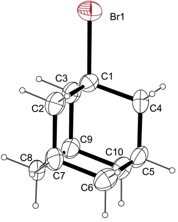
In the molecule the Br atom is bonded to one of the bridgehead positions of the carbocycle (Fig. 1). Bond lengths are
normal.
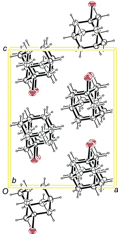
In the crystal structure, only dispersive interactions are present. No intermolecular contacts whose range falls below the
sum of the van der Waals radii of the respective atoms are existent.
A similar structure, the thiourea solvate of the compound, has been described by Chao et al. (2003) but showed disorder
among the 1-bromoadamantane moiety. However, a comparison of both molecules shows good agreement in terms of
bond lengths and angles.
The packing of the compound is shown in Fig. 2.
S2. Experimental
The compound was obtained commercially (ACROS). Crystals suitable for X-ray analysis were obtained upon free
evaporation of a solution of the compound in diethyl ether.
S3. Refinement
Carbon-bound H-atoms were placed in calculated positions (C—H 0.99 Å for methylene groups and C—H 1.00 Å for
bridgehead positions) and were included in the refinement in the riding model approximation, with U(H) set to 1.2Ueq(C).
The crystal measured is refined as a twin with a twin-plane perpendicular to [001] (Ebenenzwilling). The
Figure 1
The molecular structure of the title compound, with atom labels and anisotropic displacement ellipsoids (drawn at 50%
Figure 2
The packing of the title compound, viewed along [010].
1-Bromoadamantane
Crystal data
C10H15Br
Mr = 215.13 Monoclinic, P21/c
Hall symbol: -P 2ybc
a = 10.154 (3) Å
b = 6.8541 (11) Å
c = 13.240 (3) Å
β = 90.027 (17)°
V = 921.5 (4) Å3
F(000) = 440
Dx = 1.551 Mg m−3
Mo Kα radiation, λ = 0.71073 Å Cell parameters from 2345 reflections
θ = 3.9–26.3°
µ = 4.40 mm−1
Oxford Xcalibur diffractometer
Radiation source: fine-focus sealed tube Graphite monochromator
ω scans
Absorption correction: analytical (de Meulenaer & Tompa, 1965)
Tmin = 0.462, Tmax = 0.614
4563 measured reflections 1629 independent reflections 1313 reflections with I > 2σ(I)
Rint = 0.054
θmax = 25.3°, θmin = 3.9°
h = −12→11
k = −8→8
l = −15→14
Refinement
Refinement on F2
Least-squares matrix: full
R[F2 > 2σ(F2)] = 0.032
wR(F2) = 0.080
S = 1.02 1629 reflections 101 parameters 0 restraints
Primary atom site location: structure-invariant direct methods
Secondary atom site location: difference Fourier map
Hydrogen site location: inferred from neighbouring sites
H-atom parameters constrained
w = 1/[σ2(F
o2) + (0.0393P)2]
where P = (Fo2 + 2Fc2)/3
(Δ/σ)max < 0.001
Δρmax = 0.77 e Å−3
Δρmin = −0.38 e Å−3
Fractional atomic coordinates and isotropic or equivalent isotropic displacement parameters (Å2)
x y z Uiso*/Ueq
H102 0.1149 0.1268 0.5163 0.044*
Atomic displacement parameters (Å2)
U11 U22 U33 U12 U13 U23
Br1 0.0506 (3) 0.0472 (3) 0.0339 (2) −0.0028 (3) −0.0007 (3) 0.0092 (2) C1 0.030 (2) 0.0264 (19) 0.0282 (19) −0.0018 (18) 0.003 (4) 0.0013 (18) C2 0.027 (2) 0.040 (3) 0.042 (3) −0.006 (2) 0.007 (2) −0.002 (2) C3 0.031 (3) 0.026 (2) 0.037 (3) 0.000 (2) 0.001 (2) −0.005 (2) C4 0.029 (2) 0.037 (3) 0.033 (2) −0.008 (2) −0.001 (2) −0.009 (2) C5 0.042 (3) 0.023 (3) 0.042 (3) −0.010 (2) 0.000 (3) −0.003 (2) C6 0.050 (4) 0.029 (3) 0.046 (3) 0.001 (3) 0.006 (3) −0.002 (3) C7 0.024 (2) 0.040 (3) 0.048 (4) −0.006 (3) −0.005 (2) 0.002 (3) C8 0.035 (3) 0.036 (3) 0.033 (3) −0.012 (3) −0.009 (2) 0.002 (2) C9 0.036 (3) 0.031 (3) 0.029 (3) −0.007 (2) 0.003 (2) −0.003 (2) C10 0.033 (3) 0.035 (3) 0.043 (3) −0.008 (2) 0.004 (3) 0.001 (2)
Geometric parameters (Å, º)
Br1—C1 2.008 (4) C5—C10 1.538 (8) C1—C3 1.518 (6) C5—H5 1.0000 C1—C4 1.533 (6) C6—C7 1.540 (8) C1—C2 1.533 (6) C6—H61 0.9900 C2—C7 1.545 (7) C6—H62 0.9900 C2—H21 0.9900 C7—C8 1.523 (8) C2—H22 0.9900 C7—H7 1.0000 C3—C9 1.551 (6) C8—C9 1.533 (7) C3—H31 0.9900 C8—H81 0.9900 C3—H32 0.9900 C8—H82 0.9900 C4—C5 1.547 (7) C9—C10 1.522 (7) C4—H41 0.9900 C9—H9 1.0000 C4—H42 0.9900 C10—H101 0.9900 C5—C6 1.517 (8) C10—H102 0.9900
C1—C3—H32 110.1 C7—C8—H82 109.4 C9—C3—H32 110.1 C9—C8—H82 109.4 H31—C3—H32 108.4 H81—C8—H82 108.0 C1—C4—C5 107.1 (4) C10—C9—C8 109.5 (5) C1—C4—H41 110.3 C10—C9—C3 109.3 (4) C5—C4—H41 110.3 C8—C9—C3 108.4 (4) C1—C4—H42 110.3 C10—C9—H9 109.9 C5—C4—H42 110.3 C8—C9—H9 109.9 H41—C4—H42 108.5 C3—C9—H9 109.9 C6—C5—C10 110.5 (5) C9—C10—C5 109.4 (4) C6—C5—C4 109.7 (5) C9—C10—H101 109.8 C10—C5—C4 109.4 (4) C5—C10—H101 109.8 C6—C5—H5 109.1 C9—C10—H102 109.8 C10—C5—H5 109.1 C5—C10—H102 109.8 C4—C5—H5 109.1 H101—C10—H102 108.2